
图 1.
(A) AIE染料Pent-TMP标记细胞质膜(PM)示意图、(B) Pent-TMP和NucRed标记的原代海马神经元三维再现图像、(C) Pent-TMP标记的小鼠大脑红细胞分布的三维再现图像和(D) Pent-TMP标记的斑马鱼鳃细胞质膜图像
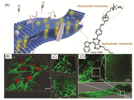
Figure 1.
(A) Schematic diagram of plasma membrane (PM) stained with AIE dye Pent-TMP, (B) the 3D reconstructed image of primary hippocampal neurons stained with Pent-TMP and NucRed, (C) the 3D reconstructed image of the distribution of erythrocytes in mouse brain stained with Pent-TMP, and (D) image of zebrafish gills stained with Pent-TMP
 下载:
下载:

 下载:
下载:


