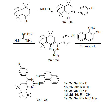
Scheme 1.
Synthesis of compounds 3a~3e
Aggregation-Induced Emission-Active Fluorescent Probe for Zn2+ Based on Isolongifolanone and Its Application in Plant-Cell Imaging
Zhonglong Wang , Jinlai Yang , Yiqin Yang , Xu Xu , Mingxin Li , Yan Zhang , Hua Fang , Haijun Xu , Shifa Wang

In recent years, various organic light-emitting materials and organic fluorescent probes have received increasing researchattention due to their potential applications such as in chemical sensors, biological markers and photoelectronic devices.[1] Many synthesized organic fluorescent molecules demonstrate efficient emission in solid state, and their excellent fluorescent properties are still retained in dilute solution.[2] The thermal stability and luminescent behaviors of these fluorophores have been greatly improved in the reported studies.[3] However, most fluorescent molecules often unavoidably suffer from aggregation-caused quenching (ACQ) effect, where their fluorescence declines dramatically in high concentrated solutions or in aggregated states.[4] As a result, an aggregation-induced emission (AIE) phenomenon has provided a preferable and straightforward pathway for overcoming the ACQ obstacle. The fluorescent molecules with AIE characteristics produce obvious fluorescence enhancement upon aggregation and hence have potential for chemical labeling and biological bio-imaging applications.[5, 6]
Molecular probes based on high selective and sensitive sensing of metal ions have been an effective tool to obtain distinct insight into the concentration and distribution of these enriched metal ions in environmental and biological systems.[7~15] In addition to rendering a remarkable fluorescence signal, a molecular probe, to be successful in detecting and imaging of metal ions in living organisms, should contain some important criteria such as excellent cell membrane permeability and more highly selective for specific metal ions than other metal ions enrich in the living cells. Zn2+ ion has been considered to be the second most abundant transition heavy metal ion in the normal human body with its concentrations varying from nanomolar (nmol·L-1) to approximately millimolar (mmol·L-1).[16] In addition to involving in various biological processes such as gene expression, [17] and neurotransmission, [18, 19] Zn2+ ion is critical for regulating the activities of numerous cellular enzymes.[20] However, because of none of the convenient magnetic signal and typical spectroscopic signature, Zn2+ can not be determined using the conventional spectroscopic techniques. The Zn2+-specific molecular probes are used as efficient methods for detecting intracellular Zn2+. Thus, Zn2+ molecular probes have been designed and developed to meet the distinct demands in recent years.[21~24]
Some natural products with preferable fluorescence properties, such as coumarin, [25] emodin, [26] and eumelanin, [27] have already been reported as examples to extend organic fluorescent material system. In particular, the optical properties of coumarin have been exploited in numerous practical applications, ranging from fluorescent probes to laser dyes.[28~30] The structural organization of eumelanin is successfully investigated by its fluorescence spectra. In addition, emodin has been used as a molecular fluorescent probe for drug-binding proteins.
Longifolene is a natural compound from turpentine and can be used for the synthesis of higher value-added products such as perfume and resin. Isolongifolanone is prepared from natural longifolene in the presence of oxidants, [31] and can be used for synthesizing a series of valuable derivatives such as α, β-unsaturated ketone, pyrazole and pyrimidine. Moreover, these isolongifolanone derivatives exhibit its decent antineoplastic and anophelifuge activities in past studies.[32] However, none of those derivatives synthesized from isolongifolanone have been used as fluorescent molecules. Owing to their great biological compatibility, the isolongifolanone derivatives can be used for bio-imaging in living organisms. Thus, we believe that we can utilize renewable and low toxic isolongifolanone to design some organic fluorescent molecules for optical devices.
Herein, we synthesized several organic fluorescent molecules comprising five electron donating and withdrawing substituents based on cheap and renewable isolongifolanone. The fluorescent properties of these molecules were efficiently controlled by their para-substituted groups on phenyl in solid state and solutions. Moreover, these derivatives with similar structures were selective and sensitive for Zn2+ ion. In addition, we also exploited the application of isolongifolanone-based derivatives in imaging intracellular Zn2+ ion in living plant tissue cells using pollen grains of Althaea rosea.
The synthetic routes of compounds 3a~3e were outlined in Scheme 1. Compounds 1a~1e were synthesized by a aldol condensation reaction of isolongifolanone with aromatic aldehydes. Then compounds 2a~2e were synthesized by the reaction of guanidine hydrochloride with compounds 1a~1e using potassium tert-butylate as a catalyst in tert-butyl alcohol. Finally, compounds 3a~3e were afforded by the reaction of 2-hydroxy-1-naphthaldehyde with compounds 2a~2e. The synthesized compounds 3a~3e were characterized by IR, NMR and HRMS.
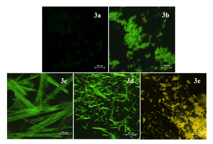
Visual fluorescent performances of solid-state compounds 3a~3e were observed under UV excitation. Compounds 3a~3e in solid state showed obvious fluorescent enhancement under 365 nm UV light. The aggregated morphologies of compounds 3a~3e were further investigated by fluorescence microscopy. The microcosmic structures were presented for displaying enhanced fluorescent variations of compounds 3a~3e using optical microscopy (Figure 1). The derivative 3e provided obvious and fluorescent enhancement with bright yellow emission, and compounds 3b, 3c and 3d emitted kelly lights and had strong fluorescence. Meanwhile, compound 3a emerged weak fluorescence enhancement and emitted a dark kelly light. From the macroscopic and microcosmic fluorescent images, the fluorescent behaviors of compounds 3a~3e were greatly affected by para-substituted groups on the phenyl in solid state.
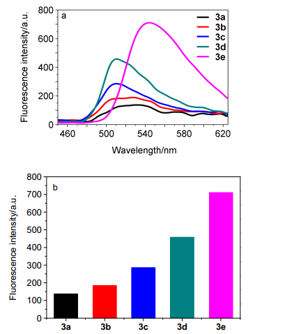
The solid-state fluorescence properties of compounds 3a~3e were also studied to further confirm the fluorescent enhancement of these synthesized derivatives with peculiar KBr pellets (0.5 μmol compound/1 g KBr). As shown in Figures 2a and 2b, compound 3e showed a maximal fluorescence emission peak at 545 nm and the fluorescence intensity of compounds 3b~3d around 510 nm was gradually enhanced, while compound 3a only offered weak fluorescence in solid state. This accorded well with the macroscopic and microcosmic fluorescent images in solid state that compound 3e exhibited the longest and strongest fluorescence emission among these similar derivatives. However, the powerful electron-withdrawing fluorin group of compound 3a could lead to serious fluorescence quenching in solid state. In addition, the fluorescence intensity of compounds 3a~3e was greatly affected by the para-substituted groups on the phenyl. Moreover, we could conclude that the fluorescent properties of these synthesized derivatives were improved by attaching electrondonating groups to the phenyl, whereas electron-with-drawing groups on the phenyl could result in self-quenching of their fluorescence in solid state.
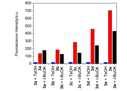
To further study the solid-state fluorescence behaviors of compounds 3a~3e towards to acids and bases, the fluorescence intensity of the mixed KBr pellets (0.5 μmol compound/1 g KBr) with the addition of 50 equiv. of p-toluenesulfonic acid and potassium tert-butoxide were investigated. As shown in Figure 3, the fluorescence intensity of compounds 3a~3e showed the uniform and obvious quenching after grinding with excessive p-toluenesulfonic acid (TsOH). The protonation of nitrogen atoms in pyrimidine groups could result in dramatic fluorescence quenching under strong acid condition. When potassium tert-butoxide was ground with compounds 3a~3e, the fluorescence intensity of compounds 3b~3e exhibited the gentle quenching, which was aroused by the deprotonation of hydroxyl in naphthol moiety under strong alkali condition. In contrast, the slight fluorescence enhancement of compound 3a was presented in grinding process. This may indicate that the fluorescence quenching effect of electron-withdrawing fluorin group seems to be inhibited by the deprotonation of hydroxyl in naphthol moiety. Thus, the strong acid and alkali conditions are adverse to fluorescence emission in solid state.
The test method of solid-state fluorescence spectra, especially those that involved the tabletting process of mixed KBr pellets, may be related to their fluorescence performance. The fluorescence intensity of compounds 3a~3e was investigated with the increasing grinding pressure. The fluorescence intensity of compounds 3a~3e exhibited lower fluorescence quenching from 5 MPa to 25 MPa. Moreover, the fluorescence intensity of compounds 3a~3e suffered from more serious quenching at the range of 25~30 MPa. It could suggest that the undue molecular stack is adverse to solid-state fluorescence emission and lead to fluorescence quenching.
The excellent photo stability is required for the long-term application of fluorescence materials, so we monitored the solid-state fluorescence intensity of compounds 3a~3e during 140 min upon continuous UV-light irradiation at room temperature. The fluorescence intensity of compounds 3a~3e had no significant decrease during the overall test time. The results showed that compounds 3a~3e had good photo stability.
In order to investigate the effect of increasing temperature on fluorescence properties in solid state, the fluorescence intensity of compounds 3a~3e was recorded within a wide window ranging from 10 to 80 ℃. The fluorescence intensity of compounds 3a~3e only showed the slight quenching during the temperature elevation process. Owing to their photo-thermal stability, compounds 3a~3e could be used as the excellent fluorescent materials under the hot conditions.
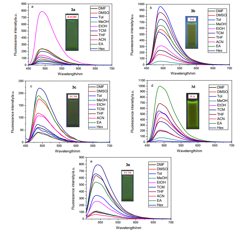
To further study the photochemical properties of the derivates, the fluorescent spectra of compounds 3a~3e (1×10⁻⁵ mol·L-1) were recorded in various solvents such as DMF, DMSO, THF, toluene, methanol, ethanol, trichloromethane, acetonitrile, ethyl acetate, and hexane. In addition, the fluorescent images of compounds 3a~3e were collected in the suitable solvents for realizing their fluorescent emission. As shown in Figure 4a, the fluorescence intensity of compound 3a was greatly affected by solvents, and it showed a better fluorescence intensity in ethanol with a clear green light. While other solvents could induce obvious fluorescence quenching. From Figure 4b, we can conclude that the solvents also offered an significant impact on the fluorescence of compound 3b. Compound 3b provided the strong fluorescence intensity in toluene and emitted a bright green light. However, its fluorescence quenched sharply in polar solvents such as ethanol, DMF, acetonitrile and methanol. The fluorescence spectra of compound 3c is shown in Figure 4c, a green fluorescent solution with the strong fluorescence intensity was obtained when it was dissolved in trichloromethane. Compound 3c exhibited a remarkable and irregular fluorescence decrease in most organic solvents. The better fluorescence intensity of compound 3d occurred in its ethyl acetate solution, and the solution emitted a strong green light (Figure 4d). Furthermore, the fluorescence intensity of compound 3d was closely related to various solvents. As shown in Figure 4e, The compound 3e had a strong fluorescence intensity and a bright green light in trichloromethane. Nevertheless, its fluorescence intensity at 490 nm quenched in polar solvents such as methanol, DMF, acetonitrile and DMSO. It may be that the fast CT process from the dimethylamino donor group to the pyrimidine core leads to quenched-fluorescence of compound 3e in polar solvents.
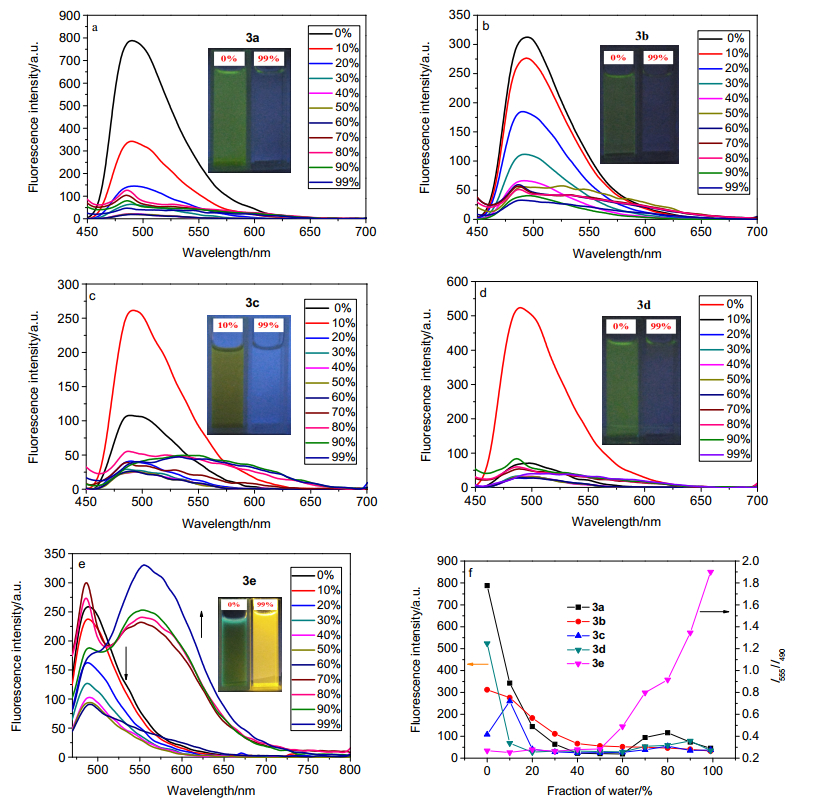
The fluorescence behaviors of compounds 3a~3e (1×10⁻⁵ mol·L-1) were studied in ethanol/water mixtures with different water volume fraction (fw=0~99%) and the results were shown in Figure 5. Compounds 3a, 3b and 3d offered very strong initial fluorescence intensity around 490 nm in pure ethanol solution and exhibited green emission. However, little fluorescence in poor solvent (fw=99%) could be observed with the disappearance of visual DMF, N, N-dimethylformamide; DMSO, dimethylsulfoxide; Tol, toluene; MeOH, methanol; EtOH, ethanol; TCM, trichloromethane; THF, tetrahydrofuran; ACN, acetonitrile; EA, ethyl acetate; Hex, hexane green emissions. As the water volume fraction increased in ethanol/water mixtures, the fluorescence intensity decreased to leveling-off state. Furthermore, the fluorescence intensity of compounds 3a and 3d was found to decrease abruptly upon the addition of more than a 20% water volume fraction, whereas the compound 3b exhibited a continuous fluorescent quenching with the increased aggregate formation. The observed fluorescent features evidently suggested the ACQ characteristics of compounds 3a, 3b and 3d. Interestingly, compound 3c showed faint fluorescence intensity in pure ethanol solution, while the temporary intense fluorescence with visible green emission was observed in 10% water fraction. Nevertheless, the ACQ effect occurred when the water fraction in ethanol/water mixtures was more than 20%. The particular fluorescent properties may indicate that compound 3c could have the short AIE characteristics in a poor solvent (fw=10%) as well as the stable ACQ characteristics in a higher water volume fraction. Interestingly, compound 3e, which had a similar structure as 3a~3d, exhibited its distinctive fluorescent performance in different ethanol/water mixtures. Compound 3e could dissolve well in pure ethanol solution and show green fluorescence. Its fluorescence intensity at 490 nm was found to reduce with increasing water volume fraction. However, a bright yellow emission was observed in poor solvent (fw=99%), which was ascribed to its superb AIE characteristics. Its fluorescence emission at 490 nm was red-shifted to the longer wavelength (550 nm) and the fluorescence intensity ratio (I550/I490) was enhanced when the water volume fraction in ethanol/water mixtures was more than 50%. Its aggregate formation was triggered by gradual increasing water volume fraction. Owing to its electron-donating ability of the para-substituted dimethylamino group on phenyl, compound 3e exhibits aggregation-induced emission (AIE) characteristics.
For the above fluorescence property studies in ethanol/water mixtures with different water volume fractions, compounds 3a~3e for sensing of Zn2+ were investigated in H2O/EtOH (V:V=99:1), H2O/EtOH (V:V=99:1), H2O/EtOH (V:V=60:40), H2O/EtOH (V:V=99:1) and H2O/EtOH (V:V=30:70), respectively. With these pale background fluorescence in trial environments, we could better evaluate the detection performances of compounds 3a~3e towards Zn2+.
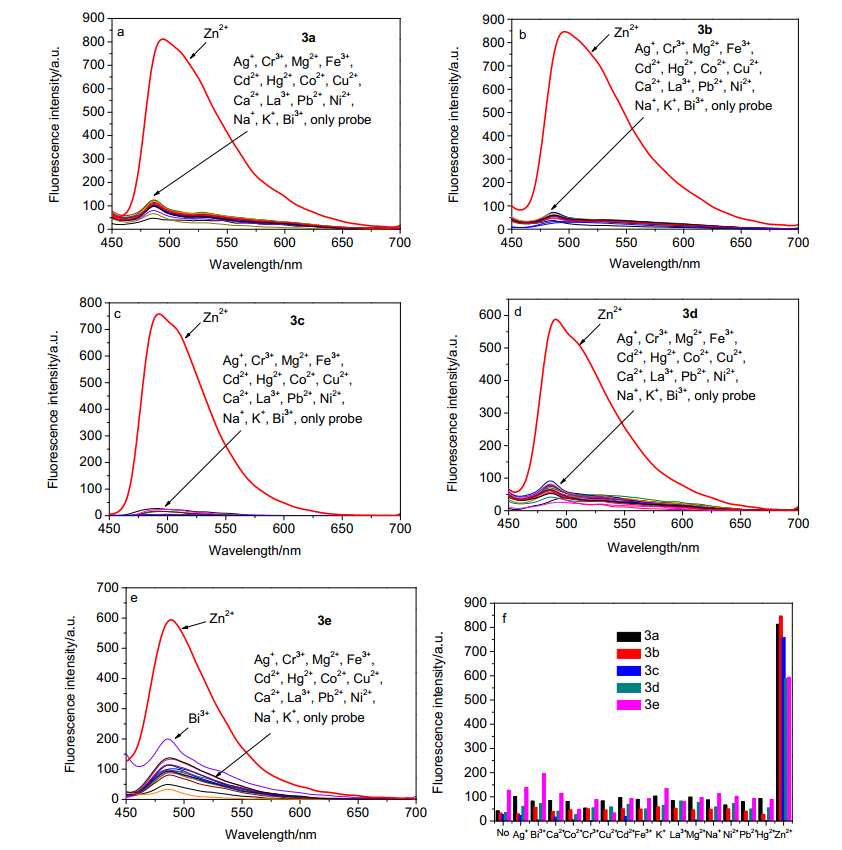
To study the optical selectivity towards various metal ions, the fluorescence spectra of compounds 3a~3e (1×10⁻⁵ mol·L-1) were investigated with different metal ions (1×10⁻⁴ mol·L-1) such as Ag+, Cr3+, Mg2+, Fe3+, Hg2+, Co2+, Cu2+, Ca2+, La3+, Pb2+, Ni2+, Na+, K+, Bi3+ and Zn2+ in corresponding ethanol/water solutions. As shown in Figure 6, other metal ions only caused negligible fluorescence changes while Zn2+ could induced dramatic fluorescence enhancement. Therefore, the significant enhanced fluorescence signals upon addition of Zn2+ indicate that compounds 3a~3e could be served as the specific Zn2+ probes in biochemical milieu.
To further investigate the optical sensitivity towards Zn2+, the fluorescence titration spectra of compounds 3a~3e (1×10⁻⁵ mol·L-1) were evaluated with gradual addition of Zn2+ in corresponding ethanol/water solutions. The fluorescence intensity of compounds 3a~3e at 495 nm was gradually enhanced with increasing concentrations of Zn2+ ion (0~1×10⁻⁴ mol·L-1). Furthermore, the addition of Zn2+ could lead to a bright green emission under 365 nm UV light. The linearity relationships of the fluorescence intensity at 495 nm versus increasing Zn2+ concentrations were also plotted to confirm efficacy of the probes in the detection of Zn2+ ion in aqueous solutions, respectively. The detection limit (LOD) is obtained using 3σbi/m, [33, 34] where σbi is the standard deviation of the blank samples and m stands for the slope of the linear calibration plot. The detection limits of compounds 3a~3e were founded to be as low as 1.07×10⁻⁸, 1.11×10⁻⁸, 1.21× 10⁻⁸, 2.14×10⁻⁸ and 2.09×10⁻⁸ mol·L-1, respectively.
The wide pH range for specific detection of ions is required for practical applications of probes, the fluorescence intensity of compounds 3a~3e (1×10⁻⁵ mol·L-1) was investigated in the absence and presence of Zn2+ ion (1×10⁻⁴ mol·L-1) in the pH range of 3~13 in corresponding ethanol-water solutions. The free compounds 3a~3e remained little fluorescence intensity over the tested pH range and only showed tiny fluorescence enhancement in the strong acidic and basic pH conditions for the detection of Zn2+. Moreover, the fluorescence intensity of compounds 3a~3e was enhanced dramatically in the presence of 10 equiv. of Zn2+ in a broad pH range of 4~11. Therefore, compounds 3a~3e could be used as the practical probes to detect Zn2+ under physiological pH conditions.
In order to investigate the coordinate effect of compounds 3a~3e with Zn2+, compound 3c has been used as an example to carry out a Job plot experiment in an ethanol-water (V/V=4/6) solution. The coordinate ratio between compound 3c and Zn2+ was found to be 1:1. The HRMS spectrum of compound 3c coordinating with Zn2+ showed the peak at m/z 589.3575, which could be interpreted as [Compound 3c+Na+Zn2+]2+.
The reversibility nature of compound 3c was evaluated by the alternating titration of EDTA into the coordinate system (3c+Zn2+), and the results showed that the fluorescence enhancement process of compound 3c induced by Zn2+ was reversible upon addition of EDTA.
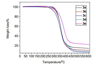
The thermal stabilities of compounds 3a~3e were also evaluated using TGA. As shown in Figure 7, compounds 3a~3e demonstrated great thermal stabilities. In addition, the 10% weight losses of compounds 3a~3e were obtained at the temperature points approaching to 321.9, 316.0, 300.5, 318.9 and 328.4 ℃. The excellent thermal stabilities enable compounds 3a~3e to be used as valuable fluorescence materials.
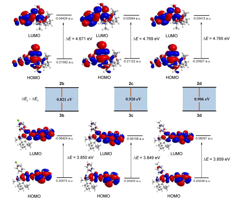
The solid-state pyrimidine derivatives 2b~2d exhibit the weak fluorescence, while their Schiff based derivates 3b~3d manifest the dramatic fluorescence enhancement in solid state. To further understand the improvements of their photophysical properties, the density functional theory (DFT) were carried out using Gaussian 09 program.[35, 36] The frontier molecular orbitals of compounds 2b~2d and 3b~3d are shown in Figure 8. The electron densities of the highest occupied molecular orbital (HOMOs) of 2b~2d are spread over the whole molecules except partial isolongifolanone groups, whereas the lowest unoccupied molecular orbitals (LUMOs) are mainly localized on the benzene and pyrimidine units. The HOMOs of compounds 3b~3d consist of the isolongifolanone parts and the naphthyl and pyrimidine groups, but their LUMOs mainly reside on the naphthyl and the pyrimidine moieties. In addition, the HOMO-LUMO energy gaps (ΔE) decrease from compounds 2b~2d to compounds 3b~3d, indicating that compounds 3b~3d are easier to be stimulated. Thus, the uniform lowering of the energy gaps (ΔE) is the intrinsic factor for the visual fluorescence enhancement of compounds 3b~3d in solid state.
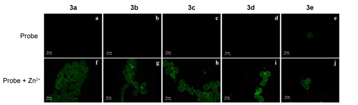
Zinc is regard as one of the most essential trace elements for biological species, which is closely related to the growth and development and metabolism of plants. To further examine the potential of these probes in imaging Zn2+ ion inside living matrices, we carried out the experiments in living plant. Althaea rosea, a common ornamental plant of the Malvaceae family was used in this experiment for detecting Zn2+ in pollen grains with the help of compounds 3a~3c. Pollen grains were firstly obtained from freshly collected mature stamens of Althaea rosea. After crushing the mature stamens a sterile Petri plate and then suspending them in distilled water, the unused debrises were carefully removed by a thin layer of perforated plastic film. The pollen grains were suspended and stirred in the solutions of compounds 3a~3e (5×10⁻⁵ mol·L-1) for 1 h, respectively. Then the suspending pollen grains were removed by repeated filtering and washing for reducing background fluorescence. After that, the pollen grains were incubated with Zn2+ water solution (2×10⁻⁴ mol·L-1) for another 1 h. As shown in Figure 9, nearly no background fluorescence was observed in the pollen grains which were incubated with compounds 3a~3d, whereas pollen grains showed decent green fluorescence when treates with compound 3e. Furthermore, the fluorescence brightness of the images was significant enhanced with the addition of Zn2+ ion under a fluorescence microscope. The results indicate that compounds 3a~3e could be used for labeling Zn2+ enrichment of plant tissues, as well as potential imaging of intracellular Zn2+ in other living organisms.
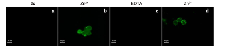
To further study the potential of these probes for labeling Zn2+ in living plant, compound 3c was used as an example to evaluate its reversibility nature in fluorescence imaging (Figure 10). With the addition of Zn2+ (2×10⁻⁴ mol·L-1), the enhanced fluorescence could be observed under a fluorescence microscope. However, the green fluorescence then disappeared from view in the presence of EDTA (2×10⁻⁴ mol·L-1). After reintroducing Zn2+ (2×10⁻⁴ mol·L-1) into the system, the bright fluorescence of the images began to recover. The results showed that the fluorescence imaging of compound 3c towards Zn2+ was reversible in living plant.
In summary, a series of simple and versatile fluorescent molecules 3a~3e based on renewable isolongifolanone were successfully synthesized. Compounds 3a~3e feature the excellent performances in solid and solutions states. The solid-state florescence intensity of compounds 3a~3e ground with excessive amount of acid and base quenches in general. In addition to good thermal stabilities, compounds 3a~3e also show the stable fluorescence intensity in solid state under long term UV irradiation, increasing operated pressure and elevated heating temperature. Compounds 3a~3d exhibit conventional aggregation-caused quenching (ACQ) characteristics. Interestingly, compound 3e, which has para-substituted dimethylamino group, displays obvious aggregation-induced emission (AIE) characteristics and results in a visible fluorescent transition from light green to bright yellow. The structural difference of 3a~3e indicates that the electron-donating ability of para-substituted groups on phenyl plays a decisive role for controlling AIE versus ACQ properties. Moreover, compounds 3a~3e prove to be highly selective and sensitive in detection of Zn2+ ion with low detection limits. The detection of compounds 3a~3e towards Zn2+ is well performed in a wide pH range (4~11) in aqueous solutions. In addition, compounds 3a~3d can used as good contrast agents for imaging Zn2+ ion in living organism. Furthermore, the synthesized fluorescence materials exploit the utilization of turpentine and provide a possible route for deep processing of forest resources.
13C NMR and 1H NMR spectra were recorded in CDCl3 and DMSO-d6 solutions on a Bruker AV 400MZ/600MZ spectrometer. Fluorescence spectra were recorded on a Perkin Elmer LS 55 fluorescence spectrophotometer. Mass spectra and infrared spectra were recorded on an America Agilent 5975c mass spectrometer and a Nicolet 380 FTIR infrared spectrometer, respectively. The purity and melting points were measured on an America Agilent 7890A gas chromatograph and an X-6 microscopic melting point apparatus, respectively. The pH values were given on a Model PHS-3C pH meter. The fluorescence images were photographed by an inverted fluorescence imaging system (LX51, OLYMPUS).
All the organic solvents and reagents were purchased from commercial sources and used without further purification. Deionized water and analytical grade solvents were used throughout the experiment. The ethanol-water solutions of different pH were prepared using HCl and NaOH solutions at the concentration of 1 mol·L-1 for pH slight adjustment. The salts used in the stock solutions of various metal ions were AgNO3, Fe(NO3)3·9H2O, CuCl2·H2O, FeCl2, KCl, ZnCl2, NaCl, Co(NO3)2·6H2O, HgSO4, CaCl2, CrCl3·6H2O, MgSO4, Pb(NO3)2, respectively.
Isolongifolanone (8 mmol), tert-butyl alcohol (30 mL), potassium tert-butoxide (5 mmol) and aromatic aldehyde (10 mmol) were successively added into a 50 mL of dried three-necked flask equipped with a stirrer, condenser and thermometer. The mixture was stirred and refluxed for 2 h until the conversion ratio of isolongifolanone exceeded 95% (monitored by GC). The reacted solution was evaporated under vacuum and extracted three times with 20 mL of ethyl acetate. The merged organic layers were then washed to neutrality with saturated salt water, dried with sodium sulfate, filtered, evaporated to afford a crude product, and then recrystallized to obtain the compounds 1a~1e with ethanol in a refrigerator.
7-(4'-Fluorobenzylidene)-isolongifolanone (1a): White crystal, 92.4% yield. m.p. 139.7~140.3 ℃; 1H NMR (600 MHz, CDCl3) δ: 0.85 (s, 3H), 0.87 (s, 3H), 1.07 (s, 3H), 1.10~1.13 (m, 1H), 1.24 (s, 3H), 1.31 (d, J=10.0 Hz, 1H), 1.48~1.55 (m, 1H), 1.63~1.68 (m, 1H), 1.78~1.81 (m, 2H), 1.83~1.87 (m, 1H), 1.99 (d, J=1.9 Hz, 1H), 2.56~2.59 (m, 1H), 2.81~2.84 (m, 1H), 7.08~7.11 (m, 2H), 7.46~7.48 (m, 3H); 13C NMR (150 MHz, CDCl3) δ: 24.14, 24.68, 25.51, 25.74, 28.37, 30.28, 31.71, 37.61, 41.65, 44.81, 48.09, 55.54, 62.94, 115.45, 115.59, 132.47, 132.53, 135.00, 135.38, 161.78, 163.44, 202.68; EIMS m/z (%): 326 (M+, 84), 311 (23), 297 (17), 283 (100), 269 (35), 257 (58), 215 (22), 161 (13), 146 (15), 134 (82), 109 (47), 91 (25), 55 (28); FT-IR (KBr) ν: 2954, 1669, 1597, 1505, 1463, 833 cm-1. Anal. calcd for C22H27FO: C 80.94, H 8.34, found C 81.05, H 8.21.
7-(4'-Chlorobenzylidene)-isolongifolanone (1b): Yellow crystal, 93.5% yield. m.p. 158.4~159.1 ℃; 1H NMR (600 MHz, CDCl3) δ: 0.85 (s, 3H), 0.87 (s, 3H), 1.06(s, 3H), 1.08~1.13 (m, 1H), 1.24 (s, 3H), 1.31~1.32 (m, 1H), 1.49~1.55 (m, 1H), 1.63~1.68 (m, 1H), 1.78~1.81 (m, 2H), 1.83~1.87 (m, 1H), 1.99 (d, J=2.0 Hz, 1H), 2.55~2.58 (m, 1H), 2.81~2.84 (m, 1H), 7.37 (d, J=8.6 Hz, 2H), 7.41 (d, J=8.6 Hz, 2H), 7.47 (s, 1H); 13C NMR (150 MHz, CDCl3) δ: 24.13, 24.67, 25.53, 25.72, 28.36, 30.28, 31.73, 37.62, 41.65, 44.83, 48.08, 55.54, 62.95, 128.65, 131.79, 134.36, 134.48, 135.19, 135.83, 202.61; EIMS m/z (%): 342 (M+, 90), 327 (23), 299 (100), 285 (35), 273 (59), 231 (20), 150 (56), 115 (60), 91 (27), 55 (30); FT-IR (KBr) ν: 2950, 2895, 2869, 1670, 1595, 1487, 1464, 825 cm-1. Anal. calcd for C22H27ClO: C 77.06, H 7.94, found C 77.12, H 7.97.
7-Benzylidene-isolongifolanone (1c): Yellow crystal, 88.1% yield. m.p. 94.1~95.0 ℃; 1H NMR (600 MHz, CDCl3) δ: 0.86 (s, 3H), 0.88 (s, 3H), 1.06 (s, 3H), 1.09~1.13 (m, 1H), 1.24 (s, 3H), 1.31 (d, J=9.9 Hz, 1H), 1.48~1.55 (m, 1H), 1.63~1.67 (m, 1H), 1.78 (d, J=4.2 Hz, 1H), 1.80~1.87 (m, 2H), 1.99 (d, J=1.9 Hz, 1H), 2.61~2.64 (m, 1H), 2.85~2.88 (m, 1H), 7.32~7.35 (m, 1H), 7.39~7.42 (m, 2H), 7.49 (s, J=7.5 Hz, 2H), 7.53 (s, 1H); 13C NMR (150 MHz, CDCl3) δ: 24.12, 24.69, 25.52, 25.73, 28.38, 30.29, 31.71, 37.62, 41.68, 44.80, 48.10, 55.59, 62.98, 128.40, 128.59, 130.63, 135.39, 135.95, 136.58, 202.82; EIMS m/z (%): 308 (M+, 100), 293 (17), 279 (4), 265 (64), 251 (21), 197 (13), 128 (10), 116 (45), 91 (35), 77 (14), 55 (15); FT-IR (KBr) ν: 2955, 2895, 2873, 1668, 1591, 1463, 1444, 755, 692 cm-1. Anal. calcd for C22H28O: C 85.66, H 9.15, found C 85.39, H 9.21.
7-(4'-Methylbenzylidene)-isolongifolanone (1d): Yellow crystal, 92.3% yield. m.p. 120.4~121.2 ℃; 1H NMR (600 MHz, CDCl3) δ: 0.85 (s, 3H), 0.88 (s, 3H), 1.06 (s, 3H), 1.09~1.13 (m, 1H), 1.24 (s, 3H), 1.30 (d, J=9.9 Hz, 1H), 1.48~1.54 (m, 1H), 1.63~1.65 (m, 1H), 1.77 (d, J=4.1 Hz, 1H), 1.80~1.87 (m, 2H), 1.98 (d, J=1.9 Hz, 1H), 2.38(s, 3H, Ar-CH3), 2.61~2.64 (m, 1H), 2.83~2.87 (m, 1H), 7.21 (d, J=8.0 Hz, 2H), 7.40 (d, J=8.0 Hz, 2H), 7.50 (s, 1H); 13C NMR (150 MHz, CDCl3) δ: 21.44, 24.13, 24.70, 25.51, 25.75, 28.40, 30.27, 31.69, 37.60, 41.81, 44.76, 48.11, 55.57, 62.96, 129.17, 130.74, 133.14, 134.58, 136.64, 138.86, 202.89; EIMS m/z (%): 322 (M+, 59), 307 (100), 279 (37), 253 (40), 240 (14), 211 (19), 130 (57), 115 (46), 105 (48), 91 (31), 79 (20), 55 (24); FT-IR (KBr) ν: 2953, 2895, 2872, 1669, 1591, 1508, 1466, 816 cm-1. Anal. calcd for C23H30O: C 85.66, H 9.38, found C 85.79, H 9.41.
7-(4'-(Dimethylamino)benzylidene)-isolongifolanone (1e): Orange crystal, 90.4% yield. m.p. 119.5~120.2 ℃; 1H NMR (600 MHz, CDCl3) δ: 0.86 (s, 3H), 0.87 (s, 3H), 1.07 (s, 3H), 1.09~1.12 (m, 1H), 1.24 (s, 3H), 1.28~1.30 (m, 1H), 1.47~1.53 (m, 1H), 1.62~1.67 (m, 1H), 1.75~1.76 (m, 1H), 1.79~1.86 (m, 2H), 1.96 (d, J=1.9 Hz, 1H), 2.63 (d, J=16.7 Hz, 1H), 2.83~2.86 (m, 1H), 3.03 (s, 6H, ArN(CH3)2), 6.74 (s, 2H), 7.46~7.49 (m, 3H); 13C NMR (150 MHz, CDCl3) δ: 24.22, 24.75, 25.49, 25.84, 28.47, 30.23, 31.63, 37.55, 40.17, 42.29, 44.62, 48.13, 55.52, 62.86, 111.65, 123.92, 130.94, 132.79, 137.33, 150.42, 202.63; EIMS m/z (%): 351 (M+, 100), 336 (5), 282 (10), 269 (22), 240 (4), 158 (28), 134 (28), 121 (11), 91 (9), 55 (10); FT-IR (KBr) ν: 2960, 2923, 2891, 2872, 1656, 1606, 1565, 1522, 1466, 1440, 822 cm-1. Anal. calcd for C24H33NO: C 82.00, H 9.46, N 3.98, found C 80.97, H 9.52, N 3.82.
7-Arylidene-isolongifolanone 1a~1e (5 mmol), guanidine hydrochloride (20 mmol), tert-butyl alcohol (60 mL) and tert-butoxide (25 mmol) were successively added into a 100 mL of dried three-necked flask equipped with a stirrer, condenser and thermometer. The mixture was stirred and refluxed for 18 h until the conversion ratio of 7-arylidene-isolongifolanone was over 95% (monitored by GC). The reacted mixture was evaporated under vacuum and extracted three times with ethyl acetate, and the combined organic layers were then washed to neutrality with saturated brines, dried with sodium sulfate, filtered, evaporated to afford a viscous liquid, and then recrystallized to obtain compounds 2a~2e with ethanol and ethyl acetate in a refrigerator.
4-(4'-Fluorophenyl)-6, 6, 10, 10-tetramethyl-5, 7, 8, 9, 10, 10a-hexahydro-6H-6a, 9-methanobenzo[h]quinazolin-2-amine (2a): Transparent crystal, 86.5% yield. m.p. 228.7~229.4 ℃; 1H NMR (600 MHz, DMSO-d6) δ: 0.59 (s, 3H), 0.75 (s, 3H), 0.94 (s, 3H), 1.09~1.10 (m, 1H), 1.19 (d, J=9.5 Hz, 1H), 1.32 (s, 3H), 1.46~1.51 (m, 1H), 1.58~1.64 (m, 1H), 1.71 (d, J=3.0 Hz, 1H), 1.78 (d, J=9.7 Hz, 1H), 1.82~1.86 (m, 1H), 2.01 (d, J=15.7 Hz, 1H), 2.19 (s, 1H), 2.80 (d, J=15.7 Hz, 1H), 6.15 (s, 2H, NH2), 7.25~7.29 (m, 2H), 7.60~7.62 (m, 2H); 13C NMR (150 MHz, CDCl3) δ: 22.93, 24.80, 25.45, 25.88, 28.38, 30.15, 32.72, 37.28, 39.58, 44.42, 48.07, 55.34, 58.01, 115.17, 116.11, 130.87, 134.78, 160.28, 162.27, 163.92, 165.13, 170.02; EIMS m/z (%): 365 (M+, 100), 364 (18), 350 (18), 336 (32), 310 (11), 283 (97), 266 (11), 242 (15), 216 (5), 198 (2), 159 (3), 115 (3), 91 (5), 55 (6); FT-IR (KBr) ν: 3486, 3272, 3149, 2957, 2872, 1616, 1554, 1511, 1454, 1378, 1226, 844 cm-1; HRMS calcd for C23H28FN3 [M+H]+366.2339, found 366.2346.
4-(4'-Chlorophenyl)-6, 6, 10, 10-tetramethyl-5, 7, 8, 9, 10, 10a-hexahydro-6H-6a, 9-methanobenzo[h]quinazolin-2-amine (2b): White crystal, 87.8% yield. m.p. 235.6~236.2 ℃; 1H NMR (600 MHz, DMSO-d6) δ: 0.59 (s, 3H), 0.75 (s, 3H), 0.94 (s, 3H), 1.09~1.10 (m, 1H), 1.18 (d, J=9.8 Hz, 1H), 1.32 (s, 3H), 1.46~1.52 (m, 1H), 1.58~1.64 (m, 1H), 1.71 (d, J=3.7 Hz, 1H), 1.78 (d, J=9.7 Hz, 1H), 1.82~1.86 (m, 1H), 2.00 (d, J=15.7 Hz, 1H), 2.19 (s, 1H), 2.79 (d, J=15.7 Hz, 1H), 6.18 (s, 2H, NH2), 7.51 (d, J=8.5 Hz, 2H), 7.58 (d, J=8.5 Hz, 2H); 13C NMR (150 MHz, CDCl3) δ: 22.92, 24.78, 25.43, 25.88, 28.38, 30.11, 32.70, 37.25, 39.47, 44.42, 48.01, 55.29, 57.94, 116.14, 128.40, 130.34, 134.93, 137.05, 160.20, 164.93, 170.09; EIMS m/z (%): 382 (M+, 31), 381 (97), 380 (18), 366 (19), 352 (33), 326 (13), 299 (100), 282 (10), 258 (17), 232 (5), 207 (5), 179 (2), 147 (6), 115 (5), 91 (6), 55 (8); FT-IR (KBr) ν: 3505, 3317, 3195, 2964, 2869, 1622, 1549, 1491, 1456, 1369, 1207, 1087, 834 cm-1; HRMS calcd for C23H28ClN3 [M+H]+ 382.2044, found 382.2017.
4-Phenyl-6, 6, 10, 10-tetramethyl-5, 7, 8, 9, 10, 10a-hexahydro-6H-6a, 9-methanobenzo[h]quinazolin-2-amine (2c): White crystal, 85.9% yield. m.p. 241.2~241.9 ℃; 1H NMR (600 MHz, DMSO-d6) δ: 0.59 (s, 3H), 0.76 (s, 3H), 0.93 (s, 3H), 1.08~1.10 (m, 1H), 1.19 (d, J=9.7 Hz, 1H), 1.33 (s, 3H), 1.45~1.51 (m, 1H), 1.58~1.64 (m, 1H), 1.72 (d, J=3.7 Hz, 1H), 1.78 (d, J=9.4 Hz, 1H), 1.82~1.86 (m, 1H), 2.03 (d, J=15.7 Hz, 1H), 2.19 (s, 1H), 2.78 (d, J=15.7 Hz, 1H), 6.12 (s, 2H, NH2), 7.42~7.46 (m, 3H), 7.52~7.54 (m, 2H); 13C NMR (150 MHz, CDCl3) δ: 22.87, 24.77, 25.37, 25.84, 28.35, 30.12, 32.63, 37.22, 39.39, 44.33, 48.03, 55.33, 57.96, 116.10, 128.09, 128.67, 128.77, 138.80, 160.33, 166.20, 169.74; EIMS m/z (%): 347 (M+, 100), 322 (18), 318 (31), 292 (11), 278 (30), 265 (91), 248 (10), 224 (13), 198 (4), 147 (4), 115 (8), 77 (8), 55 (4); FT-IR (KBr) ν: 3490, 3276, 3152, 2959, 2938, 2872, 1615, 1553, 1457, 1376, 1207, 765, 700 cm-1; HRMS calcd for C23H29N3 [M+H]+ 348.2467, found 348.2488.
4-(4'-Methylphenyl)-6, 6, 10, 10-tetramethyl-5, 7, 8, 9, 10, 10a-hexahydro-6H-6a, 9-methanobenzo[h]quinazolin-2-amine (2d): Transparent crystal, 88.2% yield. m.p. 207.1~208.0 ℃; 1H NMR (600 MHz, DMSO-d6) δ: 0.58 (s, 3H), 0.75 (s, 3H), 0.93 (s, 3H), 1.09~1.10 (m, 1H), 1.18 (d, J=9.6 Hz, 1H), 1.32 (s, 3H), 1.46~1.52 (m, 1H), 1.58~1.63 (m, 1H), 1.71 (d, J=3.5 Hz, 1H), 1.78 (d, J=9.7 Hz, 1H), 1.82~1.86 (m, 1H), 2.04 (d, J=15.7 Hz, 1H), 2.18 (s, 1H), 2.36 (s, 3H), 2.78 (d, J=15.7 Hz, 1H), 6.09 (s, 2H, NH2), 7.25 (d, J=7.9 Hz, 2H), 7.44 (d, J=7.9 Hz, 2H); 13C NMR (150 MHz, CDCl3) δ: 21.37, 22.88, 24.79, 25.41, 25.87, 28.41, 30.12, 32.68, 37.23, 39.53, 44.34, 48.02, 55.33, 57.96, 116.10, 128.81, 135.88, 138.72, 160.30, 166.20, 169.62; EIMS m/z (%): 361 (M+, 100), 346 (22), 332 (31), 306 (11), 279 (97), 262 (10), 238 (14), 212 (4), 194 (2), 165 (2), 147 (4), 118 (6), 91 (9), 55 (6); FT-IR (KBr) ν: 3507, 3314, 3197, 2963, 2868, 1623, 1550, 1457, 1374, 1206, 1177, 818 cm-1; HRMS calcd for C24H31N3 [M+H]+ 362.2590, found 362.2607.
4-(4'-(N, N-Dimethylamino)phenyl)-6, 6, 10, 10-tetrameth-yl-5, 7, 8, 9, 10, 10a-hexahydro-6H-6a, 9-methanobenzo[h]qu-inazolin-2-amine (2e): Transparent crystal, 84.2% yield. m.p. 211.3~211.9 ℃; 1H NMR (600 MHz, DMSO-d6) δ: 0.57 (s, 3H), 0.75 (s, 3H), 0.95 (s, 3H), 1.08~1.10 (m, 1H), 1.19 (d, J=9.6 Hz, 1H), 1.32 (s, 3H), 1.45~1.51 (m, 1H), 1.58~1.63 (m, 1H), 1.71 (d, J=3.4 Hz, 1H), 1.78 (d, J=9.1 Hz, 1H), 1.82~1.86 (m, 1H), 2.14 (d, J=12.9 Hz, 2H), 2.82 (d, J=15.6 Hz, 1H), 2.96 (s, 6H, N(CH3)2), 5.95 (s, 2H, NH2), 6.74 (d, J=8.8 Hz, 2H), 7.47 (d, J=8.8 Hz, 2H); 13C NMR (150 MHz, CDCl3) δ: 22.83, 24.83, 25.50, 25.83, 28.41, 30.15, 32.74, 37.24, 40.24, 40.34, 44.34, 48.09, 55.36, 58.05, 111.44, 115.79, 126.38, 130.35, 150.83, 160.32, 166.06, 169.20; EIMS m/z (%): 390 (M+, 100), 375 (19), 361 (23), 347 (12), 321 (20), 309 (18), 308 (73), 307 (24), 293 (12), 267 (13), 147 (12), 77 (4), 55 (7); FT-IR (KBr) ν: 3487, 3274, 3147, 2957, 2872, 1608, 1547, 1453, 1364, 1195, 944, 819 cm-1; HRMS calcd for C25H34N4 [M+H]+ 391.2855, found 391.2865.
2-Hydroxy-1-naphthaldehyde (1 mmol) was dissolved in dry ethanol, and dropwise added into the hot ethanol solution of compounds 2a~2e (1 mmol). The reaction mixture was vigorously stirred and refluxed overnight. The resulting precipitate was obtained by filtration and then recrystallized to obtain the compounds 3a~3e with ethanol.
1-6, 6, 10, 10-Tetramethyl-4-(4'-fluorophenyl)-5, 7, 8, 9, 10, 10a-hexahydro-6H-6a, 9-methanobenzo[h]quinazolin-2-im-ino)methyl)naphthalen-2-ol (3a): Yellow powder, 40.3% yield. m.p. 285.4~286.1 ℃; 1H NMR (400 MHz, CDCl3) δ: 0.71 (s, 3H), 0.82 (s, 3H), 1.07 (s, 3H), 1.23~1.26 (m, 1H), 1.35 (d, J=8 Hz, 1H), 1.53 (s, 3H), 1.56~1.65 (m, 1H), 1.71 (d, J=12 Hz, 1H), 1.79 (d, J=12 Hz, 1H), 1.86~1.88 (m, 1H), 1.98~2.03 (m, 1H), 2.38 (d, J=20 Hz, 1H), 2.56 (s, 1H), 2.90 (d, J=16 Hz, 1H), 6.77 (d, J=12 Hz, 1H), 7.22~7.30 (m, 3H), 7.48 (t, J=8 Hz, 1H), 7.54 (d, J=8 Hz, 1H), 7.67 (d, J=8 Hz, 1H), 7.70~7.74 (m, 2H), 9.65 (d, J=12 Hz, 1H), 14.35 (s, 1H); 13C NMR (100 MHz, CDCl3) δ: 23.20, 24.95, 25.64, 26.38, 28.68, 30.26, 33.03, 37.56, 40.21, 44.81, 48.02, 55.33, 58.30, 108.80, 115.43, 115.65, 119.07, 122.27, 124.37, 127.02, 127.30, 128.95, 129.63, 131.68, 133.91, 134.65, 141.09, 145.80, 154.76, 162.47, 164.95, 165.59, 171.05, 184.92; FT-IR (KBr) ν: 3426, 3155, 2964, 2865, 1630, 1529, 1440, 1401, 1371, 1289, 1253, 1207, 1160, 1135, 968, 838, 748 cm-1; HRMS calcd for C34H35FN3O [M+H]+ 520.2764, found 520.2767.
1-6, 6, 10, 10-Tetramethyl-4-(4'-chlorophenyl)-5, 7, 8, 9, 10, 10a-hexahydro-6H-6a, 9-methanobenzo[h]quinazolin-2-im-ino)methyl)naphthalen-2-ol (3b): Yellow powder, 42.8% yield. m.p. 271.6~272.4 ℃; 1H NMR (400 MHz, CDCl3) δ: 0.69 (s, 3H), 0.80 (s, 3H), 1.05 (s, 3H), 1.20~1.26 (m, 1H), 1.32~1.37 (m, 1H), 1.50 (s, 3H), 1.55~1.63 (m, 1H), 1.69 (d, J=12 Hz, 1H), 1.77 (d, J=8 Hz, 1H), 1.84 (s, 1H), 1.96~2.01 (m, 1H), 2.35 (d, J=16 Hz, 1H), 2.54 (s, 1H), 2.87 (d, J=16 Hz, 1H), 6.75 (d, J=8 Hz, 1H), 7.41~7.46 (m, 1H), 7.48~7.53 (m, 3H), 7.63~7.66 (m, 3H), 7.92 (d, J=8 Hz, 1H), 9.62 (d, J=12 Hz, 1H), 14.35 (s, 1H); 13C NMR (100 MHz, CDCl3) δ: 23.86, 25.60, 26.71, 27.04, 29.30, 30.93, 33.68, 38.23, 40.79, 45.49, 48.87, 56.00, 58.98, 109.52, 119.73, 122.97, 125.03, 127.71, 127.94, 129.37, 129.60, 130.27, 131.61, 135.31, 136.59, 136.98, 141.73, 146.42, 155.52, 166.13, 171.82, 185.55; FT-IR (KBr) ν: 3417, 3167, 2963, 2872, 1631, 1528, 1439, 1401, 1371, 1291, 1253, 1205, 1159, 1135, 966, 838, 747 cm-1; HRMS calcd for C34H35ClN3O [M+H]+ 536.2469, found 536.2464.
1-6, 6, 10, 10-Tetramethyl-4-phenyl-5, 7, 8, 9, 10, 10a-hexa-hydro-6H-6a, 9-methanobenzo[h]quinazolin-2-imino)methyl)naphthalen-2-ol (3c): Yellow powder, 41.2% yield. m.p. 278.3~279.0 ℃; 1H NMR (400 MHz, CDCl3) δ: 0.43 (s, 3H), 0.55 (s, 3H), 0.78 (s, 3H), 0.95~0.96 (m, 1H), 1.07 (d, J=8 Hz, 1H), 1.25 (s, 3H), 1.30~1.35 (m, 1H), 1.43~1.48 (m, 1H), 1.49 (s, 1H), 1.52 (d, J=4 Hz, 1H), 1.58 (s, 1H), 1.71~1.74 (m, 1H), 2.13 (d, J=12 Hz, 1H), 2.29 (s, 1H), 2.62 (d, J=8 Hz, 1H), 6.50 (d, J=8 Hz, 1H), 7.00 (t, J=4 Hz, 1H), 7.19 (t, J=4 Hz, 1H), 7.24 (s, 1H), 7.28~7.29 (m, 1H), 7.38~7.43 (m, 3H), 7.67 (d, J=8 Hz, 1H), 9.40 (d, J=8 Hz, 1H), 14.07 (s, 1H); 13C NMR (100 MHz, CDCl3) δ: 23.29, 25.07, 25.70, 26.49, 28.75, 30.39, 33.10, 37.69, 40.20, 44.91, 48.35, 55.51, 58.44, 108.89, 119.21, 122.57, 124.39, 126.99, 127.13, 127.43, 128.28, 128.55, 129.00, 129.61, 129.68, 129.78, 134.84, 138.11, 141.06, 146.04, 154.92, 166.85, 170.97, 184.93; FT-IR (KBr) ν: 3420, 3129, 2964, 2925, 1629, 1532, 1443, 1402, 1372, 1289, 1253, 1208, 1159, 1135, 975, 842, 750 cm-1; HRMS calcd for C34H36N3O [M+H]+ 502.2858, found 502.2860.
1-6, 6, 10, 10-Tetramethyl-4-(4'-methylphenyl)-5, 7, 8, 9, 10, 10a-hexahydro-6H-6a, 9-methanobenzo[h]quinazolin-2-im-ino)methyl)naphthalen-2-ol (3d): Yellow powder, 45.5% yield. m.p. 286.2~286.9 ℃; 1H NMR (400 MHz, CDCl3) δ: 0.71 (s, 3H), 0.82 (s, 3H), 1.06 (s, 3H), 1.23~1.28 (m, 1H), 1.34 (d, J=12 Hz, 1H), 1.53 (s, 3H), 1.57~1.63 (m, 1H), 1.70~1.85 (m, 3H), 1.98~2.03 (m, 1H), 2.41~2.46 (m, 1H), 2.49 (s, 3H), 2.56 (s, 1H), 2.91 (d, J=16 Hz, 1H), 6.78 (d, J=8 Hz, 1H), 7.29 (d, J=8 Hz, 1H), 7.36 (d, J=8 Hz, 1H), 7.47 (t, J=4 Hz, 1H), 7.54 (d, J=8 Hz, 1H), 7.61~7.68 (m, 3H), 7.95 (d, J=8 Hz, 1H), 9.68 (d, J=12 Hz, 1H), 14.34 (s, 1H); 13C NMR (100 MHz, CDCl3) δ: 21.96, 23.51, 25.31, 25.96, 26.71, 29.00, 30.62, 33.35, 37.91, 40.56, 45.12, 48.58, 55.73, 58.66, 109.05, 119.44, 122.72, 124.59, 127.34, 127.71, 129.22, 129.49, 129.88, 129.92, 135.10, 135.48, 140.23, 141.27, 146.31, 155.06, 167.05, 171.04, 185.20; FT-IR (KBr) ν: 3430, 3135, 2965, 2864, 1629, 1529, 1441, 1402, 1371, 1288, 1254, 1208, 1161, 1134, 967, 838, 749 cm-1; HRMS calcd for C35H38N3O [M+H]+ 516.3015, found 516.3038.
1-6, 6, 10, 10-Tetramethyl-4-(4'-(N, N-dimethylamino)phe-nyl)-5, 7, 8, 9, 10, 10a-hexahydro-6H-6a, 9-methanobenzo[h]-quinazolin-2-imino)methyl)naphthalen-2-ol (3e): Yellow powder, 46.3% yield. m.p. 293.8~294.4 ℃; 1H NMR (400 MHz, CDCl3) δ: 0.66 (s, 3H), 0.78 (s, 3H), 1.05 (s, 3H), 1.17~1.21 (m, 1H), 1.24 (s, 2H), 1.31 (d, J=8 Hz, 1H), 1.48 (s, 3H), 1.53~1.59 (m, 1H), 1.66~1.69 (m, 3H), 1.75~1.81 (m, 3H), 1.94~1.99 (m, 1H), 2.50 (t, J=8 Hz, 2H), 2.93 (d, J=16 Hz, 1H), 3.06 (s, 6H), 6.75 (d, J=12 Hz, 1H), 6.80 (d, J=12 Hz, 2H), 7.45 (t, J=8 Hz, 1H), 7.51 (d, J=8 Hz, 1H), 7.63 (d, J=8 Hz, 1H), 7.69 (d, J=12 Hz, 2H), 7.93 (d, J=8 Hz, 1H), 9.68 (d, J=12 Hz, 1H), 14.31 (s, 1H); 13C NMR (100 MHz, CDCl3) δ: 23.12, 25.00, 25.71, 26.32, 28.69, 29.90, 30.25, 33.05, 37.54, 40.43, 40.71, 44.71, 48.24, 55.33, 58.33, 108.47, 111.43, 119.04, 121.73, 124.10, 125.15, 126.92, 127.47, 128.85, 129.57, 131.18, 134.87, 140.82, 146.17, 151.45, 154.45, 166.38, 170.09, 184.85; FT-IR (KBr) ν: 3448, 3058, 2963, 2876, 1631, 1544, 1463, 1403, 1378, 1286, 1252, 1211, 1184, 1133, 976, 839, 747 cm-1; HRMS calcd for C36H41N4O [M+H]+ 545.3280, found 545.3278.
Supporting Information The Supporting Information of optical data, NMR, HRMS is available free of charge via the Internet at http://sioc-journal.cn/.
Mei, J.; Leung, N. L.; Kwok, R. T.; Lam, J. W.; Tang, B. Z. Chem. Rev. 2015, 115, 11718. doi: 10.1021/acs.chemrev.5b00263
Owens, R. M.; Malliaras, G. G. MRS Bull. 2010, 35, 449. doi: 10.1557/mrs2010.583
Hong., Y.; Lam., J. W. Y.; Tang., B. Z. Chem. Soc. Rev. 2011, 40, 5361. doi: 10.1039/c1cs15113d
Sekkat, N.; van den Bergh, H.; Nyokong, T.; Lange, N. Molecules 2011, 17, 98. doi: 10.3390/molecules17010098
Sathiyan, G.; Sakthivel, P. Dyes Pigm. 2017, 143, 444. doi: 10.1016/j.dyepig.2017.04.065
Zhang, F.; Di, Y.; Li, Y.; Qi, Q.; Qian, J.; Fu, X.; Xu, B.; Tian, W. Dyes Pigm. 2017, 142, 491. doi: 10.1016/j.dyepig.2017.04.004
Chen, Y.; Bai, Y.; Han, Z.; He, W.; Guo, Z. Chem. Soc. Rev. 2015, 44, 4517. doi: 10.1039/C5CS00005J
Ponnuvel, K.; Kumar, M.; Padmini, V. Sens. Actuators B:Chem. 2016, 227, 242. doi: 10.1016/j.snb.2015.12.017
Ghosh, S. K.; Kim, P.; Zhang, X. A.; Yun, S. H.; Moore, A.; Lippard, S. J.; Medarova, Z. Cancer Res. 2010, 70, 6119. doi: 10.1158/0008-5472.CAN-10-1008
Zhou, Y.; Kim, H. N.; Yoon, J. Bioorg. Med. Chem. Lett. 2010, 20, 125. doi: 10.1016/j.bmcl.2009.11.028
Zhao, Q.; Huang, C.; Li, F. Chem. Soc. Rev. 2011, 40, 2508. doi: 10.1039/c0cs00114g
王少静, 李长伟, 李锦, 陈邦, 郭媛, 化学学报, 2017, 75, 383. http://kns.cnki.net/KCMS/detail/detail.aspx?filename=hxxb201704007&dbname=CJFD&dbcode=CJFQWang, S.; Li, C.; Li, J.; Chen, B.; Guo, Y. Acta Chim. Sinica 2017, 75, 383(in Chinese). http://kns.cnki.net/KCMS/detail/detail.aspx?filename=hxxb201704007&dbname=CJFD&dbcode=CJFQ
杨立敏, 刘波, 李娜, 唐波, 化学学报, 2017, 75, 1047. http://kns.cnki.net/KCMS/detail/detail.aspx?filename=hxxb201711003&dbname=CJFD&dbcode=CJFQYang, L.; Liu, B.; Li, N.; Tang, B. Acta Chim. Sinica 2017, 75, 1047(in Chinese). http://kns.cnki.net/KCMS/detail/detail.aspx?filename=hxxb201711003&dbname=CJFD&dbcode=CJFQ
Zhou, J.; Zhang, J.; Ren, H.; Dong, X.; Zheng, X.; Zhao, W. Chin. J. Chem. 2016, 34, 715. doi: 10.1002/cjoc.v34.7
王延宝, 赵宝祥, 有机化学, 2016, 36, 1539. http://d.wanfangdata.com.cn/Periodical/yjhx201607007Wang, Y.; Zhao, B. Chin. J. Org. Chem. 2016, 36, 1539(in Chinese). http://d.wanfangdata.com.cn/Periodical/yjhx201607007
Kimura, E.; Koike, T. Chem. Soc. Rev. 1998, 27, 179. doi: 10.1039/a827179z
Falchuk, K. H. Mol. Cell. Biochem. 1998, 188, 41. doi: 10.1023/A:1006808119862
Cuajungco, M. P.; Lees, G. J. Neurobiol. Dis. 1997, 4, 137. doi: 10.1006/nbdi.1997.0163
Choi, D. W.; Koh, J. Y. Annu.Rev. Neurosci. 1998, 21, 347. doi: 10.1146/annurev.neuro.21.1.347
Maret, W.; Jcob, C.; Vallee, B. L.; Fisher, E. H. Proc. Natil. Acad. Sci. U. S. A. 1999, 96, 1936. doi: 10.1073/pnas.96.5.1936
Liu, X.; Fu, C.; Ren, X.; Liu, H.; Li, L.; Meng, X. Biosens. Bioelectron. 2015, 74, 322. doi: 10.1016/j.bios.2015.06.034
Yuan, C.; Liu, X.; Wu, Y.; Lu, L.; Zhu, M. Spectrochim. Acta, Part A 2016, 154, 215. doi: 10.1016/j.saa.2015.10.035
Qu, L.; Yin, C.; Huo, F.; Chao, J.; Zhang, Y.; Cheng, F. Sens. Actuators, B 2014, 191, 158. doi: 10.1016/j.snb.2013.09.114
Li, N.; Xiang, Y.; Chen, X.; Tong, A. Talanta 2009, 79, 327. doi: 10.1016/j.talanta.2009.03.057
Young, D. M.; Welker, J. J. C.; Doxsee, K. M. J. Chem. Educ. 2011, 88, 319. doi: 10.1021/ed1004883
Vargas, F.; Rivas, C.; Medrano, M. Toxicol. Mech. Methods 2004, 14, 227. doi: 10.1080/15376520490434467
Perna, G.; Palazzo, G.; Mallardi, A.; Capozzi, V. J.Lumin. 2011, 131, 1584. doi: 10.1016/j.jlumin.2011.03.055
Shiraishi, Y.; Nakamura, M.; Kogure, T.; Hirai, T. New J.Chem. 2016, 40, 1237. doi: 10.1039/C5NJ02873F
Orrego-Hernandez, J.; Nunez-Dallos, N.; Portilla, J. Talanta 2016, 152, 432. doi: 10.1016/j.talanta.2016.02.020
Yu, S.-Y.; Hsu, C.-Y.; Chen, W.-C.; Wei, L.-F.; Wu, S.-P. Sens. Actuators, B 2014, 196, 203. doi: 10.1016/j.snb.2014.01.121
Nayak, U. R.; Dev, S. Tetrahedron 1960, 8, 42. doi: 10.1016/S0040-4020(01)93329-0
芮坚, 杨金来, 黄建峰, 王佳瑜, 徐徐, 徐海军, 王石发, 有机化学, 2016, 36, 2183.Jian, R.; Jianlai, Y.; Jianfeng, H.; Jiayu, W.; Xu, X.; Haijun, X.; Shifa, W. Chin. J. Org. Chem. 2016, 36, 2183(in Chinese).
Kar, C.; Adhikari, M. D.; Datta, B. K.; Ramesh, A.; Das, G. Sens. Actuators, B 2013, 188, 1132. doi: 10.1016/j.snb.2013.08.005
Chen, X.; Xiang, Y.; Li, Z.; Tong, A. Anal. Chim.Acta 2008, 625, 41. doi: 10.1016/j.aca.2008.07.016
Becke, A. D. J. Chem. Phys. 1993, 98, 5648. doi: 10.1063/1.464913
Lee, C.; Yang, W.; Parr, R. G. Physic. Rev. B 1988, 37, 785. doi: 10.1103/PhysRevB.37.785

Figure 2 (a) Fluorescence spectra of solid-state compounds 3a~3e, and (b) fluorescence intensity of 3a~3e in solid state (λex=325 nm)

Figure 3 Fluorescence intensity of solid-state compounds 3a~3e (0.5 μmol/g) in the presence of 50 equiv. of p-toluenesulfonic acid and potassium tert-butoxide

Figure 5 Fluorescence spectra of compounds 3a~3e in H2O/EtOH mixtures with different water volume fractions (λex=425 nm)

Figure 6 Fluorescence spectra of compounds 3a~3e (1×10⁻⁵ mol·L-1) in the presence of various metal ions (1×10⁻⁴ mol·L-1) in corresponding ethanol-water solutions (λex=425 nm)

Figure 9 Fluorescence microscopic photographs of Althaea rosea pollen grains treated with (a~e) compounds 3a~3e (50 μmol·L-1), and (f~j) Zn2+ (200 μmol·L-1) followed by 3a~3e (λex=460 nm)

 扫一扫看文章

扫一扫关注我们
 下载:
下载:

 下载:
下载:


