Table 1.
Summary of Crystal Data and Structure Refinements for 1~3
Citation:
Kun ZHU, Jin-Xia YANG, Ye-Yan QIN, Yuan-Gen YAO. Three Novel Luminescent Zinc(II) Compounds Constructed by Employing Mixed-ligand Strategy[J]. Chinese Journal of Structural Chemistry,
2021, 40(9): 1152-1160.
doi:
10.14102/j.cnki.0254–5861.2011–3129

Three Novel Luminescent Zinc(II) Compounds Constructed by Employing Mixed-ligand Strategy
English
Three Novel Luminescent Zinc(II) Compounds Constructed by Employing Mixed-ligand Strategy
Abstract:
Three novel coordination polymers, [Zn(suc)(o-bix)]n (1), [Zn(suc)(m-bix)·H2O]n (2) and [Zn2(suc)2(p-bix)2·4H2O]n (3) (H2suc = succinic acid, o-bix = 1, 2-bis(imidazol-1-ylmethyl)-benzene, m-bix = 1, 3-bis(imidazol-1-ylmethyl)-benzene, p-bix = 1, 4-bis(imidazol-1-ylmethyl)-benzene), have been synthesized and structurally characterized by elemental analysis, IR spectroscopy, thermogravimetric analysis, powder X-ray diffraction, and single-crystal X-ray diffraction. These three coordination polymers present various structures originated from auxiliary N-donor ligands with different configurations. Compound 1 shows a 2D network with 44-sql topology. Compound 2 exhibits an infinite chain, and the adjacent chains are extended into a 2D sheet by π-π stacking interactions. Changing the conformation of the N-donor ligand leads to 3 featuring a 3D framework with a novel 4-connected (65·8) topology. In addition, the solid-state photoluminescent properties of compounds 1~3 are investigated.
-
Key words:
- zinc(II) compounds
- / hydrothermal reaction
- / aliphatic dicarboxylates
- / mix-ligand
- / luminescence
-
1. INTRODUCTION
Coordination polymers have been recognized as an excellent material due to their interesting network structures and potential applications such as gas storage, magnetism, luminescent sensing, electrochemistry, catalysis, and so on[1-11]. Herein, the rational design and construction of CPs have attracted significant interest. To date, many interesting CPs have been designed and synthesized by using molecular design and crystal engineering methods. However, the accurate control of structural dimensionality of CPs with predictable potential properties has been highly influenced by various factors and is still a significant challenge fraught with difficulties. Among these factors, the mixed polycarboxylate and N-donor ligands have been proven to an effective approach to achieve novel CPs[12-18]. Inspired by this consideration, our synthetic strategy is reasonable selection of polycarboxylate and N-donor ligands to construct desired CPs with novel structures and unique properties. For polycarboxylate ligand, a common selection is aromatic carboxylate ligands which have been justified as efficient and versatile candidates. Compared with aromatic carboxylate ligands, little attention has been paid to aliphatic carboxylate ligands even though their flexibility and conformational freedoms can offer a greater degree of structural diversity. In our previous reports[19, 20], we investigated a series of Zn(II)/Cd(II) CPs constructed using different aliphatic dicarboxylates and auxiliary N-donor ligands. As a continuation of this attractive subject, we chose aliphatic dicarboxylate ligand-succinic acid (H2suc) as a flexible linear dicarboxylate to construct novel coordination networks due to their flexibility and conformational freedoms[21]. As we know, in the generation of ternary CPs, N-donor ligands play a dual role of building units and templates. Therefore, here we selected three semirigid isomers of bis(1, 2, 4-triazol-1-ylmethyl)benzene (o-bix, m-bix, p-bix) ligands viewing the influence of different configurations on the final structures. The bix ligand has more advantages in the assembly of versatile structures, mainly because it possesses cis and trans conformations as a result of the free rotation of two methylene groups, which facilitates the assembly of various CPs[22-25]. When adopting a cis-conformation, the bix ligands prefer to form a M2(bix)2 metallocyclic ring to generate the polyrotaxane and polycatenate nets, whereas it will lead to higher dimensional networks when adopting the trans conformations.
Fortunately, we applied this strategy and obtained three new CPs, namely, [Zn(suc)(o-bix)]n (1), [Zn(suc)(m-bix)·H2O]n (2), and [Zn2(suc)2(p-bix)2·4H2O]n (3). As expected, the self-assembly of mixed aliphatic dicarboxylate and N-donor ligands is an effective method to generate unique architectures and topologies. In a word, a systematic study was carried out in this work by using varied carboxylate ligands, and CPs with different structures and topology were obtained. The results provide a nice example of the construction of CPs using mix ligand strategy, and their syntheses, crystal structures, topologies, thermal stabilities, and photoluminescence properties are reported in this paper.
2. EXPERIMENTAL
2.1 Materials and equipments
The bix ligands were synthesized by the literature method[26]. All other starting materials used in this work were commercially purchased and used without further purification. Elemental analyses of C, H and N were performed on an EA1110 CHNS-0 CE elemental analyzer. Infrared spectra of solid samples were recorded on a Nicolet Magna 750 FT-IR spectrometer in the range of 400~4000 cm−1. PXRD patterns were performed on a Rigaku Dmax2500 X-ray diffractometer with CuKα radiation (λ = 1.54056 Å) with a step size of 0.05°. Thermogravimetric analyses were carried out on a NetzschSTA499C integration thermal analyzer under a nitrogen atmosphere from 30 to 900 ºC at a heating rate of 10 ºC/min. The luminescence spectra were recorded on an Edinburgh FLS1000 TCSPC fluorescence spectrophotometer at room temperature.
2.2 Synthesis of [Zn(suc)(o-bix)]n (1)
A mixture of Zn(NO3)2·6H2O (0.120 g, 0.4 mmol), H2suc (0.078 g, 0.4 mmol), o-bix (0.096 g, 0.4 mmol), NaHCO3 (0.065 g, 0.8 mmol) and H2O (15 mL) was sealed in a 23 mL Teflon-lined stainless-vessel under autogenous pressure at 100 ºC for 72 h and then cooled to room temperature slowly. Colorless prism crystals yield in 45%. Anal. Calcd. (%) for 1 C18H18N4O4Zn: C, 51.45; H, 4.23; N, 13.34. Found (%): C, 51.27; H, 4.26; N, 13.17. IR (KBr pellet, cm–1) for 1: 3013(w), 2954(w), 1620(s), 1593(s), 1525(m), 1429(m), 1396(s), 1294(m), 1240(m), 1108(s), 1026(w), 949(m), 881(w), 852(w), 781(w), 715(m), 655(m).
2.3 Synthesis of [Zn(suc)(m-bix)·H2O]n (2)
The preparation of 2 was similar to that of 1 except that m-bix was used instead of o-bix. Colorless block crystal yield in 51%. Anal. Calcd. (%) for 2 C18H20N4O5Zn: C, 49.34; H, 4.57; N, 12.79. Found (%): C, 49.24; H, 4.64; N, 12.98. IR (KBr pellet, cm–1) for 2: 3115(w), 1595(s), 1523(w), 1394(s), 1283(m), 1232(w), 1097(m), 1027(w), 953 (w), 893(w), 752(w), 729(w), 658(w).
2.4 Synthesis of [Zn2(suc)2(p-bix)2·4H2O]n (3)
The preparation of 3 was similar to that of 1 except that p-bix was used instead of o-bix. Colorless block crystal yield in 39%. Anal. Calcd. (%) for 3 C36H44N8O12Zn2: C, 47.39; H, 4.83; N, 12.29. Found (%): C, 47.51; H, 4.62; N, 12.07. IR (KBr pellet, cm–1) for 3: 3450 (s), 1597 (s), 1572 (s), 1518 (m), 1427 (m), 1401 (m), 1298 (m), 1232 (w), 1181 (w), 1106 (m), 1028(w), 979 (w), 954 (m), 867 (w), 768 (m), 722 (m), 655 (m).
2.5 Structure determination
X-ray crystal structure determination and reflection data of compound 1 were collected on a Bruker Smart Apex CCD diffractometer with graphite-monochromated MoKα radiation (λ = 0.71073 Å), 2 on an Oxford Xcalibur E diffractometer, and 3 on a SuperNova (Dual source) diffractometer with graphic-monochromatic CuKα radiation (λ = 1.54178 Å) at room temperature. Absorption corrections were applied using program SADABS[27]. The structure was solved by direct methods using SHELXS-2014 and refined on F2 by full-matrix least-squares with SHELXL-2014[28]. All non-hydrogen atoms were refined anistropically, and all hydrogen atoms attached to carbon and nitrogen atoms were placed at their ideal positions. Crystallographic data and structure refinement parameters for compounds 1~3 are collated in Table 1. Selected bond lengths and bond angles are listed in Table 2.
Table 1

 DownLoad:
CSV
DownLoad:
CSV
Compound 1 2 3 Empirical formula C18H18N4O4Zn C18H20N4O5Zn C36H44N8O12Zn2 Formula eight 419.75 437.77 911.57 Crystal system Monoclinic Triclinic Monoclinic Space group P21/n P $ \overline 1 $ P21/n a (Å) 10.9336(6) 7.9557(4) 13.0955(2) b (Å) 9.2900(3) 11.0995(7) 14.3912(2) c (Å) 18.3100(10) 11.5245(8) 21.1992(4) α (°) 90 85.841(5) 90 β (°) 90.035(5) 72.628(6) 93.4790(10) γ (°) 90 70.996(5) 90 V (Å3) 1859.80(16) 917.96(11) 3987.84(11) Z 4 2 4 Dc (mg·m-3) 1.499 1.584 1.518 F(000) 864.0 452.0 1888.0 GOF on F2 1.068 1.023 1.030 R, wR (I > 2σ(I))a 0.0635, 0.1692 0.0619, 0.1582 0.0299, 0.0790 R, wR (all data)b 0.0845, 0.1925 0.0728, 0.1675 0.0329, 0.0811 a R = ∑||Fo| – |Fc||/∑|Fo|, b wR = [Σw(Fo2 – Fc2)2/Σw(Fo2)2]1/2 Table 2
Table 2. Selected Bond Lengths and Bond Angles for 1~3DownLoad:
CSV
Compound 1 Bond Dist Bond Dist Bond Dist Zn(1)–O(3)a 1.935(3) Zn(1)–N(1) 2.028(4) Zn(1)–N(3)b 2.006(4) Zn(1)–O(1) 2.054(6) Angle (o) Angle (o) Angle (o) O(3)a–Zn(1)–N(1) 117.5(2) O(3)a–Zn(1)–N(3)b 115.9(2) N(1)–Zn(1)–N(3)b 101.57(17) O(3)a–Zn(1)–O(1) 105.04(17) N(1)–Zn(1)–O(1) 90.27(19) N(3)b–Zn(1)–O(1) 124.43(17 Compound 2 Bond Dist. Bond Dist. Bond Dist. Zn(1)–O(1) 1.948(4) Zn(1)–N(1) 2.011(4) Zn(1)–N(3)a 2.031(4) Zn(1)–O(3)b 2.032(6) Angle (o) Angle (o) Angle (o) O(1)–Zn(1)–N(1) 114.25(17) O(1)–Zn(1)–N(3)a 112.34(18) N(1)–Zn(1)–N(3)a 111.60(17) O(1)–Zn(1)–O(3)b 107.99(19) N(1)–Zn(1)–O(3)b 117.7(2) N(3)a–Zn(1)–O(3)b 90.7(2) Compound 3 Bond Dist Bond Dist. Bond Dist. Zn(1)–O(1) 1.9536(16) Zn(1)–O(7)a 1.9929(16) Zn(1)–N(7) 2.0107(19) Zn(1)–N(5) 2.0310(19) Zn(2)–O(5) 1.9675(16) Zn(2)–O(3) 1.9699(15) Zn(2)–N(3)b 1.9869(19) Zn(2)–N(1) 2.0122(19) Angle (o) Angle (o) Angle (o) O(1)–Zn(1)–O(7)a 111.06(7) O(1)–Zn(1)–N(7) 133.18(7) O(7)a–Zn(1)–N(7) 102.99(7) O(1)–Zn(1)–N(5) 103.57(7) O(7)a–Zn(1)–N(5) 100.94(7) N(7)–Zn(1)–N(5) 100.33(8) O(5)–Zn(2)–O(3) 100.48(6) O(5)–Zn(2)–N(3)b 119.25(7) O(3)–Zn(2)–N(3)b 103.34(7) O(5)–Zn(2)–N(1) 102.65(7) O(3)–Zn(2)–N(1) 117.32(7) N(3)b–Zn(2)–N(1) 113.76(8) Symmetry codes: compound 1: (a): 1.5–x, 0.5+y, 0.5–z; (b) 0.5+x, –0.5–y, –0.5+z; compound 2: (a): 1–x, 1–y, 1–z; (b): 2–x, –y, –z; compound 3: (a): 1+x, y, z; (b) 0.5+x, 0.5–y, –0.5+z 3. RESULTS AND DISCUSSION
3.1 Description of structure 1
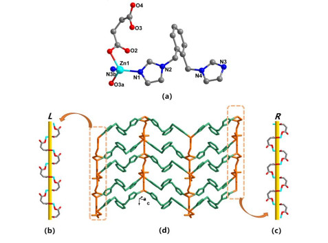
X-ray structural analysis reveals that compound 1 crystallizes in monoclinic space group P21/n, the asymmetric unit of which consists of one crystallographically independent Zn(II) cation, one suc2− anion, and one o-bix ligand (Fig. 1a). The Zn(II) center displays a slightly distorted tetrahedral [ZnO2N2] geometry via coordinated by two carboxylate oxygen atoms and two imidazole nitrogen atoms. Bond lengths and bond angles within the coordination sphere are listed in Table 2. The suc2- ligands are in a μ2-η1: η0: η1: η0 monodentate-bridging fashion to link Zn(II) cations to form interesting left- and right-handed helical [Zn(suc)]n chains with a pitch of 9.299 Å (Fig. 1b and 1c). These chain motifs are connected through o-bix ligands to give a typical 2D layer (Fig. 1d), which span a Zn⋅⋅⋅Zn contact distance of 11.928 Å. Topological analysis of compound 1 reveals it to be a 4-connected 44-sql network.
Figure 1
 Figure 1. (a) Coordination environment of the Zn(II) atom in 1 (symmetry codes: a = 1.5–x, 0.5+y, 0.5–z; b = 0.5+x, –0.5–y, –0.5+z). (b) and (c) Perspective views of 1D left- and right-handed helical chains constructed by Zn(II) cations and suc2- ligands. (d) Perspective view of the 2D layer of compound 1
3.2 Description of structure 2
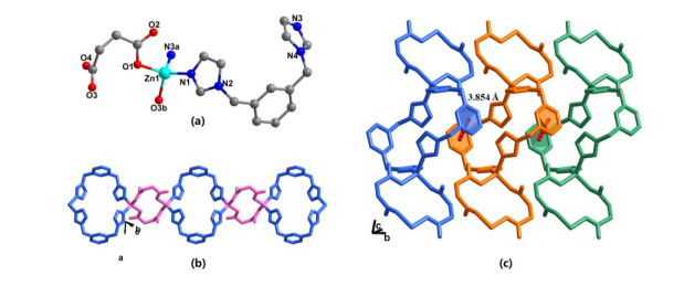
Single-crystal X-ray structural analysis of compound 2 shows that the structure is constructed by one Zn(II) cation, one suc2- anion, one m-bix ligand, and one lattice water molecule (Fig. 2a). Each Zn(II) atom is four-coordinated and exhibits a distorted tetrahedral geometry surrounded by two carboxylic oxygen atoms from two suc2- ligands and two nitrogen atoms from two m-bix ligands. Similar to compound 1, the suc2- ligand in 2 adopts a μ2-η1: η0: η1: η0 monodentate-bridging mode. On the basis of this connection mode, two suc2- ligands bridge two Zn cations forming a cyclic closed [Zn2(suc)2] structure. And gauche conformational m-bix ligands candle two Zn(II) atoms to generate a [Zn2(m-bix)2] hexagonal ring. Two types of loop structures are combined together into an infinite 1D chain (Fig. 2b). Furthermore, the adjacent chains are extended into a 2D sheet by π-π stacking interactions (centroid-to-centroid and perpendicular distances: 3.854 and 3.553 Å, dihedral angel: 0.00º) among aromatic cycles of m-bix ligands (Fig. 2c).
Figure 2
 Figure 2. (a) Coordination environment of the Zn(II) atom in 2 (symmetry codes: a = 1–x, 1–y, 1–z; b = 2–x, –y, –z). (b) View of 1D chain constructed by two types of cyclic closed structures. (c) 2D layer formed by π-π stacking interactions of neighboring chains
3.3 Description of structure 3
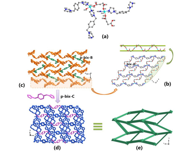
A single-crystal X-ray diffraction study reveals that 3 crystallizes in monoclinic system with P21/n space group. As shown in Fig. 3a, the asymmetric unit of 3 is composed of two Zn(II) cations, two suc2− anions, one and two halves of p-bix ligands, and four coordinated water molecules. Both Zn(II) cations possess a distorted {ZnN2O2} tetrahedral coordination environment, which are occupied by two carboxylate oxygens from two suc2− ligands and two nitrogen donors from two p-bix ligands. For convenience, the p-bix ligands containing N(1), N(5), and N(7) are designated p-bix-A, p-bix-B, and p-bix-C, respectively.
Figure 3
 Figure 3. (a) Coordination environment of the Zn(II) atom in 3 (symmetry codes: a = 1+x, y, z; b = 0.5+x, 0.5–y, –0.5+z; c = 1–x, 1–y, –z; d = 2–x, –y, –z), (b) Perspective view of the meso-helical chain (top); 2D (4, 4) layer formed by Zn(II) cations, suc2- and p-bix-A ligands (bottom), (c) View of the 2D layers are pillared by p-bix-B ligands into a 3D structure, (d) Perspective view of the 3D framework of 3 along the b axis, highlighting the channels occupied by bidentate pillared coordinated p-bix-C ligands, (e) Schematic description of the 3D framework with (65·8) topology
Each suc2− anion bridges two Zn(II) atoms with its two carboxylates in μ1-η1: η0 fashions to form an infinite 1D meso-helical chain with a pitch of 13.095 Å (Fig. 3b top). These adjacent meso-helical chains are extended to a 2D undulated 44 layer with the aid of the p-bix-A ligands (Fig. 3b bottom). Furthermore, the 2D layers are pillared by p-bix-B ligands to generate a 3D open structure. Finally, the p-bix-C molecules further join the Zn1 atoms to finish the coordination sphere of the metal atoms and give a more stabilized 3D framework (Fig. 3d). A better insight into such an involved framework can be accessed by the application of a topological approach, if each Zn(II) cation is considered as a four-connected node and suc2− anions and p-bix ligands are linkers. This framework can be simplified into a four-connected net with short and long Schlafl̈i symbols 65·8 and 6.6.6.6.62.82, which is clearly different from the CdS topology and represents a new topological prototype (Fig. 3e).
A close inspection of the structure discloses that the 3D framework contains a small solvent-accessible void space of 11.6% (461.1 Å3 per unit cell) of the total crystal volume occupied by the lattice water molecules. The water molecules are connected with the host framework by O–H···O hydrogen bonds between water molecules and the carboxyl groups of suc2- (Table 3), which further stabilizes the 3D structure of 3.
Table 3
Table 3. Hydrogen-bonding Geometrical Parameters (Å, °) of Compound 3DownLoad:
CSV
D–H···A D–H H···A D···A D–H···A O(1w)–H(1wA)···O(2w)c 0.85 1.95 2.783(2) 167 O(1w)–H(1wB)···O(6) 0.85 1.89 2.717(2) 164 O(2w)–H(2wA)···O(4) 0.85 1.88 2.723(2) 172 O(2w)–H(3wA)···O(4w)d 0.85 1.96 2.803(3) 171 O(3w)–H(3wA)···O(2) 0.85 2.10 2.869(2) 150 O(4w)–H(4wA)···O(7) 0.85 2.04 2.884(2) 176 O(4w)–H(4wB)···O(2w)c 0.85 1.98 2.825(3) 174 Symmetry codes: (c): –0.5+x, 0.5–y, –0.5+z; (d) 0.5–x, –0.5+y, 0.5–z 3.4 Influence of organic ligands on the structures
Form the experimental results and structure discussions above, we can see that all Zn(II) atoms in 1~3 are located in a tetrahedral four-coordinated environment, and suc2- ligands show the same μ2-η1: η0: η1: η0 coordinated mode. All the suc2- and different bix ligands act as uniformly 2-connected linkers, so the resulting structural differences of compound 1~3 are evidently associated with the different positions of 2-methylimidazolyl groups and conformations of the three isomeric bis(2-methyl-imidazole) ligands. The geometrical parameters of suc2- ligands and bis(imidazole) ligands in compounds 1~3 are listed in Table 4. In compound 1, the o-bix ligands exhibit trans conformation and extend the 1D [Zn(suc)]n chains into a 2D sheet; while in compound 2, the m-bix ligands show cis conformation and cradle the Zn(II) atoms to create metallocycles which inhibit the formation of a higher dimensional structure, thus resulting in an infinite 1D chain. Both trans and cis conformations of p-bix ligand coexist in compound 3, which leads to a 3D complicated structure. As a result, the difference in conformation of the bis(imidazole) ligands causes the structural difference of compounds 1~3. Structural comparisons indicate that the positions of the coordinated groups in the ligand backbone and the ligand conformation play an important role in governing the structural dimension and topologies of the final compounds.
Table 4
Table 4. Geometrical Parameters of Bis(imidazole) and suc2- Ligands in Compounds 1~3DownLoad:
CSV
Compound No. Bis(imidazole) ligands suc2- ligands Ligands Conformation of bix ligands Dihedral angles between the two imidazole rings (°) Coordination mode Separation between Zn atomsa (Å) 1 o-bix trans 19.721 μ2-η1: η0: η1: η0 5.410 2 m-bix cis 88.648 5.472 3 p-bix cis, trans, trans 24.345, 0, 0 6.400, 7.045 a The separation between Zn(II) atoms linked by suc2- ligands 3.5 PXRD patterns
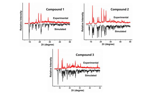
The powder X-ray diffractions (PXRD) for compounds 1~3 were performed to characterize their purity. As depicted in Fig. 4, the peak positions of simulated and experimental patterns are in good agreement, demonstrating the phase purity. The difference in reflection intensities was due to the variation in preferred orientation of the powder samples during the collection of the experimental PXRD data.
Figure 4
 Figure 4. XRD patterns of compounds 1~3
3.6 Thermal analysis
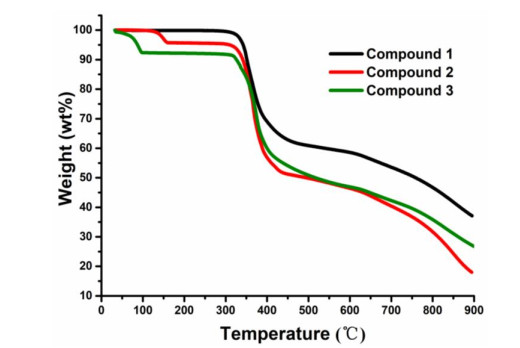
The thermostability of these compounds is explored by thermogravimetric analysis (TGA) in nitrogen gas from 30 to 900 ℃(Fig. 5). Compound 1 does not contain guest molecules, so there is no obvious weight loss before 310 ℃. Then the framework begins to collapse, accompanying the loss of organic ligands. The TGA curve of 2 shows a one-step weight loss process from 120 to 160 ℃, corresponding to the release of lattice water molecules (obsd. 4.22%, calcd. 4.11%). The overall framework of 2 begins to collapse at 310 ℃. For 3, the weight loss due to the departure of lattice water molecule is observed from 30 to 105 ℃(obsd. 7.74%, calcd. 7.90%). Then the compound reaches a plateau with no further weight loss up to 310 ℃. Upon further heating, the host framework begins to decompose.
Figure 5
 Figure 5. TGA curves of compounds 1~3
3.7 Luminescence property
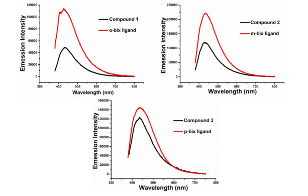
CPs based on Zn(II) ions are of great interest due to their potential applications in photochemistry, chemical optical sensors and electroluminescent devices. Therefore, the photoluminescence spectra of compounds 1~3 are measured at room temperature. In order to understand the origin of emissions, the solid-state luminescence of free ligands o-bix, m-bix, p-bix, as well as H2suc are investigated. As shown in Fig. 6, all as-synthesized compounds exhibit broad and strong luminescence emission. Excitation at 360 nm leads to intense violet luminescent emissions with the maxima at 434 nm for 1, 431 nm for 2, and 435 nm for 3, respectively. The free o-bix, m-bix, and p-bix ligands show intense emission bands at 426 nm (λex = 360 nm), 435 nm (λex = 360 nm), and 434 nm (λex = 360 nm), respectively. And there was no obvious emission observed for free H2suc ligands under the same experimental conditions[29]. As we know, the Zn(II) ion is difficult to oxidize or reduce due to its d10 configuration[30]. Therefore, compared to the emission spectra between compounds 1~3 and the free bix ligands, the similar adsorption peak positions indicate that their most possible luminescent mechanism originates from the π*→π transitions of o-bix, m-bix, and p-bix ligands. In addition, further investigation indicates that the luminescent intensities of compounds 1~3 dramatically decrease compared with that of corresponding bix ligands. It may be attributed to the coordination action of N-donor ligands to the Zn(II) ions, which lowers the rigidity of the ligands and increase the loss of energy via vibration motions.
Figure 6
 Figure 6. Solid-state luminescence emission spectra of compounds 1~3 and the bix ligands
4. CONCLUSION
In summary, three novel luminescent Zn(II) compounds have been successfully synthesized and structurally characterized. They display various structural motifs, including the 2D layer (1), 1D chain (2), and 3D network with novel (65·8) topology (3). The structural differences of the compounds demonstrate that variations of the conformations of auxiliary ligands are critical to the assembly of CPs in some particular systems.
-
-
[1]
Noro, S.; Kitaura, R.; Kondo, M.; Kitagawa, S.; Ishii, T.; Matsuzaka, H.; Yamashita, M. Framework engineering by anions and porous functionalities of Cu(II)/4, 4΄-bpy coordination polymers. J. Am. Chem. Soc. 2002, 124, 2568–2583. doi: 10.1021/ja0113192
-
[2]
Yoon, M.; Srirambalaji, R.; Kim, K. Homochiral metal-organic frameworks for asymmetric heterogeneous catalysis. Chem. Rev. 2012, 112, 1196–1231. doi: 10.1021/cr2003147
-
[3]
Hu, Z.; Deibert, B. J.; Li, J. Luminescent metal-organic frameworks for chemical sensing and explosive detection. Chem. Soc. Rev. 2014, 43, 5815–5840. doi: 10.1039/C4CS00010B
-
[4]
Manna, P.; Das, S. K. Perceptive approach in assessing rigidity versus flexibility in the construction of diverse metal-organic coordination networks: synthesis, structure, and magnetism. Cryst. Growth Des. 2015, 15, 1407–1421. doi: 10.1021/cg501787m
-
[5]
Noh, T. H.; Jung, O. S. Recent advances in various metal-organic channels for photochemistry beyond confined spaces. Acc. Chem. Res. 2016, 49, 1835–1843. doi: 10.1021/acs.accounts.6b00291
-
[6]
Yang, X. G.; Lin, X. Q.; Zhao, Y. B.; Zhao, Y. S.; Yan, D. P. Lanthanide metal-organic framework microrods: colored optical waveguides and chiral polarized emission. Angew. Chem. Int. Ed. 2017, 56, 7961–7965.
-
[7]
Wu, Y. P.; Tian, J. W.; Liu, S.; Li, B.; Zhao, J.; Ma, L. F.; Li, D. S.; Lan, Y. Q.; Bu, X. Bi-microporous metal-organic frameworks with cubane [M4(OH)4] (M = Ni, Co) clusters and pore-space partition for electrocatalytic methanol oxidation reaction. Angew. Chem. Int. Ed. 2019, 58, 12185–12189. doi: 10.1002/anie.201907136
-
[8]
Wang, K. B.; Wang, X.; Zhang, D.; Wang, H. J.; Wang, Z. K.; Zhao, M. Y.; Xi, R.; Wu, H.; Zheng, M. B. Interpenetrated nano-MOFs for ultrahigh-performance supercapacitors and excellent dye adsorption performance. CrystEngComm. 2018, 20, 6940–6949. doi: 10.1039/C8CE01067F
-
[9]
Chen, C. X.; Wei, Z. W.; Cao, C. C.; Yin, S. Y.; Qiu, Q. F.; Zhu, N. X.; Xiong, Y. Y.; Jiang, J. J.; Pan, M.; Su, C. Y. All roads lead to Rome: tuning the luminescence of a breathing catenated Zr-MOF by programmable multiplexing pathways. Chem. Mater. 2019, 31, 5550–5557. doi: 10.1021/acs.chemmater.9b01258
-
[10]
Jiang, W.; Yang, J. Q.; Yan, G. S.; Zhou, S.; Liu, B.; Qiao, Y.; Zhou, T. Y.; Wang, J. J.; Che, G. B. A novel 3-fold interpenetrated dia metal-organic framework as a heterogeneous catalyst for CO2 cycloaddition. Inorg. Chem. Commum. 2020, 113, 107770. doi: 10.1016/j.inoche.2020.107770
-
[11]
Ren, S. S.; Jiang, W.; Wang, Q. W.; Li, Z. M.; Qiao, Y.; Che, G. B. Synthesis, structures and properties of six lanthanide complexes based on a 2-(2-carboxyphenyl)imidazo(4, 5-f)-(1, 10)phenanthroline ligand. RSC Adv. 2019, 9, 3102–3112. doi: 10.1039/C8RA09207A
-
[12]
Steel, P. J. Ligand design in multimetallic architectures: six lessons learned. Acc. Chem. Res. 2005, 38, 243–250. doi: 10.1021/ar040166v
-
[13]
Stock, N.; Biswas, S. Synthesis of metal-organic frameworks (MOFs): routes to various MOF topologies, morphologies, and composites. Chem. Rev. 2012, 112, 933–969 doi: 10.1021/cr200304e
-
[14]
Tan, Y. X.; He, Y. P.; Zhang, Y.; Zheng, Y. J.; Zhang, J. Solvent controlled assembly of four Mn(II)-2, 5-thiophenedicarboxylate frameworks with rod-packing architectures and magnetic properties. CrystEngComm. 2013, 15, 6009–6014. doi: 10.1039/c3ce40677f
-
[15]
Huang, S. Y.; Li, J. Y.; Li, J. Q.; Xu, W. Y.; Luo, M. B.; Zhu, Y.; Luo, F. Exceptional temperature-dependent coordination sites from acylamide groups. Dalton Trans. 2014, 43, 5260–5264. doi: 10.1039/c3dt53123f
-
[16]
Zhang, J. W.; Kan, X. M.; Liu, B. Q.; Liu, G. C.; Tian, A. X.; Wang, X. L. Systematic investigation of reaction-time dependence of three series of copper-lanthanide/lanthanide coordination polymers: syntheses, structures, photoluminescence, and magnetism. Chem. Eur. J. 2015, 21, 16219–16228. doi: 10.1002/chem.201502203
-
[17]
Zhao, J.; Liu, X.; Wu, Y.; Li, D. S.; Zhang Q. Surfactants as promising media in the field of metal-organic frameworks. Coord. Chem. Rev. 2019, 391, 30–43. doi: 10.1016/j.ccr.2019.04.002
-
[18]
Ghosh, A. K.; Hazra, A.; Mondal, A.; Banerjee, P. Weak interactions: the architect behind the structural diversity of coordination polymer. Inorg. Chim. Acta 2019, 488, 86–119. doi: 10.1016/j.ica.2019.01.008
-
[19]
Yang, J. X.; Qin, Y. Y.; Ye, R. P.; Zhang, X.; Yao, Y. G. Employing mixed-ligand strategy to construct a series of luminescent Cd(II) compounds with structural diversities. CrystEngComm. 2016, 18, 8309–8320. doi: 10.1039/C6CE01607C
-
[20]
Yang, J. X.; Zhai, J. Q.; Zhang, X.; Qin, Y. Y.; Yao, Y. G. Tuning different kinds of entangled metal-organic frameworks through modifying the spacer group of aliphatic dicarboxylate ligands and reactant ratio. Dalton Trans. 2016, 45, 711–723. doi: 10.1039/C5DT03731J
-
[21]
Yang, J. X.; Qin, Y. Y.; Cheng, J. K.; Yao, Y. G. Construction of a series of Zn(II) compounds with different entangle motifs by varying flexible aliphatic dicarboxylic acids. Cryst. Growth Des. 2015, 15, 2223−2234. doi: 10.1021/cg501879w
-
[22]
Liu, Y. H.; Zhang, F. J.; Wu, P. Y.; Deng, C. F.; Yang, Q. M.; Xue, J. J.; Shi, Y. H.; Wang, J. Cobalt(II)-based metal-organic framework as bifunctional materials for Ag(I) detection and proton reduction catalysis for hydrogen production. Inorg. Chem. 2019, 58, 924−931. doi: 10.1021/acs.inorgchem.8b03046
-
[23]
Wang, Z. X.; Ren, Y. X.; Cao, J.; Tang, L.; Zhang, M. L.; Zhou, S. H. Structural assembly from 1D to 3D motivated by the linear co-ligands, and the magnetic and photocatalytic properties of five NiII coordination polymers with 5-(4΄-carboxylphenyl)nicotinic acid. New J. Chem. 2018, 42, 17991–18000. doi: 10.1039/C8NJ02921K
-
[24]
He, X.; Lu, X. P.; Li, M. X.; Morris, R. E. Tuning different kinds of entangled networks formed by isomers of bis(1, 2, 4-triazol-1-ylmethyl)benzene and a flexible tetracarboxylate ligand. Cryst. Growth Des. 2013, 13, 1649−1654. doi: 10.1021/cg3018562
-
[25]
Zhao, F. H.; Huang, L. W.; He, Y. C.; Yan, X. Q.; Li, Z. L.; Jia, X. M.; Feng, R.; Li, J. X.; You, J. M. Two entangled Cd(II) MOFs of sebacic acid and bis(2-methyl-imidazole) ligands for selective sensing of Fe3+. Inorg. Chim. Acta 2020, 499, 119184. doi: 10.1016/j.ica.2019.119184
-
[26]
Hoskins, B. F.; Robson, R.; Slizys, D. A. An infinite 2D polyrotaxane network in Ag2(bix)3(NO3)2 (bix = 1, 4-bis(imidazol-1-ylmethyl)benzene). J. Am. Chem. Soc. 1997, 119, 2952−2953. doi: 10.1021/ja9642626
-
[27]
Sheldrick, G. M. SADABS, Institute for Inorganic Chemistry. University of Göttingen. Göttingen, Germany 1996.
-
[28]
Sheldrick, G. M. SHELXL-2014, Program for Crystal Structure Refinement. University of Gottingen, Germany 2014.
-
[29]
Uebler, J. W.; Wilson, J. A.; LaDuca, R. L. A. Donor disposition and aliphatic conformation effects on structure in luminescent zinc dicarboxylate coordination polymers with isomeric dipyridylamide coligands. CrystEngComm. 2013, 15, 1586−1596. doi: 10.1039/c2ce26929e
-
[30]
Allendorf, M. D.; Bauer, C. A.; Bhakta, R. K.; Houk, R. J. T. Luminescent metal-organic frameworks. Chem. Soc. Rev. 2009, 38, 1330−1352.
-
[1]
-
Figure 1 (a) Coordination environment of the Zn(II) atom in 1 (symmetry codes: a = 1.5–x, 0.5+y, 0.5–z; b = 0.5+x, –0.5–y, –0.5+z). (b) and (c) Perspective views of 1D left- and right-handed helical chains constructed by Zn(II) cations and suc2- ligands. (d) Perspective view of the 2D layer of compound 1
Figure 2 (a) Coordination environment of the Zn(II) atom in 2 (symmetry codes: a = 1–x, 1–y, 1–z; b = 2–x, –y, –z). (b) View of 1D chain constructed by two types of cyclic closed structures. (c) 2D layer formed by π-π stacking interactions of neighboring chains
Figure 3 (a) Coordination environment of the Zn(II) atom in 3 (symmetry codes: a = 1+x, y, z; b = 0.5+x, 0.5–y, –0.5+z; c = 1–x, 1–y, –z; d = 2–x, –y, –z), (b) Perspective view of the meso-helical chain (top); 2D (4, 4) layer formed by Zn(II) cations, suc2- and p-bix-A ligands (bottom), (c) View of the 2D layers are pillared by p-bix-B ligands into a 3D structure, (d) Perspective view of the 3D framework of 3 along the b axis, highlighting the channels occupied by bidentate pillared coordinated p-bix-C ligands, (e) Schematic description of the 3D framework with (65·8) topology
Figure 6 Solid-state luminescence emission spectra of compounds 1~3 and the bix ligands
Table 1. Summary of Crystal Data and Structure Refinements for 1~3
Compound 1 2 3 Empirical formula C18H18N4O4Zn C18H20N4O5Zn C36H44N8O12Zn2 Formula eight 419.75 437.77 911.57 Crystal system Monoclinic Triclinic Monoclinic Space group P21/n P $ \overline 1 $ P21/n a (Å) 10.9336(6) 7.9557(4) 13.0955(2) b (Å) 9.2900(3) 11.0995(7) 14.3912(2) c (Å) 18.3100(10) 11.5245(8) 21.1992(4) α (°) 90 85.841(5) 90 β (°) 90.035(5) 72.628(6) 93.4790(10) γ (°) 90 70.996(5) 90 V (Å3) 1859.80(16) 917.96(11) 3987.84(11) Z 4 2 4 Dc (mg·m-3) 1.499 1.584 1.518 F(000) 864.0 452.0 1888.0 GOF on F2 1.068 1.023 1.030 R, wR (I > 2σ(I))a 0.0635, 0.1692 0.0619, 0.1582 0.0299, 0.0790 R, wR (all data)b 0.0845, 0.1925 0.0728, 0.1675 0.0329, 0.0811 a R = ∑||Fo| – |Fc||/∑|Fo|, b wR = [Σw(Fo2 – Fc2)2/Σw(Fo2)2]1/2  下载: 导出CSV
Table 2. Selected Bond Lengths and Bond Angles for 1~3
Compound 1 Bond Dist Bond Dist Bond Dist Zn(1)–O(3)a 1.935(3) Zn(1)–N(1) 2.028(4) Zn(1)–N(3)b 2.006(4) Zn(1)–O(1) 2.054(6) Angle (o) Angle (o) Angle (o) O(3)a–Zn(1)–N(1) 117.5(2) O(3)a–Zn(1)–N(3)b 115.9(2) N(1)–Zn(1)–N(3)b 101.57(17) O(3)a–Zn(1)–O(1) 105.04(17) N(1)–Zn(1)–O(1) 90.27(19) N(3)b–Zn(1)–O(1) 124.43(17 Compound 2 Bond Dist. Bond Dist. Bond Dist. Zn(1)–O(1) 1.948(4) Zn(1)–N(1) 2.011(4) Zn(1)–N(3)a 2.031(4) Zn(1)–O(3)b 2.032(6) Angle (o) Angle (o) Angle (o) O(1)–Zn(1)–N(1) 114.25(17) O(1)–Zn(1)–N(3)a 112.34(18) N(1)–Zn(1)–N(3)a 111.60(17) O(1)–Zn(1)–O(3)b 107.99(19) N(1)–Zn(1)–O(3)b 117.7(2) N(3)a–Zn(1)–O(3)b 90.7(2) Compound 3 Bond Dist Bond Dist. Bond Dist. Zn(1)–O(1) 1.9536(16) Zn(1)–O(7)a 1.9929(16) Zn(1)–N(7) 2.0107(19) Zn(1)–N(5) 2.0310(19) Zn(2)–O(5) 1.9675(16) Zn(2)–O(3) 1.9699(15) Zn(2)–N(3)b 1.9869(19) Zn(2)–N(1) 2.0122(19) Angle (o) Angle (o) Angle (o) O(1)–Zn(1)–O(7)a 111.06(7) O(1)–Zn(1)–N(7) 133.18(7) O(7)a–Zn(1)–N(7) 102.99(7) O(1)–Zn(1)–N(5) 103.57(7) O(7)a–Zn(1)–N(5) 100.94(7) N(7)–Zn(1)–N(5) 100.33(8) O(5)–Zn(2)–O(3) 100.48(6) O(5)–Zn(2)–N(3)b 119.25(7) O(3)–Zn(2)–N(3)b 103.34(7) O(5)–Zn(2)–N(1) 102.65(7) O(3)–Zn(2)–N(1) 117.32(7) N(3)b–Zn(2)–N(1) 113.76(8) Symmetry codes: compound 1: (a): 1.5–x, 0.5+y, 0.5–z; (b) 0.5+x, –0.5–y, –0.5+z; compound 2: (a): 1–x, 1–y, 1–z; (b): 2–x, –y, –z; compound 3: (a): 1+x, y, z; (b) 0.5+x, 0.5–y, –0.5+z
下载: 导出CSV
Table 3. Hydrogen-bonding Geometrical Parameters (Å, °) of Compound 3
D–H···A D–H H···A D···A D–H···A O(1w)–H(1wA)···O(2w)c 0.85 1.95 2.783(2) 167 O(1w)–H(1wB)···O(6) 0.85 1.89 2.717(2) 164 O(2w)–H(2wA)···O(4) 0.85 1.88 2.723(2) 172 O(2w)–H(3wA)···O(4w)d 0.85 1.96 2.803(3) 171 O(3w)–H(3wA)···O(2) 0.85 2.10 2.869(2) 150 O(4w)–H(4wA)···O(7) 0.85 2.04 2.884(2) 176 O(4w)–H(4wB)···O(2w)c 0.85 1.98 2.825(3) 174 Symmetry codes: (c): –0.5+x, 0.5–y, –0.5+z; (d) 0.5–x, –0.5+y, 0.5–z
下载: 导出CSV
Table 4. Geometrical Parameters of Bis(imidazole) and suc2- Ligands in Compounds 1~3
Compound No. Bis(imidazole) ligands suc2- ligands Ligands Conformation of bix ligands Dihedral angles between the two imidazole rings (°) Coordination mode Separation between Zn atomsa (Å) 1 o-bix trans 19.721 μ2-η1: η0: η1: η0 5.410 2 m-bix cis 88.648 5.472 3 p-bix cis, trans, trans 24.345, 0, 0 6.400, 7.045 a The separation between Zn(II) atoms linked by suc2- ligands
下载: 导出CSV
-


 扫一扫看文章
计量
- PDF下载量: 1
- 文章访问数: 1144
- HTML全文浏览量: 8
