Table 1.
Crystal and Experiment Data for Crystals 1~3
Citation:
Shu-Wang GE, Bai-Wang SUN. Synthesis, Crystal Structure, Spectroscopy and Hirshfeld Analysis of 4, 6-Diamino-2-cyclopropylaminopyrimidine-5-carbonitrile with Different Solvents: N, N-dimethylformamide, Methanol and Water[J]. Chinese Journal of Structural Chemistry,
2022, 41(3): 220327.
doi:
10.14102/j.cnki.0254-5861.2011-3359

Synthesis, Crystal Structure, Spectroscopy and Hirshfeld Analysis of 4, 6-Diamino-2-cyclopropylaminopyrimidine-5-carbonitrile with Different Solvents: N, N-dimethylformamide, Methanol and Water
English
Synthesis, Crystal Structure, Spectroscopy and Hirshfeld Analysis of 4, 6-Diamino-2-cyclopropylaminopyrimidine-5-carbonitrile with Different Solvents: N, N-dimethylformamide, Methanol and Water
Abstract:
Three new solvates of 4, 6-diamino-2-cyclopropylamino-5-pyrimidine carbonitrile (DCL): crystal 1 (DCL with N, N-dimethylformamide (DMF)), crystal 2 (DCL with methanol) and crystal 3 (DCL with water) were synthesized and characterized by single-crystal X-ray diffraction, thermal behaviour, Hirshfeld surface and powder X-ray powder diffraction (PXRD). 1H and 13C NMR spectra confirm solvent molecules existing in the crystal lattice. Crystal 1 forms a 1:1 DCL: DMF crystal; crystal 2 gives a 1:0.5 DCL: methanol crystal and crystal 3 gets a 1:1.5 DCL: H2O crystal. The three crystals are all primarily stabilized by a strong N–H⋅⋅⋅N hydrogen bonding interaction between DCL and the solvents. The structures are stabilized by H⋅⋅⋅H, N–H⋅⋅⋅O, N–H⋅⋅⋅N and O–H⋅⋅⋅O intermolecular interactions. When crystal 2 is dried at 150 ℃, the new polymorph with no solvate is obtained.
-
Key words:
- dicyclanil
- / crystal
- / spectroscopy
- / Hirshfeld surfaces
-
1. INTRODUCTION
So far, the solvate of active pharmaceutical ingredients is a hot topic and it achieves remarkable development in "crystal engineering"[1-6]. Polymorphism and hydrate formation are especially powerful means to alternate crystal forms of drugs[7]. If the crystals of pharmaceuticals are engineered, the properties like bioavailability, stability, and processibility could be optimized[8, 9].
Dicyclanil (DCL)[10, 11] is one of the most important growth regulators for insect of the newer generation. DCL can kill dipster and flea and prevent fly, mosquito becoming pupa or imago. Especially, it can prevent animal from the disoperation of mosquito and Aedes aegypti. DCL's toxicity is very low, and therefore its safety is better.
However, we can complicate the manufacturing process of solid DCL due to its ability to afford polymorphs, hydrates and other solvates. It is very important for pharmaceutical industry to determine the optimal and controlled conditions to obtain the solid with known properties[12].
Up to now, eight polymorphic unsolvated crystals, A, B, C, D, E, F, G and H, have been patented[13, 14]. The most stable polymorph D crystallizes from octanol, and there is still no report about solvate. The crystal structures of three solvates of DCL: N, N-dimethylformamide (DMF) (1:1), DCL: methanol (2:1) and DCL: H2O (1:1) are present in this paper. These crystals were characterized by TGA, DSC, IR, and single-crystal diffraction. Furthermore, molecular Hirshfeld surfaces of DCL in crystals were performed. The N–H⋅⋅⋅N hydrogen bond between DCL and solvents plays a key role.
2. EXPERIMENTAL
2.1. Materials and methods
Methanol, DMF was commercially available from J & K SCIENTIFIC LTD, and DCL provided by Changzhou Yabang Pharmaceutical & Chemical Co., LTD. All of the reagent grade solvents from Sinopharm Chemical Reagent Co., Ltd were used directly without further purification. Infrared spectra were recorded on a SHIMADZU IR prestige-21 FTIR-8400S spectrometer from 4000 to 400 cm–1 with the samples in potassium bromide pellets. DSC and TGA measurements were carried out by a Mettler-Toledo TGA/ DSC STARe System at the 10 K·min-1 heating rate under a dry N2 atmosphere flowing at 20 cm3·min-1 over a range of 40~400 ℃. We analysized the TGA/DSC data with STARe Software. NMR spectra were recorded on a Varian FT-400 MHz instrument. Also, by taking a solvent peak as reference, we recorded the chemical shifts in parts per million (ppm) on the scale.
2.2 Preparation of the crystals (1~3)
Crystal 1: DCL (400 mg) was stirred in DMF (30 mL), which was stirred for about 30 minutes before evaporating at 25 ℃. Colorless needle-like crystals of crystal 1 were separated from the liquor a few days later.
Crystal 2: DCL (300 mg) was suspended in 30 mL of methanol/water (95:5). After heating to 64 ℃ and stirring for about 35 mintures before evaporation at 25 ℃, colorless crystals 2 in plate form were separated several days later.
Crystal 3: DCL (1 g) was stirred in DMF (50 mL) for approximately half an hour. The distilled water (50 mL) was added in the solution. After slowly evaporating the solvent at 25 ℃, colorless block-like crystals of crystal 3 were separated from the liquor after one month.
2.3 X-ray crystallographic study
The single crystal X-ray diffraction data of crystals 1, 2 and 3 were obtained at 293 K by using the Rigaku SCXmini diffractometer with an ω-scan mode using a graphitemonochromated MoKα radiation (λ = 0.071073 nm)[15]. We integeated the lattice parameters using vector analysis and refined them from the diffraction matrix. The absorption correction was finished based on the Bruker SADABS program with multi-scan method. Crystal data and refinements for these three crystals are listed in Table 1. The structure solution and refinement were respectively performed with SHELXS-97 and SHELXL-97[16]. All the nonhydrogen atoms were anisotropically refined, while the hydrogen ones were inserted in calculated positions and fixed during the least-squares[17]. The molecular graphics were drawn with mercury[18] and diamond program[19].
Table 1

 DownLoad:
CSV
DownLoad:
CSV
Compound 1 2 3 Formula C11H17N7O C17H24N12O C16H26N12O3 Formula weight 263.32 412.48 434.49 Crystal system Monoclinic Triclinic Monoclinic Space group P21/n P $ \overline 1 $ C2/c a (Å) 7.5744(15) 8.9850(18) 9.1220(18) b (Å) 17.305(4) 11.359(2) 27.311(6) c (Å) 11.150(2) 11.559(2) 18.234(4) α (º) 90 109.31(3) 90 β (º) 97.73(3) 100.10(3) 104.35(3) γ (º) 90 107.46(3) 90 V (Å3) 1448.2(5) 1011.4(4) 4400.9(15) Z 4 2 8 Dc (Mg·m-3) 1.208 1.355 1.312 T (K) 293(2) 293(2) 293(2) μ(mm-1) 0.085 0.095 0.097 Crystal dimensions (mm3) 0.3 × 0.2 × 0.11 0.3 × 0.22 × 0.17 0.3 × 0.2 × 0.1 No. of reflns collected 3311 4614 4029 No. of unique reflns 1823 2542 2725 S 1.020 1.038 1.006 CCDC No. 1003933 1003932 1019413 2.4 X-ray powder diffraction
X-ray powder diffraction measurements of crystals 1~3 and DCL were recorded on a D8 ADVANCE XRD (Bruker, Germany) with CuKα radiation (λ = 1.54056 Å) at 40 mA and 45 kV. Into a glass holder the sample was packed to collect the diffraction patterns over a 5~45° 2θ range at the scan rate of 3 °·min-1.
2.5 Hirshfeld surface calculations
We performed Molecular Hirshfeld surface calculations by using the CrystalExplorer program[20], and the principles have been reported in literature[21-25]. When reading cif files of crystals 1~3 into the CrystalExplorer program for analysis, all the hydrogen bond lengths were modified automatically to typical neutron-derived values (N–H = 1.009, C–H = 1.083 Å). Displayed by using the standard 0.6~2.6 Å with de and di distance scales, the 2-D fingerprint plots are shown on the graph axes.
3. RESULTS AND DISCUSSION
3.1 Crystal structures of crystals 1~3
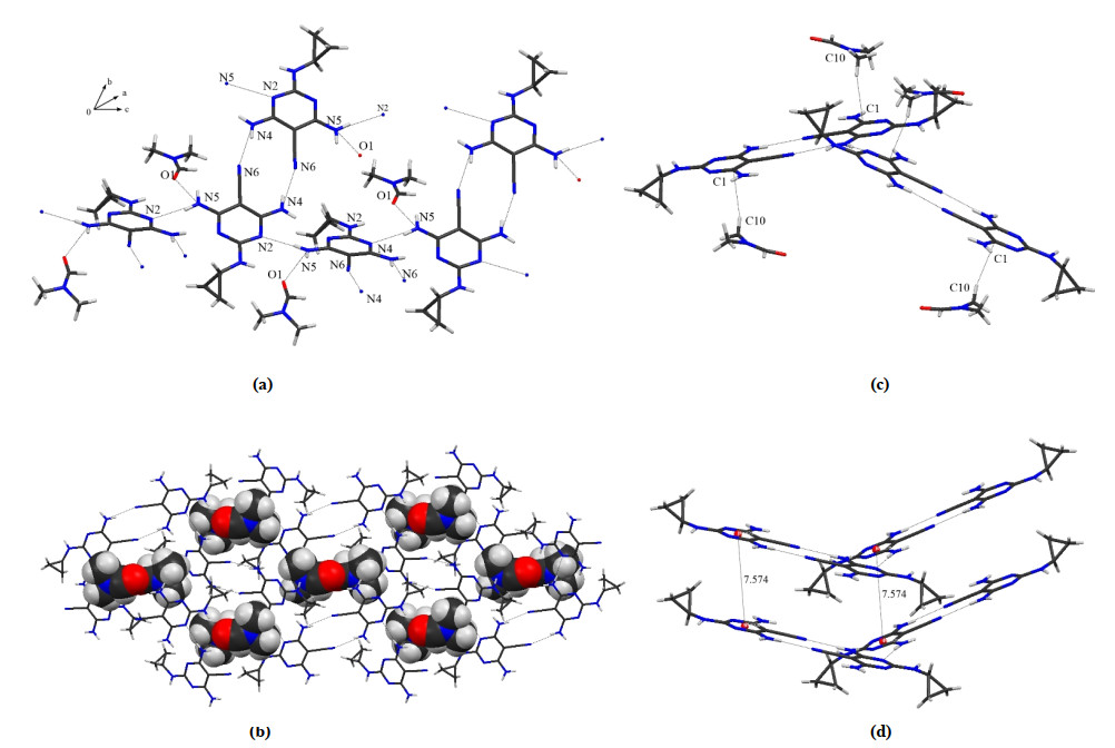
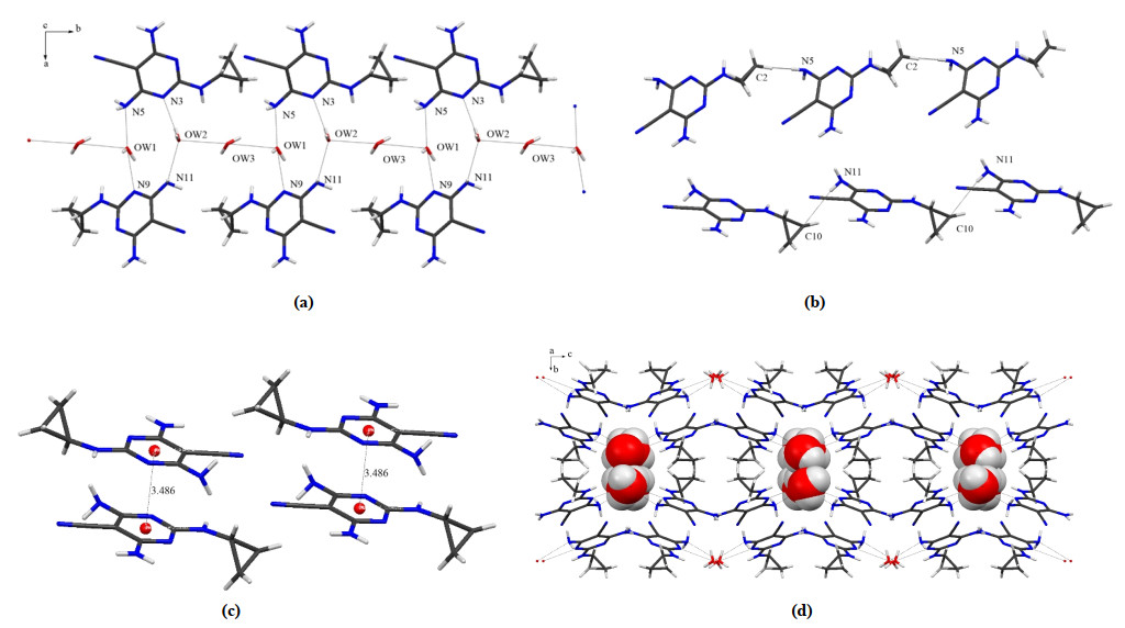
DCL forms a 1:1 solvate with DMF (crystal 1) and crystallizes in space group P21/n with Z = 4. The DCL molecules in crystal 1 are self-assembled through various kinds of weak interactions. The important interactions are as follows: N(5)–H(5A)⋅⋅⋅N(2) (dD⋅⋅⋅A (Å) = 2.993 Å, ∠D–H⋅⋅⋅A = 170°), N(5)–H5B⋅⋅⋅O(1) (dD⋅⋅⋅A (Å) = 2.929 Å, ∠D–H⋅⋅⋅A = 136°), and N(4)–H(4B)⋅⋅⋅N(6) (dD⋅⋅⋅A (Å) = 3.011 Å, ∠D–H⋅⋅⋅A = 164°) (Fig. 1a). The hydrogen bonds of crystals 1~3 are all given in Table 2. The oxygen atom (O1) of DMF participates in hydrogen bonding with N(5)–H from a neighbor molecule, getting a hydrogen-bonded dimeric motif of DCL molecules with DMF molecules. Scuh dimeric motifs connecte with each other through N–H⋅⋅⋅N hydrogen bonds to get hydrogenbonded sheets along plane (100) (Fig. 1b). Besiedes strong hydrogen bonds, weak interactions like C–H⋅⋅⋅π also make contribution to the stability of the crystal. One C–H bond of the methyl groups of DMF is involved in C–H⋅⋅⋅π (dD⋅⋅⋅A (Å) = 3.780 Å) interaction (Fig. 1c) which is much weaker compared to the related hydrogen bonds. The distance of two sixmembered rings of DCL with a neighboring DCL molecule is 7.574 Å (Fig. 1d), so no significant π⋅⋅⋅π intermolecular interaction appears. The N–H⋅⋅⋅N and N–H⋅⋅⋅O interactions contribute the most in crystal 1.
Figure 1
 Figure 1. (a) Hydrogen-bonded dimers, (b) Packing of the molecules of crystal 1 viewed along the (100) plane, (c) C–H···π interactions and (d) π···π interactions
Table 2
Table 2. Hydrogen Bonds in Crystals 1, 2 and 3DownLoad:
CSV
D–H···A D–H (Å) H···A (Å) D···A (Å) ∠D–H···A (°) Symmetry operation Crystal 1 N(4)–H(4A)···N(3) 0.86 2.30 3.158(6) 174 x+1/2, –y+1/2, z+1/2 N(4)–H(4B)···N(6) 0.86 2.18 3.011(6) 164 –x, –y+1, –z+2 N(5)–H(5A)···N(2) 0.86 2.14 2.993(5) 170 x–1/2, –y+1/2, z–1/2 N(5)–H(5B)···O(1) 0.86 2.25 2.929(6) 136 –x+1/2, y+1/2, –z+3/2 N(1)–H(1C)···O(1) 0.82 2.38 3.165(6) 161 –x+1, –y, –z+2 Crystal 2 N(12)–H(12A)···N(3) 0.86 2.13 2.988(7) 172 x, y–1, z N(12)–H(12B)···N(1) 0.86 2.33 3.113(7) 152 x+1, y–1, z N(11)–H(11A)···N(2) 0.86 2.42 3.284(7) 178 x+1, y, z+1 N(11)–H(11B)···N(5) 0.86 2.53 3.388(9) 164 –x+2, –y, –z+2 N(10)–H(10A)···N(7) 0.86 2.33 3.189(8) 176 x, y+1, z N(10)–H(10B)···N(5) 0.86 2.35 3.137(11) 153 x–1, y+1, z N(9)–H(9A)···N(6) 0.86 2.63 3.372(6) 172 x–1, y, z–1 N(9)–H(9B)···N(1) 0.86 2.63 3.372(7) 145 –x–1, –y+1, –z+1 O(1)–H(1)···O(1) 0.82 2.31 2.995(6) 141 –x–1, –y+1, –z+1 N(4)–H(4A)···O(1) 1.00 2.25 3.188(5) 156 –x+1, –y+1, –z+1 Crystal 3 N(5)–H(5B)···O(W1) 0.86 2.13 2.939(8) 156 –x+1, –y+1, –z+1 N(5)–H(5B)···N(6) 0.86 2.49 3.251(7) 147 –x+5/2, –y+3/2, –z+2 N(11)–H(11A)···O(W2) 0.86 2.13 2.941(9) 156 x+1/2, y–1/2, z N(11)–H(11B)···C(10) 0.86 2.49 3.245(8) 147 –x+2, –y, –z N(4)–H(4A)···N(8) 0.86 2.34 3.170(9) 162 –x+1, –y+1, –z+1 N(4)–H(4B)···N(12) 0.86 2.34 3.146(7) 155 –x+2, –y+1, –z+1 N(10)–H(10A)···N(2) 0.86 2.33 3.163(7) 162 –x+1, –y+1, –z+1 N(10)–H(10B)···N(6) 0.86 2.34 3.145(7) 156 –x+2, –y+1, –z+1 O(W1)–H(W1A)···N(9) 1.04 1.81 2.821(9) 164 x–1/2, y+1/2, z O(W2)–H(W2A)···N(3) 1.07 1.82 2.828(6) 155 –x+1, –y+1, –z+1 O(W3)–H(W3A)···O(W2) 0.71 2.55 3.089(6) 134 x–1/2, y+1/2, z O(W1)–H(W1B)···O(W3) 0.68 2.50 3.095(5) 148 –x+1/2, y–1/2, –z+1/2 O(W2)–H(W2A)···O(W3) 0.71 2.59 3.211(6) 148 –x+1/2, y–1/2, –z+1/2 O(W3)–H(W3B)···O(W2) 0.91 2.35 3.089(8) 139 x–1/2, y+1/2, z Crystal 2 forms a 2:1 (DCL: methanol) crystal in space group P
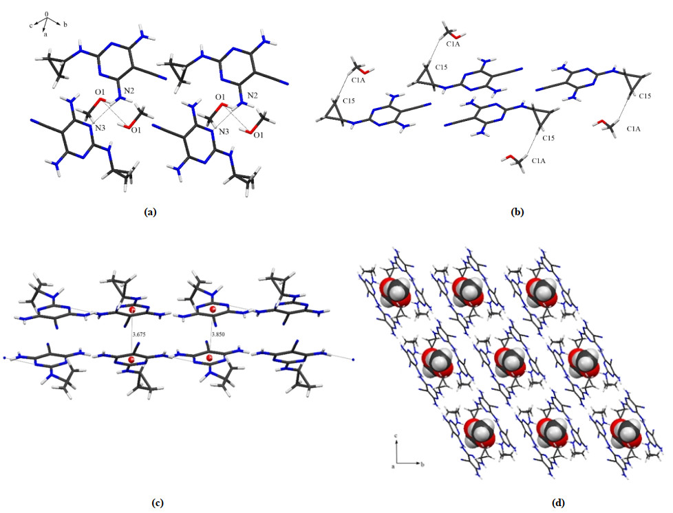
$ \overline 1 $ with Z = 2. Different from crystal 1, the DCL molecules self-assemble into dimers only by N(12)–H(12A)⋅⋅⋅N(3) interactions (dD⋅⋅⋅A (Å) = 2.988 Å, ∠D–H⋅⋅⋅A = 172°) and the methanol molecules form self-assembled dimers through O(1)–H(1)⋅⋅⋅O(1) interactions (dD⋅⋅⋅A (Å) = 2.995 Å, ∠D– H⋅⋅⋅A = 140°) (Fig. 2a). Similarity appears in the packing of DCL molecules in crystal 2 with 1. Also, C–H⋅⋅⋅π interactions help stabilize the lattice. One C–H bond of the methyl groups of the methanol molecule is involved in C–H⋅⋅⋅π (dD⋅⋅⋅A (Å) = 3.551 Å) interaction (Fig. 2b). But the strength of π⋅⋅⋅π interaction exists in crystal 2. The distances of two sixmembered rings of DCL with a neighboring DCL molecule are 3.675 or 3.850 Å (Fig. 2c). The guest methanol molecules are embedded through O–H⋅⋅⋅O interactions in cavities caused by the assembly of DCL molecules along the a-axis (Fig. 2d).Figure 2
 Figure 2. (a) Hydrogen-bonded dimers, (b) C–H…π interactions and (c) π…π interactions, (d) Packing of crystal 2 viewed along the a-axis
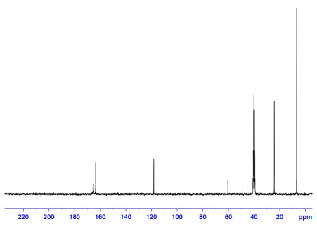
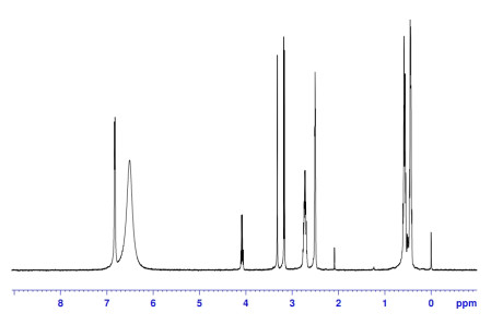
Although the crystal data of 2 are obtained, it is still hard to confirm the existence of water or methanol molecule in the crystal lattice. More work and efforts were required to answer this question. Solid-state 13C spectrum can be used to identify whether the crystal is hydrate or non-hydrate, which can be characterized by 13C peak of the solvent in lattice of crystal 2 (Fig. 3). The chemical shift (161.3 ppm) observed suggests the existence of methanol in 2. It is also verified by 1H NMR spectra that the hydrogen of CH3OH corresponds to the chemical shifts 3.3 and 2.8 ppm (3:1) in Fig. 4.
Figure 3
 Figure 3. 13C NMR spectra of crystal 1 and the solvate is DMSO
Figure 4
 Figure 4. 1H NMR spectra of crystal 1 and the solvate is DMSO
Crystal 3 is of C2/c space group, and an asymmetric unit contains 3 water molecules and 2 DCL molecules. This structure is not an exception to 2 according to the dimers formed by N(5)–H(5B)⋅⋅⋅O(W1) (dD⋅⋅⋅A (Å) = 2.939 Å, ∠D– H⋅⋅⋅A = 156°), O(W2)–H(W2A)⋅⋅⋅N(3) (dD⋅⋅⋅A (Å) = 2.828 Å, ∠D–H⋅⋅⋅A = 155°), O(W1)–H(W1A)⋅⋅⋅N(9) (dD⋅⋅⋅A (Å) = 2.821 Å, ∠D–H⋅⋅⋅A = 164°), and N(11)–H(11B)⋅⋅⋅ O(W2) (dD⋅⋅⋅A (Å) = 2.941 Å, ∠D–H⋅⋅⋅A = 146°) interactions. Howerer, the dimeric assemblies here are connected one another by bridging water molecules (Fig. 5a). The oxygen atom O(W3)–H of H2O participates in intermolecular hydrogen bonding with O(W2) or O(W1) of a neighboring molecule. One N–H bond of amino group of DCL molecule takes part in N–H⋅⋅⋅π (dD⋅⋅⋅A (Å) = 3.722 or 3.734 Å) interactions (Fig. 5b). The π⋅⋅⋅π interation (dD⋅⋅⋅A (Å) = 3.486 Å) (Fig. 5c) is stronger than that in crystals 1 and 2. The water assisted assembly in crystal 3 makes a modified host system with a butterfly-like structure (Fig. 5d).
Figure 5
 Figure 5. (a) Hydrogen-bonded dimers, (b) N–H…π interactions and (c) π…π interactions, (d) Packing of the molecules (viewed along the a-axis) of crystal 3
3.2 Thermogravimetric analysis and dynamic scanning calorimetry crystals 1~3 and DCL (4)
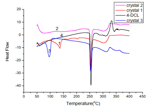
Thermal behaviors of crystals 1, 2 and 3 together with their raw materials (4-DCL) were studied on a STARe System (from Mettler-Toledo) at a heating rate of 10 ℃ per minute in a 50~400 ℃ range, as shown in Fig. 6. The melting points of 4-DCL, crystals 1, 2 and 3 are found to be 253.2, 254.5, 251.3 and 252.1 ℃ in turn. No significant differences are observed among the samples tested at the meting points. This indicates that the heating process does not result in the formation of primary forms, which is confirmed by the XRPD measurement. But the solvents lose at different temperature ranges: 80~120, 60~100 and 50~ 100 ℃ with the mass loss of about 28.4%, 8.9% and 12.6% (w/w) for crystals 1~3, respectively. In fact, the theoretical contents of solvents for 1~3 should be 27.8%, 8.4% and 12.4% (w/w) for DMF, methanol and water correspondingly. The results indicate that the theoretical data are in good agreement with the experimental ones.
Figure 6-1
 Figure 6-1. TGA profiles of 1~3 and 4-DCL
Figure 6-2
 Figure 6-2. DSC profiles of crystals 1~3 and4-DCL
3.3. IR of crystals 1~3 and DCL (4)
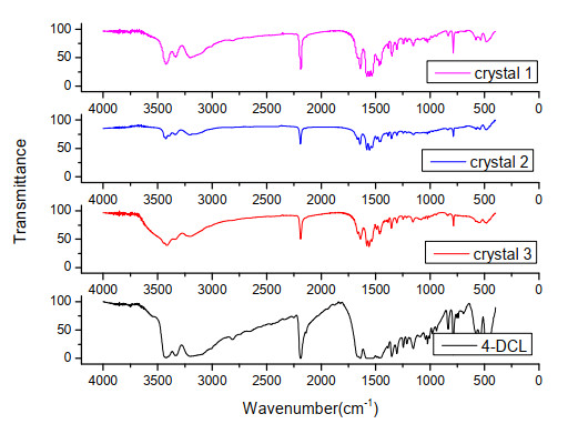
IR spectra of crystals 4-DCL and crystals 1~3 are shown in Fig. 7 in the 4000~400 cm-1 region. The absorption peaks around 3420 and 3160 cm-1 can be attributed to the stretching vibration of NH2 and NH groups, and those around 2194 cm-1 to that of C≡N. The aromatic C=C stretching of DCL molecule occurs at 1570 cm-1. The strong peak at 1460 cm-1 is due to the C–H deformation. The peaks at 1660 and 1150 cm-1 result from the C=O stretching vibration of carboxylic group in crystal 1. The broad peak observed at 3410 cm-1 is caused by the presence of OH stretching vibration in crystals 2 and 3. There is no significant differences in the four curves.
Figure 7
 Figure 7. IR spectra of crystals 1~3 and dicyclanil (4-DCL)
3.4 Powder X-ray diffraction analysis of 1~3 and DCL (4)
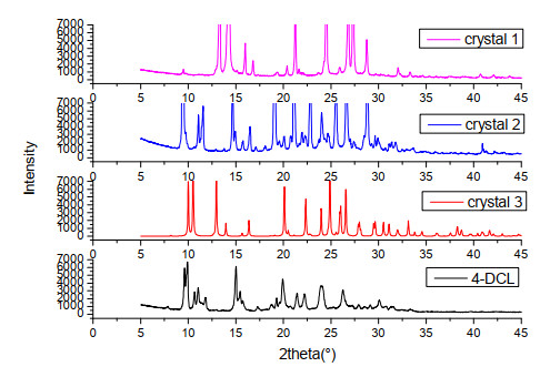
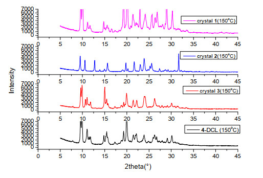
The crystals 1~3 and 4-DCL show unique powder X-ray diffraction patterns (Figs. 8 and 9), and such diffraction is remarkably useful for the identification of structural relationships between different solvated crystal forms of DCL. The PXRD patterns of crystals 1~3 and 4-DCL are distinguishable in Fig. 8. The results show that when slovates are different, the positon of peak will change. When crystals 1~3 and 4-DCL were dried at 150 ℃ for 1 h, the data of PXRD are the same in crystals 1 and 3 (Fig. 9), which is similar to 4-DCL before heating. But in crystal 2, there are no gross structural changes accompanying the desolvation process. This is a new polymorph with no solvate.
Figure 8
 Figure 8. XPRD of crystals 1~3 and dicyclanil (4-DCL)
Figure 9
 Figure 9. XPRD of crystals 1~3 and dicyclanil (4-DCL) after heating to 150 ℃
3.5 Hirshfeld surface analysis of crystals 1~3
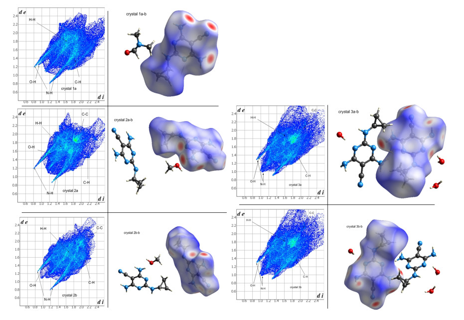
The 2D fingerprint plots and 3D Hirshfeld surfaces, unique for all crystal structures and polymorph, of DCL in 1~3 are depected in Fig. 11, clearly showing similarities and differences of the effections on different coformers on intermolecular interactions of DCL. Two types of dicyclanil molecules in crystals 1 and 3 are identified as 1a, 1b and 3a, 3b. 1a represents the dicyclanil molecule which connects with the near methanol molecule (Fig. 11-1a-b), while 1b is the dicyclanil molecule that connects with the far methanol molecule (Fig. 11-1b-b). 3a shows the dicyclanil molecule connected with two water molecules (Fig. 11-3a-b), while 3b stands for the dicyclanil molecule connecting one water molecule (Fig. 11-3b-b). In crystals 1~3, the main intermolecular interactions around dicyclanil are H⋅⋅⋅H, O⋅⋅⋅H, C⋅⋅⋅H, N⋅⋅⋅H, C⋅⋅⋅N and N⋅⋅⋅N and contacts. For the "butterfly", the widths result from the contributions to the total Hirshfeld surfaces (Table 3). In Table 3, we cannot find C–C contact in crystal 1, which is confirmed by its crystal structure.
Figure 11
 Figure 11. 2-D fingerprint plots of DCL (left) and Hirshfeld surface analysis of DCL (right) in crystals 1~3
Table 3
Table 3. Contacts Contributions to the Hirshfeld Surfaces of DCL in Crystals 1~3DownLoad:
CSV
Crystal 1a Crystal 2a Crystal 2b Crystal 3a Crystal 3b O···H 5.7 0.8 2.4 1.6 1.9 N···H 31.1 31.2 32.6 31.2 31.1 H···H 46.7 46.2 45.9 47.7 47.3 C···H 14.2 11, 7 10.8 10.2 10.2 C···C 0 3.6 2.3 2.0 2.1 C···N 0.6 3.8 3.9 3.8 3.7 C···O 0 0.1 0 0 0 N···N 0.8 1.1 1.8 0.8 0.8 N···O 0 1.3 0.3 1.9 2.0 O···O 0 0 0 0 0 Fig. 11-1a-b displays the Hirshfeld surface analysis for DCL in crystal 1. The deep red large circular depressions which are visible on the side of 3D Hirshfeld surfaces correspond to the obvious hydrogen bonding contacts; small red cycles on the surfaces show the C−H⋅⋅⋅π interactions, while in the 2D fingerprint plots, blue color points indicate the short contacts of H⋅⋅⋅H, H⋅⋅⋅O, and H⋅⋅⋅N interactions. The H⋅⋅⋅H interactions reflected in the middle of scattered points (in the 2D fingerprint plot, Fig. 11-1a) occupy 46.7% of total Hirshfeld surfaces. Also, N⋅⋅⋅H interactions make a relatively remarkable contribution, 31.1%, to the total Hirshfeld surfaces of 1.
The Hirshfeld surface analysis for DCL in crystal 2 (Fig. 11-2a-b) is different from Fig. 11-1a-b and Fig.11-1b-b. The C⋅⋅⋅C interactions have significant contribution to the total Hirshfeld surfaces of crystal 2, which comprises 3.6%, differrnt from the O⋅⋅⋅H hydrogen bonding interactions comprising 0.8%. The other interactions are similar to crystal 1.
The Hirshfeld surface analysis for DCL in crystal 3 is similar to that in crystal 2. The H···H hydrogen bonds still contribute most significantly to the total Hirshfeld surfaces (47.7%), a bit more than the value in 2a-b, and a longer contact follows the N⋅⋅⋅H interactions (31.2%), a little less than that in 2b-b. Besides, the C⋅⋅⋅C, C⋅⋅⋅O, and N⋅⋅⋅O intermolecular interactions are found and summarized in Table 3.
4. CONCLUSION
we have studied in detail the crystal structures, IR, thermal behaviour, Hirshfeld surface and PXRD of three new crystals: 1 (Dicyclanil (DCL) with N, N-dimethylformamide (DMF)), 2 (DCL with methanol) and 3 (DCL with water). 1H and 13C NMR spectra can be used to comfirm the slovate of methanol in cryatal 2. The Hirshfeld surface together with fingerprint plot analysis, acting as a novel method to visualize intermolecular interactions, shows that in these three crystals the close contacts of DCL are dominated by H⋅⋅⋅H, N⋅⋅⋅H, H⋅⋅⋅O and π⋅⋅⋅π interactions. Detailed structural investigations of such three crystals suggest that the N–H⋅⋅⋅N and N–H⋅⋅⋅O hydrogen bonds between DCL and solvents play essential roles in forming the elementary structure of the crystals. The new polymorph without solvate was obtained by drying the crystal 2 at 150 ℃.
-
-
[1]
Krivoshein, A. V. Molecular pharmaceutics and solid-state chemistry of drugs. Curr. Pharm. Des. 2016, 22, 4881–4882. doi: 10.2174/1381612822666160826115401
-
[2]
Morissette, S. L.; Soukasene, S.; Levinson, D.; Cima, M. J.; Almarsson, O. Elucidation of crystal form diversity of the HIV protease inhibitor ritonavir by high-throughput crystallization. PNAS 2003, 100, 2180–2184. doi: 10.1073/pnas.0437744100
-
[3]
Trotta, J. T.; Zeidan, T. A.; Tilak, P. A.; Foxman, B. M.; Almarsson, O.; Oliveira, M. A.; Chiarella, R. A.; Hickey, M. B.; Remenar, J. F. Aripiprazole and dehydro-aripiprazole solid solutions: crystalline combinations of drug and active metabolite in tailored compositions. Cryst. Growth Des. 2020, 20, 3944–3956. doi: 10.1021/acs.cgd.0c00263
-
[4]
Springuel, G.; Leyssens, T. Innovative chiral resolution using enantiospecific co-crystallization in solution. Cryst. Growth Des. 2012, 12, 3374–3378.
-
[5]
Alieva, A.; Boyes, M.; Vetter, T.; Casiraghi, C. Selective polymorphism of alpha-glycine by acoustic levitation. CrystEngComm. 2020, 22, 7075–7081. doi: 10.1039/D0CE00856G
-
[6]
Luo, Y. H.; Sun, B. W. Co-crystallization of pyridine-2-carboxamide with a series of alkyl dicarboxylic acids with different carbon chain: crystal structure, spectroscopy and Hirshfeld analysis. Spectrochimi. Acta A 2014, 120, 228–236. doi: 10.1016/j.saa.2013.09.144
-
[7]
Safari, F.; Olejniczak, A.; Katrusiak, A. Pressure-promoted solvation of resorcinol. Cryst. Growth Des. 2020, 20, 3112–3118. doi: 10.1021/acs.cgd.9b01732
-
[8]
Bucar, D. K.; Lancaster, R. W.; Bernstein, J. Disappearing polymorphs revisited. Angew. Chem. Int. Ed. 2015, 54, 6972–6993.
-
[9]
Wawrzycka-Gorczyca, I.; Borowski, P.; Osypiuk-Tomasik, J.; Mazur, L.; Koziol, A. E. Crystal structure of olanzapine and solvates. Part 3. Two and three-component solvates with water, ethanol, butan-2-ol and dichloromethane. J. Mol. Struct. 2007, 830, 188–197. doi: 10.1016/j.molstruc.2006.07.017
-
[10]
Food safety commission of Japan. Dicyclanil. Food Safety 2018, 6, 136–138.
-
[11]
Matthew, W.; Richard, W. Evaluation of dicyclanil (CLiKZiN®) treatment for the early-season protection of ewes against blowfly strike. Vet Parasitol. 2012, 188, 200–202. doi: 10.1016/j.vetpar.2012.03.016
-
[12]
Luo, Y. H.; Wang, J. W.; Li, Y. J.; Chen, C.; An, P. J.; Wang, S. L.; You, C. Q.; Sun, B. W. Selective separation of aqueous sulphate anions via crystallization of sulphate-water clusters. CrystEngComm. 2017, 19, 3362–3369.
-
[13]
Marti, E.; Oechslein, W.; Geoffroy, A. J. Dicyclanil polmorphs and hydrates and their prepration. Patent WO99/10333. 1998-08-25.
-
[14]
Lau, K. K. Vetrinary compsiton. Patent. AU2010100438. 2010-05-07.
-
[15]
Rigaku. Anonymous. X-ray Spectrom. 2002, 30, 195–195.
-
[16]
Sheldrick, G. M. SHELXS97: Programs for Crystal Structure Analysis. University of Göttingen. Germany 1997.
-
[17]
Wang, J. W.; Zhang, Y. W.; Wang, M. X.; Luo, Y. H.; Sun, B. W. Assembly of 6-aminonicotinic acid and inorganic anions into different dimensionalities: crystal structure, absorption properties and Hirshfeld surface analysis. Polyhedron 2017, 124, 243–250.
-
[18]
Mercury 2.3 Supplied with Cambridge Structural Database, CCDC: Cambridge, U. K.
-
[19]
Brandenburg, K. DIAMOND: Crystal and Molecular Structure Visualization. Version 3.1b. Crystal Impact GbR. Bonn. Germany 2006.
-
[20]
Wolff, S. K.; Grimwood, D. J.; McKinnon, J. J.; Jayatilaka, D.; Spackman, M. A. Crystal Explorer 1.5, University of Western Australia, Perth, Australia.
-
[21]
Luo, Y. H.; Sun, B. W. Pharmaceutical Co-crystals of pyrazinecarboxamide (PZA) with various carboxylic acids: crystallography, Hhirshfeld surfaces, and dissolution study. Cryst. Growth Des. 2013, 13, 2098–2106. https://www.sciencedirect.com/science/article/pii/S0022354916319621
-
[22]
Fabbiani, F. P. A.; Byrne, L. T.; McKinnon, J. J.; Spackman, M. A. Solvent inclusion in the structural voids of form II carbamazepine: single-crystal X-ray diffraction, NMR spectroscopy and Hirshfeld surface analysis. CrystEngComm. 2007, 9, 728–731. https://pubs.rsc.org/en/content/articlelanding/2007/ce/b708303n#!
-
[23]
Parkin, A.; Barr, G.; Dong, W.; Gilmore, C. J.; Jayatilaka, D.; McKinnon, J. J.; Spackman, M. A.; Wilson, C. C. Comparing entire crystal structures: structural genetic fingerprinting. CrystEngComm. 2007, 9, 648–652. https://pubs.rsc.org/en/content/articlelanding/2007/ce/b704177b#!
-
[24]
Luo, Y. H.; Zhang, C. G.; Xu, B.; Sun, B. W. A cocrystal strategy for the precipitation of liquid 2, 3-dimethyl pyrazine with hydroxyl substituted benzoic acid and a Hirshfeld surfaces analysis of them. CrystEngComm. 2012, 14, 6860–6868. https://pubs.rsc.org/en/content/articlelanding/2012/ce/c2ce25767j#!
-
[25]
Luo, Y. H.; Wang, Y.; Zhao, J.; Wang, Y. H. A thermally labile copper(II) complex with hetero N- and O-donor ligands: crystal structure, Hirshfeld surfaces, thermal and luminescent properties. Spectrochimi. Acta A 2014, 122, 246–252. https://www.sciencedirect.com/science/article/pii/S1386142513014078
-
[1]
-
Figure 1 (a) Hydrogen-bonded dimers, (b) Packing of the molecules of crystal 1 viewed along the (100) plane, (c) C–H···π interactions and (d) π···π interactions
Figure 2 (a) Hydrogen-bonded dimers, (b) C–H…π interactions and (c) π…π interactions, (d) Packing of crystal 2 viewed along the a-axis
Figure 5 (a) Hydrogen-bonded dimers, (b) N–H…π interactions and (c) π…π interactions, (d) Packing of the molecules (viewed along the a-axis) of crystal 3
Figure 11 2-D fingerprint plots of DCL (left) and Hirshfeld surface analysis of DCL (right) in crystals 1~3
Table 1. Crystal and Experiment Data for Crystals 1~3
Compound 1 2 3 Formula C11H17N7O C17H24N12O C16H26N12O3 Formula weight 263.32 412.48 434.49 Crystal system Monoclinic Triclinic Monoclinic Space group P21/n P $ \overline 1 $ C2/c a (Å) 7.5744(15) 8.9850(18) 9.1220(18) b (Å) 17.305(4) 11.359(2) 27.311(6) c (Å) 11.150(2) 11.559(2) 18.234(4) α (º) 90 109.31(3) 90 β (º) 97.73(3) 100.10(3) 104.35(3) γ (º) 90 107.46(3) 90 V (Å3) 1448.2(5) 1011.4(4) 4400.9(15) Z 4 2 8 Dc (Mg·m-3) 1.208 1.355 1.312 T (K) 293(2) 293(2) 293(2) μ(mm-1) 0.085 0.095 0.097 Crystal dimensions (mm3) 0.3 × 0.2 × 0.11 0.3 × 0.22 × 0.17 0.3 × 0.2 × 0.1 No. of reflns collected 3311 4614 4029 No. of unique reflns 1823 2542 2725 S 1.020 1.038 1.006 CCDC No. 1003933 1003932 1019413  下载: 导出CSV
Table 2. Hydrogen Bonds in Crystals 1, 2 and 3
D–H···A D–H (Å) H···A (Å) D···A (Å) ∠D–H···A (°) Symmetry operation Crystal 1 N(4)–H(4A)···N(3) 0.86 2.30 3.158(6) 174 x+1/2, –y+1/2, z+1/2 N(4)–H(4B)···N(6) 0.86 2.18 3.011(6) 164 –x, –y+1, –z+2 N(5)–H(5A)···N(2) 0.86 2.14 2.993(5) 170 x–1/2, –y+1/2, z–1/2 N(5)–H(5B)···O(1) 0.86 2.25 2.929(6) 136 –x+1/2, y+1/2, –z+3/2 N(1)–H(1C)···O(1) 0.82 2.38 3.165(6) 161 –x+1, –y, –z+2 Crystal 2 N(12)–H(12A)···N(3) 0.86 2.13 2.988(7) 172 x, y–1, z N(12)–H(12B)···N(1) 0.86 2.33 3.113(7) 152 x+1, y–1, z N(11)–H(11A)···N(2) 0.86 2.42 3.284(7) 178 x+1, y, z+1 N(11)–H(11B)···N(5) 0.86 2.53 3.388(9) 164 –x+2, –y, –z+2 N(10)–H(10A)···N(7) 0.86 2.33 3.189(8) 176 x, y+1, z N(10)–H(10B)···N(5) 0.86 2.35 3.137(11) 153 x–1, y+1, z N(9)–H(9A)···N(6) 0.86 2.63 3.372(6) 172 x–1, y, z–1 N(9)–H(9B)···N(1) 0.86 2.63 3.372(7) 145 –x–1, –y+1, –z+1 O(1)–H(1)···O(1) 0.82 2.31 2.995(6) 141 –x–1, –y+1, –z+1 N(4)–H(4A)···O(1) 1.00 2.25 3.188(5) 156 –x+1, –y+1, –z+1 Crystal 3 N(5)–H(5B)···O(W1) 0.86 2.13 2.939(8) 156 –x+1, –y+1, –z+1 N(5)–H(5B)···N(6) 0.86 2.49 3.251(7) 147 –x+5/2, –y+3/2, –z+2 N(11)–H(11A)···O(W2) 0.86 2.13 2.941(9) 156 x+1/2, y–1/2, z N(11)–H(11B)···C(10) 0.86 2.49 3.245(8) 147 –x+2, –y, –z N(4)–H(4A)···N(8) 0.86 2.34 3.170(9) 162 –x+1, –y+1, –z+1 N(4)–H(4B)···N(12) 0.86 2.34 3.146(7) 155 –x+2, –y+1, –z+1 N(10)–H(10A)···N(2) 0.86 2.33 3.163(7) 162 –x+1, –y+1, –z+1 N(10)–H(10B)···N(6) 0.86 2.34 3.145(7) 156 –x+2, –y+1, –z+1 O(W1)–H(W1A)···N(9) 1.04 1.81 2.821(9) 164 x–1/2, y+1/2, z O(W2)–H(W2A)···N(3) 1.07 1.82 2.828(6) 155 –x+1, –y+1, –z+1 O(W3)–H(W3A)···O(W2) 0.71 2.55 3.089(6) 134 x–1/2, y+1/2, z O(W1)–H(W1B)···O(W3) 0.68 2.50 3.095(5) 148 –x+1/2, y–1/2, –z+1/2 O(W2)–H(W2A)···O(W3) 0.71 2.59 3.211(6) 148 –x+1/2, y–1/2, –z+1/2 O(W3)–H(W3B)···O(W2) 0.91 2.35 3.089(8) 139 x–1/2, y+1/2, z
下载: 导出CSV
Table 3. Contacts Contributions to the Hirshfeld Surfaces of DCL in Crystals 1~3
Crystal 1a Crystal 2a Crystal 2b Crystal 3a Crystal 3b O···H 5.7 0.8 2.4 1.6 1.9 N···H 31.1 31.2 32.6 31.2 31.1 H···H 46.7 46.2 45.9 47.7 47.3 C···H 14.2 11, 7 10.8 10.2 10.2 C···C 0 3.6 2.3 2.0 2.1 C···N 0.6 3.8 3.9 3.8 3.7 C···O 0 0.1 0 0 0 N···N 0.8 1.1 1.8 0.8 0.8 N···O 0 1.3 0.3 1.9 2.0 O···O 0 0 0 0 0
下载: 导出CSV
-


 扫一扫看文章
计量
- PDF下载量: 5
- 文章访问数: 616
- HTML全文浏览量: 79
