
Scheme 1.
Synthesis of Schiff-base L
A Novel Salicylaldehyde Schiff-base Fluorescent Probe for Selective Detection of Cu2+Ion
Yuan-Da LI , Jin-Hao KUANG , Hai-Bo QIN , Ke-Xiang LIU , Xu FAN , Zhong-Yan LI , Lin YUAN

Cu2+ ion is a very important transition metal ion and has been widely used in the manufacturing industry. In animals and plants, Cu2+ ion also plays a very important physiological role[1-3]. For example, in the human body, Cu2+ ion is an essential trace metal ion, which is the second only to zinc ion and iron ion. Absence or excess of Cu2+ ion can cause various diseases, such as osteoporosis, Parkinson's disease, Alzheimer's disease, et al.[4-7]. Therefore, quantitative detection of Cu2+ ion is of great significance. Among many detection methods, the fluorescence sensor has attracted much attention due to its advantages such as good selectivity, high sensitivity, simple operation and real-time online detection. At present, the most researched fluorescent probes are usually divided into rhodamines, fused ring aromatic hydrocarbons, naphthimides, quinolines and borofluorodipyrrole[8-10]. Some of these probes are applied to the detection of Cu2+ ion[11-14]. However, most of the fluorescent probes have the disadvantages of difficulty in synthesis or poor biocompatibility, which limits their further practical application.
Owing to the simple synthesis and good coordination ability, Schiff-base derivatives are often designed as fluorescent probes for detecting metal cations[15, 16]. In this work, we have synthesized a novel Schiff-base fluorescent probe L from the 2:1 M condensation of 4-(diethylamino)-2-hydroxybenzaldehyde with ethylenediamine, as shown in Scheme 1. In addition, the possible fluorescent sensors and photochemical properties for certain metal ions have been explored for this Schiff-base L. The designed L exhibited strong yellow fluorescence (excitation: 393 nm, emission: 519 nm), and the fluorescence was quenched clearly by adding Cu2+ ion. And L showed high selectivity and sensitivity for Cu2+ without interference by many other common metal ions.
All reagents and solvents for the synthesis and analysis were of analytical grade and used without further purification. Infrared (IR) spectra in the 4000~400 cm-1 region were recorded on a Niconet AVATAR-360 spectrometer using KBr pellets. 1H NMR and 13C NMR were measured at 400 MHz using a Bruker AV400 spectrometer with CDCl3 as the solvent and tetramethylsilane (TMS) as the internal standard. The crystallographic data were collected with a Bruker APEX II CCD area detector diffractometer using Mo-Kα radiation (λ = 0.71073 Å).
A mixture of 4-(diethylamino)-2-hydroxybenzaldehyde (2 mmol), ethylenediamine (1 mmol) and anhydrous ethanol (20 mL) was heated at 80 ℃ and monitored by TLC. After the reaction is over, the reaction mixture was cooled to room temperature and a large amount of solid precipitates appeared. The crude product was filtered from the solvent, and then recrystallized from absolute ethanol to get light yellow crystal of L (0.35 g, yield: 85%). 1H NMR(CDCl3, 400MHz) δ (ppm): 13.577 (br, 2H), 7.928 (s, 2H), 6.893 (d, J = 8.703 Hz, 2H), 6.053 (d, J = 8.785 Hz, 2H), 6.006 (s, 2H), 3.668 (s, 4H), 3.276 (q, J = 21.163 Hz, 8H), 1.085 (t, J = 14.080 Hz, 12H). 13C NMR (CDCl3, 100MHz) δ (ppm): 165.034, 163.259, 150.522, 132.026, 107.198, 102.007, 97.077, 58.971, 43.432, 11.682. FT-IR: Selected IR data (KBr, cm-1): 3423.2, 2971.9, 1614.2, 1452.7, 1284.5, 1036.8, 857.1, 703.3.
The spectral analyses were done in ethanol solution at 25 ℃. The concentration of L was 16 μmol·L-1. Solutions of Ag+, K+, Na+, Ba2+, Ca2+, Co2+, Cu2+, Zn2+, Ni2+, Pb2+, Sn2+, Cr3+, Al3+ and Fe3+ ions were prepared with nitrate or hydrochloride salts in water (2 mmol·L-1). The excitation wavelength was 362 nm and the fluorescence emission spectra were recorded within the scope of 400~600 nm.
In order to study the binding mode of L and Cu2+ ion, the crystal of complex L-Cu2+ was prepared. The crystallographic data for L-Cu2+ were collected with a Bruker APEX II CCD area detector diffractometer using Mo-Kα radiation (λ = 0.71073 Å). Data collections, cell refinements, data reductions and absorption corrections were performed using multi-scan methods with Bruker software[17]. The structures were solved by direct methods using SIR2004[18]. The non-hydrogen atoms were refined anisotropically by full-matrix least-squares method on F2 using SHELXL[19]. Molecular graphics were prepared with the Olex2 program[20].
As shown in Scheme 1, compound L was obtained by the condensation reaction between tris base with 4-(diethylamino)-2-hydroxybenzaldehyde in ethanol at 80 ℃ with a satisfactory yield. The Schiff-base L is light yellow powder, stable in air and soluble in the most common organic solvents such as MeOH, EtOH, CHCl3, DMSO and DMF. In the 1H NMR spectra, the signal for the imine proton in L appears at 7.928 ppm, as a singlet peak. The 1H NMR spectra of L exhibit -OHphenolic proton resonances at 13.577 ppm in turn, which suggests that here the -OHphenolic takes along some positive charges.
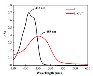
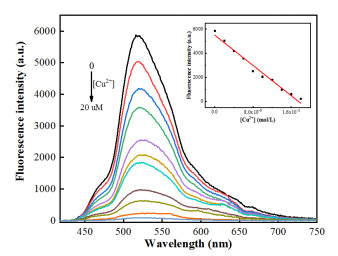
The UV-Vis spectra of L and L-Cu2+ were measured. As shown in Fig. 1, the maximum absorption wavelength of L was red-shifted from 413 to 455 nm, with adding Cu2+ ion. To get insight into fluorescence intensity changes with the increase of Cu2+ concentration, the fluorescence spectra changes of L towards Cu2+ were measured. As shown in Fig. 2, upon the addition of Cu2+ to the solution of L, the fluorescence spectra at 519 nm clearly quenched under the excitation of 393 nm. Moreover, there was a good linear relationship between the fluorescence intensity of probe L and the concentration of Cu2+ in the range of 0 to 20 μmol·L-1. According to the reported definition (LOD = 3σ/k), the detection limit of L for Cu2+ was found to be 19 nmol·L-1. The fluorescence spectra results proved that L has a high sensitivity toward Cu2+.
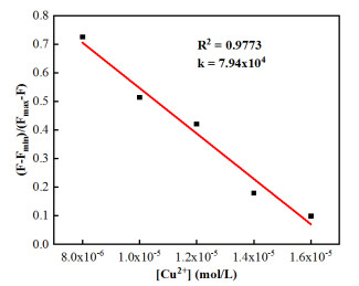
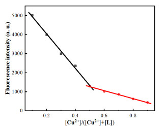
The binding constant K of L-Cu2+ was calculated according to the Benesi-Hildebrand equation. Depending on the slope, the results obtained were KL = 7.94 × 10⁴ L·mol-1, indicating that L had a great binding affinity to Cu2+ (Fig. 3). In order to study the binding of L with Cu2+, the Job plot was measured by using a total concentration of 16 μmol·L-1 L and Cu2+, and the result indicated that the combination of L and Cu2+ is 1:1 stoichiometry (Fig. 4).
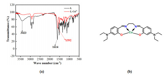
The infrared spectra of L and L-Cu2+ were measured. As shown in Fig. 5a, the absorption peak of C=N group in L was shifted from 1614 to 1592 cm-1 with adding Cu2+ ion because the electron cloud density of N atom in C=N group decreased after L complexed with Cu2+. And the absorption signal of OH group in L disappeared from 3423 cm-1, with adding the Cu2+ ion, which indicated that OH was deprotonated. By combining the above information, the binding mode of L and Cu2+ was speculated, as shown in Fig. 5b.
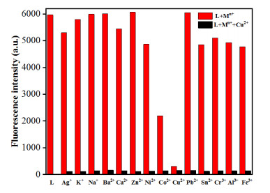
To further understand the fluorescent property of L with Cu2+, the fluorescence response behaviors of L to various metal ions were investigated (Fig. 6). The results showed no obvious changes in the fluorescence intensity after adding other metal ions to L solution, including Ag+, K+, Na+, Ba2+, Ca2+, Zn2+, Ni2+, Pb2+, Sn2+, Cr3+, Al3+ and Fe3+. However, fluorescence intensities declined with adding Co2+ possibly because of the heavy metal effect. Moreover, the competitive experiments proved that the presence of metal ions, such as Ag+, K+, Na+, Ba2+, Ca2+, Co2+, Zn2+, Ni2+, Pb2+, Sn2+, Cr3+, Al3+ and Fe3+ ions, did not interfere with the quenched fluorescence. The results indicated that L has good sensibility and selectivity toward Cu2+.
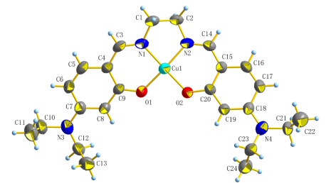
The crystal of Schiff-base L-Cu2+ was cultivated to further confirm the real binding mode of L and Cu2+ ion. The molecular structure of L-Cu2+ is shown in Fig. 7. It is obvious that L and Cu2+ ion are combined according to the ratio of 1:1. Cu and N(1), N(2), O(1), O(2) form four coordinate bonds, with the H atom of the phenol group disappearing.
In summary, one novel Schiff-base probe L was developed as efficient chemical sensor for the highly selective detection of Cu2+. The maximum absorption wave length of L was red-shifted from 413 to 455 nm, with adding Cu2+ ion. After the addition of Cu2+ ion, the fluorescence quenching of L was observed at 519 nm under the excitation of 393 nm, which did not take effect for other metal ions. Probe L is highly selective for Cu2+ detection, which may be attributed to the fact that the Jahn-Teller deformation of Cu2+ complexes provides excellent thermodynamic stability among all metal cations. The job plot revealed that the binding ratio of probe L to Cu2+ ion is 1:1, which could be further confirmed by the crystal structure of the complex L-Cu2+. Cu2+ ion detection limit of probe L was measured to be 19 nmol·L-1. Our group job herein could provide certain new insights for exploring highly selective and sensitive Schiff-base fluorescent sensors.
Zhu, Y. M.; Wang, Z. L.; Yang, J.; Xu, X.; Wang, S. F.; Cai, Z. C.; Xu, H. J. N, N-bis(2-pyridylmethyl)amine-based truxene derivative as a highly sensitive fluorescence sensor for Cu2+ and Ni2+ ion. Chin. J. Org. Chem. 2019, 39, 427–433. doi: 10.6023/cjoc201807042
Tachapermpon, Y.; Chaneam, S.; Charoenpanich, A.; Sirirak, J.; Wanichacheva, N. Highly Cu2+ sensitive and selective colorimetric and fluorescent probes: utilizations in batch, flow analysis and living cell imaging. Sensor Actuat. B-Chem. 2017, 241, 868–879. doi: 10.1016/j.snb.2016.11.009
Meng, X. J.; Zhao, J. Z.; Ma, W. B. Progress in fluorescent probes for Cu2+ and anions, neutral molecules sequential recognition. Chin. J. Org. Chem. 2020, 40, 276–283. doi: 10.6023/cjoc201908039
Xu, J. B.; Liu, N.; Hao, C. W.; Han, Q. Q.; Duan, Y. L.; Wu, J. A novel fluorescence "on-off-on" peptide-based chemosensor for simultaneous detection of Cu2+, Ag+ and S2–. Sensor Actuat. B-Chem. 2019, 280, 129–137. doi: 10.1016/j.snb.2018.10.038
Barnham, K. J.; Masters, C. L.; Bush, A. L. Neurodegenerative diseases and oxidative stress. Nat. Rev. Drug Discov. 2004, 3, 205–214. doi: 10.1038/nrd1330
Zeng, L.; Miller, E. W.; Pralle, A.; Isacoff, E. Y.; Chang, C. J. A selective turn-on fluorescent sensor for imaging copper in living cells. J. Am. Chem. Soc. 2006, 128, 10–11. doi: 10.1021/ja055064u
Carter, K. P.; Young, A. M.; Palmer, A. E. Fluorescent sensors for measuring metal ions in living systems. Chem. Rev. 2014, 114, 4564–4601. doi: 10.1021/cr400546e
Zhang, B. B.; Qin, F. Y.; Niu, H. W.; Liu, Y.; Zhang, D.; Ye, Y. A highly sensitive and fast responsive naphthalimide-based fluorescent probe for Cu2+ and its application. New J. Chem. 2017, 41, 14683–14688. doi: 10.1039/C7NJ02813J
Tang, J.; Ma, S. G.; Zhang, D.; Liu, Y. Q.; Zhao, Y. F.; Ye, Y. Highly sensitive and fast responsive ratiometric fluorescent probe for Cu2+ based on a naphthalimide-rhodamine dyad and its application in living cell imaging. Sensor Actuat. B-Chem. 2016, 236, 109–115. doi: 10.1016/j.snb.2016.05.144
Cai, F. J.; Xia, F.; Guo, Y. X.; Zhu, W. H.; Fu, B.; Liang, X.; Wang, S. F.; Cai, Z. C.; Xu, H. J. "Off-on-off" type of selectively pH-sensing 8-hydroxyquinoline-substituted gallium(III) corrole. New J. Chem. 2019, 43, 18012–18017. doi: 10.1039/C9NJ04544A
Tao, J. Y.; Song, W. T.; Sun, L.; Li, Z. Z.; Fu, B.; Cai, Z. C.; Xu, H. J. Synthesis, photo-physical properties and cell imaging of meso-2, 6-dichlorophenyl boron-dipyrromethene derivatives. Chin. J. Struct. Chem. 2019, 38, 1503–1510.
Saura, A. V.; Burguete, M. I.; Galindo, F.; Luis, S. V. Novel fluorescent anthracene-bodipy dyads displaying sensitivity to pH and turn-on behaviour towards Cu(II) ions. Org. Biomol. Chem. 2017, 15, 3013–3024. doi: 10.1039/C7OB00274B
Song, Y. T.; Tao, J. Y.; Wang, Y.; Cai, Z. C.; Fang, X. Y.; Wang, S. F.; Xu, H. J. A novel dual-responsive fluorescent probe for the detection of copper(II) and nickel(II) based on BODIPY derivatives. Inorg. Chim. Acta 2021, 516, 120099–7. doi: 10.1016/j.ica.2020.120099
Chen, W. T. Structure, photophysical and electrochemical properties of a copper porphyrin with a three-dimensional framework. Acta Crystallogr. C 2020, 76, 133–138.
Lodeiro, C.; Pina, F. Luminescent and chromogenic molecular probes based on polyamines and related compounds. Coord. Chem. Rev. 2009, 253, 1353–1383.
Li, Z. Y.; Jia, G. K.; Yuan, L.; Bai, P. F.; He, H.; Zhou, Q. Syntheses, crystal structures and biological activities of three new Schiff bases derived from substituted salicylaldehyde and tris base. Chin. J. Struct. Chem. 2017, 36, 1792–1802.
Bruker: APEX3, SAINT-Plus, XPREP. Bruker AXS Inc., USA 2016.
Burla, M. C.; Caliandro, R.; Camalli, M.; Carrozzini, B.; Cascarano, G. L.; De Caro, L.; Giacovazzo, C.; Polidori, G.; Spagna, R. SIR2004: an improved tool for crystal structure determination and refinement. J. Appl. Crystallogr. 2005, 38, 381–388.
Sheldrick, G. M. SHELXS-97, Program for X-ray Crystal Structure Solution. University of Göttingen, Germany 1997.
Dolomanov, O. V.; Bourhis, L. J.; Gildea, R. J.; Howard, J. A. K.; Puschmann, H. OLEX2: a complete structure solution, refinement and analysis program. J. Appl. Crystallogr. 2009, 42, 339–341.

Figure 2 Fluorescence spectra of L (16 μmol/L) in the presence of different concentrations of Cu2+ (from 0 to 20 μmol·L-1)

Figure 6 Relative emission of L (16 μmol/L) and its complexation with Cu2+ (32 μmol/L) in the presence of different metal ions (32 μmol/L). The response of L on its own is used as a control

 扫一扫看文章

扫一扫关注我们
 DownLoad:

 下载:
下载:


