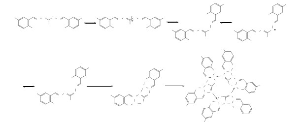
Scheme 1.
Ketone-enol and its dehydrogenation to anion interconversion of 1,5-disubstituted carbohydrazide and metal coordination; R = Cl(T1), Me(T2); Butyl was omitted for clarity
Two Novel Dibutyltin Complexes with Trimers and Hexanuclear Based on the Bis(5-Cl/Me-salicylaldehyde) Carbohydrazide: Syntheses, Structures, Fluorescent Properties and Herbicidal Activity
Yong-Lan FENG , Fu-Xing ZHANG , Dai-Zhi KUANG , Chun-Lin YANG

The 1,5-bis-salicylaldehyde-carbazide compound formed by the condensation reaction of salicylaldehyde and its derivatives with carbazide not only has good biological activity[1, 2], but also is a good ligand containing multiple coordination points of O and N. The C=N double bond produces cis- and trans-isomerization, the C–N and N–N bonds rotate to produce spatial conformation, and the amide (Ar-CH=N-NH-C(O/S)-NH-N=CH-Ar) chain often undergoes ketone and enol conversion[3, 4]. Oxygen and nitrogen anions are produced by the cleavage and dehydrogenation of O–H and N–H bonds of ligands. This carbonyl (> C=O), ammonia (> C=N), or phenolic hydroxyl (Ar-OH) oxygen and amino (> N–H) nitrogen have lone pair electrons, and so on. Therefore, ligands and metals have many kinds of coordination forms, resulting in metal complexes with various structures. Also, organotin compounds have good biological activity[5], and tin atoms have special bonding effect. They not only form organotin compounds RnSnX4-n (n: 1~4) containing Sn–C covalent bonds with carbon atoms, but also form organotin complexes RnSnX4-nLx containing N, O, S and other atoms to coordinate with Sn. Organotin compounds[6-8] with rich and peculiar structures and properties. Therefore, the combination of two biologically active bis(substituted salicylaldehyde) carbazides with organotin can be helpful for the study of organotin's biological activity and pharmaceuticals[9-11]. In this paper, the ligands bis(5-chlorosalicylaldehyde) carbazide (L1) and bis(5-methylsalicylaldehyde) carbazide (L2) were prepared by the reaction of carbazide with 5-chlorosalicylaldehyde and 5-methylsalicylaldehyde, and two hexanuclear triangular dibutyltin complexes (T) were synthesized by the reaction of dibutyltin oxide with ligands in microwave methanol solvothermal environment, respectively. The fluorescence properties of the complexes in DMF and DMF-water mixed solvents, the herbicidal activities of the ligands and their complexes against Portulaca oleracea L., Amaranthus spinosus L., Cassia tora L., Brassica campestris L. ssp. chinensis var. utilis Tsen et Lee and Amaranthus tricolor L. were preliminarily studied.
MicroSYNT Lab station for microwave assisted (ltaly). The melting points were obtained on an X-4 microscopic melting point apparatus and uncorrected. Elemental analyses of C, H and N were performed with a Perkin-Elmer 2400 Ⅱ elemental analyzer. IR spectra (KBr discs) were recorded on a Shimadzu FTIR 8700 spectrometer (4000~400 cm-1 range). 1H and 13C NMR spectra were measured on a Bruker Avance 500 spectrometer (Switzerland) with CDCl3 as solvent and TMS as an internal standard. Crystal structure was determined on a Bruker Smart Apex Ⅱ CCD diffractometer (Germany). F-7000 Fluorescence Spectrometer (Shimadzu). MGC-HP intelligent artificial climate box (Shanghai Yiheng Scientific Instrument Co., Ltd.).
All reagents were purchased from commercial supplies (Energy chemical reagent Co., Lid; grade: CP) without further purification.
According to reference[12], a mixture of 5-chlorosalicylaldehyde (40 mmol, 6.26 g) or 5-methylsalicylaldehyde (40 mmol, 5.446 g), carbohydrazide (20 mmol, 1.80 g) and ethanol-acetic acid (50 mL, V/V = 7:3) was placed in a 100 mL reactor, then the mixture was stirred and heated to reflux for 5 h. After cooling down to room temperature, the solution was filtered and the solvent was removed by vacuum evaporation. The crude product was recrystallized from appropriate solvent to yield bis(5-chlorosalicylaldehyde) carbazide (L1) and bis(5-methylsalicylaldehyde) carbazide (L2)
L1, white powder 4.544 g, yield: 61.88%, m.p.: 102~103 ℃. Anal. Calcd. (%) for C15H12Cl2N4O3: C, 49.07; H, 3.29; N, 15.26. Found (%): C, 49.05; H, 3.29; N, 15.27. IR: 3239(m, νO-H, νN-H), 3044(m, νAr-H), 1680(s, νC=O, νC=N), 1622, 1594, 1485(s, νC=C) cm-1. 1H NMR (CDCl3) δ (ppm): 11.05~10.89(m, 4H, O-H, N-H), 9.01~8.94(m, 2H, H-C=N), 6.90~8.51(m, 6H, Ar-H).
L2, pale yellow powder 4.508 g, Yield: 69.07%, m.p.: 148~149 ℃. Anal. Calcd. (%) for C17H18N4O3: C, 62.56; H, 5.66; N, 17.17. Found (%): C, 62.56; H, 5.64; N, 17.15. IR: 3254(m, νO-H, νN-H), 3032(m, νAr-H), 2911(m, νC-H), 1653(s, νC=O, νC=N), 1624, 1568(s, νC=C) cm-1. 1H NMR (CDCl3) δ (ppm): 10.85~10.51(m, 4H, O-H, N-H), 8.29~8.39(m, 2H, H-C=N), 7.30~6.70(m, 6H, Ar-H), 2.25(s, 6H, -CH3).
A mixture of 1 mmol ligand, 2 mmol dibutyl tin oxide, and 15 mL anhydrous methanol was placed in a 50 mL Teflon-lined reactor and set on the microwave power 800W of MicroSYNT Lab station for microwave assisted synthesis. The mixture was heated by microwave radiation for two hours. The reactants were naturally cooled down to room temperature. Then the solution was obtained by filtration, and the filtrate was removed by evaporation in vacuo. Crystals of the complexes were obtained by recrystallization from methanol.
T1, pale yellow crystal, 1.967 g. Yield: 76.83%, m.p.: 212~213 ℃. Anal. Calcd. (%) for C96H139O10N13Cl6Sn6: C, 45.04; H, 5.47; N, 7.11. Found (%): C, 45.09; H, 5.48; N, 7.08. IR: 2955(m, νAr-H), 1663(s, νC=N), 1638, 1603, 1420(s, νC=C), 546(s, νSn-O), 507(s, νSn-N), 449(s, νSn-C) cm-1. 1H NMR (CDCl3) δ (ppm): 8.39~7.73(m, 6H, H-C=N), 7.31~6.60(m, 18H, Ar-H), 2.96~2.89(m, 24H, SnCH2-), 1.66~0.85(m, 84H, methyl and methylene of butyl). 13C NMR (CDCl3) δ (ppm): 165.75, 163.54, 162.59 (C=O), 154.52, 153.94(C=N), 146.12~117.27(Ar-C), 36.55~13.60 (-CH2-, -CH3).
T2, yellow crystal, 1.334 g. Yield: 56.42%, m.p.: 224~246 ℃. Anal. Calcd. (%) for C99H150O9N12Sn6: C, 50.29; H, 6.39; N, 7.11. Found (%): C, 50.29; H, 6.38; N, 7.12. IR: 2953(m, νAr-H), 1614(s, νC=N), 1590, 1554, 1481(s, νC=C), 561(s, νSn-O), 525(s, νSn-N), 446(s, νSn-C) cm-1. 1H NMR (CDCl3) δ (ppm): 8.36~8.05(m, 6H, H-C=N), 7.09~6.68 (m, 18H, Ar-H), 2.26~2.25(m, 24H, SnCH2-), 1.67~0.84(m, 102H, methyl and methylene of butyl). 13C NMR (CDCl3) δ (ppm): 162.93, 162.81(C=O), 155.91, 147.41, 144.35(C=N), 134.68~116.69 (Ar-C), 27.48~13.60(-CH2-, -CH3).
The crystal data were collected by a Bruker Smart Apex Ⅱ CCD diffractometer (MoKα radiation, λ = 0.71073 Å). The structure was solved by direct methods with SHELXS program and refined by full-matrix least-squares technique using the SHELXL-2014/7. Hydrogen atoms were placed in calculated positions or located from difference Fourier maps, and refined isotropically with isotropic vibration parameters related to the non- hydrogen atom to which they are bound. All calculations were performed with SHELXL[12] program within WINGX.
The crystal of complex T1 with dimensions of 0.23mm × 0.21mm × 0.20mm was selected. The data were collected by the diffractometer in the ranges of 2.47≤θ≤26.00º, –18≤h≤18, –19≤k≤19 and –31≤l≤31. A total of 59973 reflections were collected and 21893 were independent (Rint = 0.0328), of which 15739 observed reflections with I > 2σ(I) were used in the succeeding refinements. Empirical formula, C96H139Cl6N13O10Sn6, 2560.03 g/mol, triclinic system, space group P
The crystal of complex T2 with dimensions of 0.20mm × 0.20mm × 0.20mm was selected. The data were collected by the diffractometer in the ranges of 1.37≤θ≤25.10º, –27≤h≤34, –20≤k≤15 and –51≤l ≤51. A total of 53509 reflections were collected and 19424 were independent (Rint = 0.0383), of which 13924 with I > 2σ(I) were used in the succeeding refinements. Empirical formula, C99H150N12O9Sn6, 2364.44 g/mol, monoclinic system, space group C2/c, a = 29.287(3), b = 17.2756(16), c = 43.446(4) Å, β = 92.4030(10)°, V = 21962(3) Å3, T = 296(2) K, Z = 8, Dc = 1.430 Mg/m3, µ = 1.400 mm-1 and F(000) = 9600. A full-matrix least-squares refinement gave the final R = 0.1141, wR = 0.2909 (w = 1/[σ2(Fo2) + (0.1201P)2 + 986.0775P], where P = (Fo2 + 2Fc2)/3), S = 1.001, (Δρ)max = 2.058 and (Δρ)min = –1.999 e/Å3.
The herbicidal activities of compounds were determined by plating method[13, 14]. The root and stem lengths of the target plants, such as Portulaca oleracea L., Amaranthus spinosus L., Cassia tora L., Brassica campestris L. ssp. chinensis var. utilis Tsen et Lee and Amaranthus tricolor L., were determined. Each treatment was repeated thrice and the growth inhibition rate (I) of target plants was calculated by comparing the difference between the average plant growth length (l0) of water as a reference solution and the average plant growth length (l1) of the treated plants compared with the l0 value. If I is positive, the complex has inhibitory effect on target plants, while when I is negative, it can promote the target plants.
|
|
Comparing the characteristic peaks of infrared spectra of ligands and their complexes, the association of ligands led to the shift of stretching vibration absorption peaks of O–H radicals to low wave numbers (3239 (L1) cm–1, 3254 (L2) cm–1) and overlapped with the stretching vibration peaks of N–H radicals. In the complexes, the infrared characteristic peaks of ν(O–H) and ν(N–H) disappeared, and weak characteristic peaks of ν(Sn–O) and_ν(Sn–N) appeared in 546 (T1), 561 (T2) cm–1 and 507 (T1), 525 (T2) cm–1[15, 16], respectively. The ν(C=O) and ν(C=N) peaks of ligands almost overlap. In the complexes, the characteristic peaks shift to low wavenumber 17 cm–1 (T1) and 39 cm–1 (T2), respectively. These two bonds may be weakened by the co-coordination of keto-enol oxygen and C=N and tin. In addition, the coordination between the ligand and tin also affects the shift of ν(C–H) characteristic peaks of benzene rings to low wavenumbers 88.8 cm–1 (T1) and 79.1 cm–1 (T2).
In the complexes, the chemical shifts of N=C–H and Ar–H protons shift to higher fields. Moreover, the chemical shifts of hydroxyl and amino H protons of the ligands disappeared from 11.05 to 10.89 (L1) and 10.85 to 10.51 (L2) ppm. The results are supported by infrared spectroscopy. The carbonyl group (C=O) of the complexes and the carbon of Schiff base (N=C) are affected by oxygen and nitrogen, and the characteristic lines are formed at the high chemical shift in 13C NMR. For example, the spectral lines of carbonyl carbon are 165.75, 163.54, 162.59 ppm (T1) and 162.93, 162.81 ppm (T2), and Schiff base carbon are 154.52, 153.94 ppm (T1) and 155.91, 147.41, 144.35 ppm (T2). There are also carbon spectra of benzene ring carbon and methyl and methylene, characterizing and supporting the structures of complexes.
IR and 1H NMR analyses of the ligands and their complexes show that the ligands not only convert ketone (Ⅰ) to enol (Ⅱ)[17], but also form (Ⅲ) by C–N rotation. Then amino nitrogen dehydrogenation produces nitrogen anion (Ⅳ) and three-atom four-electron conjugation system (Ⅴ) with adjacent N=C bond. Coordination with dibutyltin forms a unit structure (To), three of which coordinate with each other through the nitrogen atom of C=N to produce the title complex (T), as shown in Scheme 1.
Theoretical optimization and single point calculation of energy difference[18], ΔE1(EⅡ – EⅠ), ΔE2 (EⅣ – EⅡ), and ΔE3 (EⅣ – EⅠ), for different R systems by G03W/hf/6-31g were carried out. When R is chlorine, ΔE1, ΔE2 and ΔE3 are –138.51, 1603.10 and 1741.62 kJ·mol–1; when R is Me, they are –125.73, 1655.32 and 1781.05 kJ·mol-1, respectively. It can be seen that the system energy of conversion from ketone (Ⅰ) to enol (Ⅱ) is lower and more stable, but the conversion of enol (Ⅱ) to anion (Ⅳ) increases the system energy by 1740~1780 kJ-1. The stabilization energy can be formed by the formation of complexes through (Ⅳ) coordination with tin.
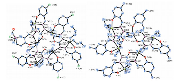
Crystal molecular structure (Fig. 1) and bond parameters (Table 1) show that a phenoxy, imine N and dehydrogenated nitrogen coordinate with dibutyltin to form a unit structure. The bond lengths and bond angles of tin and ligand atoms are different. Tin and ligand atoms form a five-coordinated deformed triangular bipyramidal configuration. Another phenoxy, imino N and enoxy of the ligand and the enol of the adjacent ligand produce imino nitrogen which coordinates with another dibutyltin. The bond lengths and bond angles of the ligand are also different. Tin and the coordination atoms form a hexacoordinate distorted octahedron configuration. Because the ligand produces enoltype conjugated azo with imine and participates in the coordination of adjacent tin, a trihexanuclear dibutyltin complex is constructed by cross-coordination of three unit structures. The three six-coordinated tin atoms Sn(2), Sn(4) and Sn(6) are located in the core to form a small triangle, and the coordination oxygen, nitrogen and carbon atoms form a 12-membered ring. Three five-coordinated tin atoms Sn(1), Sn(3) and Sn(5) form a large triangle at the outer. The small triangle is inverted in the large triangle to form a spatial skeleton structure. The plane angles of the two triangles are 7.87° (T1) and 0.58° (T2), respectively, as shown in Fig. 2. The edge lengths of each triangle are unequal, and the corresponding edges of complex T2 are slightly longer than those of the triangle of T1. It is possible that the spatial effect of methyl on six benzene rings is larger than that of the chlorine atoms.
 DownLoad:
CSV
DownLoad:
CSV
| T1 | T2 | T1 | T2 | |||
| Bond | Dist. | Bond | Bond | Dist. | Dist. | |
| O(1)–Sn(1) | 2.089(8) | 2.101(12) | N(11)–Sn(6) | 2.423(7) | 2.449(11) | |
| O(2)–Sn(2) | 2.104(6) | 2.087(9) | N(1)–N(2) | 1.389(9) | 1.383(16) | |
| O(3)–Sn(2) | 2.365(6) | 2.339(10) | N(3)–N(4) | 1.398(9) | 1.402(16) | |
| O(4)–Sn(3) | 2.098(7) | 2.061(11) | N(5)–N(6) | 1.391(9) | 1.388(16) | |
| O(5)–Sn(4) | 2.098(7) | 2.062(11) | N(7)–N(8) | 1.393(9) | 1.385(19) | |
| O(6)–Sn(4) | 2.341(6) | 2.337(10) | N(9)–N(10) | 1.397(10) | 1.370(15) | |
| O(7)–Sn(5) | 2.063(7) | 2.080(10) | N(11)–N(12) | 1.400(9) | 1.399(15) | |
| O(8)–Sn(6) | 2.093(6) | 2.082(10) | Sn(1)…Sn(3) | 10.490 | 10.933 | |
| O(9)–Sn(6) | 2.393(5) | 2.352(11) | Sn(1)…Sn(5) | 10.846 | 10.892 | |
| N(2)–Sn(1) | 2.238(6) | 2.221(12) | Sn(3)…Sn(5) | 10.858 | 10.921 | |
| N(4)–Sn(1) | 2.229(7) | 2.163(13) | Sn(1)…Sn(2) | 5.503 | 5.504 | |
| N(3)–Sn(2) | 2.423(6) | 2.419(12) | Sn(2)…Sn(3) | 5.481 | 5.432 | |
| N(5)–Sn(2) | 2.416(7) | 2.375(13) | Sn(3)…Sn(4) | 5.552 | 5.490 | |
| N(6)–Sn(3) | 2.224(7) | 2.199(13) | Sn(4)…Sn(5) | 5.577 | 5.439 | |
| N(8)–Sn(3) | 2.190(7) | 2.188(11) | Sn(5)…Sn(6) | 5.437 | 5.486 | |
| N(7)–Sn(4) | 2.467(7) | 2.409(15) | Sn(1)…Sn(6) | 5.423 | 5.414 | |
| N(9)–Sn(4) | 2.353(7) | 2.352(10) | Sn(2)…Sn(6) | 5.059 | 5.174 | |
| N(10)–Sn(5) | 2.247(7) | 2.220(11) | Sn(2)…Sn(4) | 5.129 | 5.138 | |
| N(12)–Sn(5) | 2.179(7) | 2.157(11) | Sn(4)…Sn(6) | 5.079 | 5.112 | |
| N(1)–Sn(6) | 2.360(7) | 2.354(12) | ||||
| Angle | (°) | (°) | Angle | (°) | (°) | |
| O(1)–Sn(1)–N(4) | 83.6(3) | 82.5(5) | O(5)–Sn(4)–N(9) | 79.4(2) | 80.6(4) | |
| O(1)–Sn(1)–N(2) | 154.8(3) | 151.3(5) | O(6)–Sn(4)–N(7) | 132.1(2) | 132.4(4) | |
| N(4)–Sn(1)–N(2) | 71.3(2) | 71.3(4) | O(6)–Sn(4)–N(9) | 68.6(2) | 67.8(3) | |
| O(2)–Sn(2)–O(3) | 144.2(2) | 145.9(4) | N(9)–Sn(4)–N(7) | 159.2(2) | 159.5(4) | |
| O(2)–Sn(2)–N(3) | 81.9(2) | 82.9(4) | O(7)–Sn(5)–N(10) | 152.4(3) | 154.9(4) | |
| O(2)–Sn(2)–N(5) | 77.4(2) | 79.5(4) | O(7)–Sn(5)–N(12) | 83.2(3) | 83.5(4) | |
| O(3)–Sn(2)–N(3) | 133.6(2) | 130.8(4) | N(12)–Sn(5)–N(10) | 71.2(3) | 71.9(4) | |
| O(3)–Sn(2)–N(5) | 67.0(2) | 67.7(4) | O(8)–Sn(6)–O(9) | 147.5(2) | 145.9(4) | |
| N(5)–Sn(2)–N(3) | 159.3(2) | 160.7(4) | O(8)–Sn(6)–N(1) | 79.4(2) | 78.9(4) | |
| O(4)–Sn(3)–N(6) | 151.4(3) | 153.3(4) | O(8)–Sn(6)–N(11) | 81.5(2) | 82.1(4) | |
| O(4)–Sn(3)–N(8) | 82.3(3) | 83.3(4) | O(9)–Sn(6)–N(1) | 68.1(2) | 67.2(4) | |
| N(8)–Sn(3)–N(6) | 70.8(3) | 70.7(4) | O(9)–Sn(6)–N(11) | 131.0(2) | 132.0(4) | |
| O(5)–Sn(4)–O(6) | 147.7(2) | 148.2(4) | N(1)–Sn(6)–N(11) | 160.2(2) | 160.8(4) | |
| O(5)–Sn(4)–N(7) | 80.2(2) | 79.3(5) |
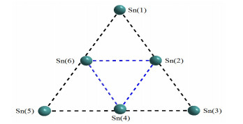
The geometric data of Sn···Sn are shown in Table 1
In crystal stacking, some weak interactions are formed between adjacent molecules. The intermolecular hydrogen bond geometry data of complexes are shown in Table 2. For example, in complex T1, H(44) forms a hydrogen bond with adjacent O(10ⅰ) atoms, ∠C(44)–H(44)⋅⋅⋅O(10ⅰ): 0.2380 nm, 158.44º. In addition, some hydrogen bonds such as H(60B)⋅⋅⋅Cl(1ⅱ), H(82A)⋅⋅⋅Cl(4ⅲ), H(84B)⋅⋅⋅Cl(4ⅲ) and H(70B)⋅⋅⋅Cl(6ⅳ) are formed by the interaction of H(60B), H(82A), H(84B) and H(70B) atoms with adjacent Cl(1ⅱ), Cl(4ⅲ) and Cl(6ⅳ) atoms, respectively. Here, there are also C–H⋅⋅⋅H–C actions. The three-dimensional supramolecular structure is formed by these weak interactions between adjacent molecules.
DownLoad:
CSV
| Comp. | D–H…A | D–A | H…A | D…A | ∠DHA | Symmetry code |
| T1 | C(44)–H(44)…O(10ⅰ) | 0.931 | 2.380 | 3.264 | 158.44 | ⅰ1+x, y, z |
| C(60)–H(60B)…Cl(1ⅱ) | 0.970 | 2.914 | 3.426 | 114.09 | ⅱ1–x, 1–y, 1–z | |
| C(82)–H(82A)…Cl(4 iii) | 0.971 | 2.819 | 3.563 | 134.02 | ⅲ–1+x, y, z | |
| C(84)–H(84B)…Cl(4ⅲ) | 0.969 | 2.926 | 3.661 | 132.52 | ⅲ–1+x, y, z | |
| C(70)–H(70B)…Cl(6ⅳ) | 0.970 | 2.859 | 3.815 | 168.52 | ⅳ1–x, –y, 2–z | |
| T2 | C(13)–H(13)…O(7ⅰ) | 0.930 | 2.782 | 3.208 | 113.66 | ⅰx, 1+y, z |
DMF-H2O (V: V) was used to prepare complex solutions with concentrations of 0.3906 (T1) and 0.4229 (T2) μM·L-1, respectively. At room temperature, the T1 (or T2)-DMF-H2O solution system with a volume ratio of DMF to water of 7:3 was used to determine the ultraviolet-visible absorption spectra. The maximum absorption peaks of T1-DMF-H2O and T2-DMF-H2O solution systems were found at 414 and 288 nm, respectively, and further confirmed by 3D fluorescence scanning. The fluorescence emission spectra of T1-DMF-H2O and T2-DMF-H2O systems with different volume fractions of water were measured by fluorescence spectrometer with 414 and 288 nm as excitation wavelength, respectively. The effect of water on the fluorescence intensity of the solution system was investigated, as shown in Fig. 3. The DFM-water solution system of complex T1 emits strong fluorescence at 456 to 510 nm, which may be caused by the transfer of electrons from tin to ligand. The fluorescence intensity varies with the change of water content. When the water content reaches 10%, the fluorescence intensity reaches the maximum value[19]. This indicates that the system has aggregation-enhanced emission (AEE) characteristics in the range of 0~10% water content[20]. With further increasing the water content, the fluorescence intensity decreases. When the water content gets to 50%, the fluorescence almost quenches. It may be that the instability of solution system leads to the loss of agglomeration fluorescence enhancement effect. In addition, the wavelength of fluorescence emission peaks shifted red first and then purple with the increase of water content in the solution system, suggesting that the water may also affect the luminescence of T1-DMF-H2O system.
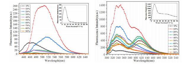
The DFM-water solution system of complex T2 emits strong fluorescence from ligand π-π transition at 332~348 nm and from electron transfer to ligand at 392~414 nm under 288 nm excitation. The fluorescence intensity of emission spectra at 332~348 nm with different volume fractions of water is similar to that of T1-DMF-H2O system. However, the emission of 392~414 nm varies with the volume fraction of water in the solution system. When the volume fraction of water is 0, 20, 40 and 90%, the emission of the system is not obvious. When the volume fraction of water is 10, 30, 50, 60, 70 and 80%, the emission of the spectrum appears, and the final fluorescence quenching is weakened with the increase of water content in solution system. This phenomenon needs further study, which can provide reference for the study of Bi (5-R-salicylaldehyde) carbazide organotin aggregation-induced luminescent materials.
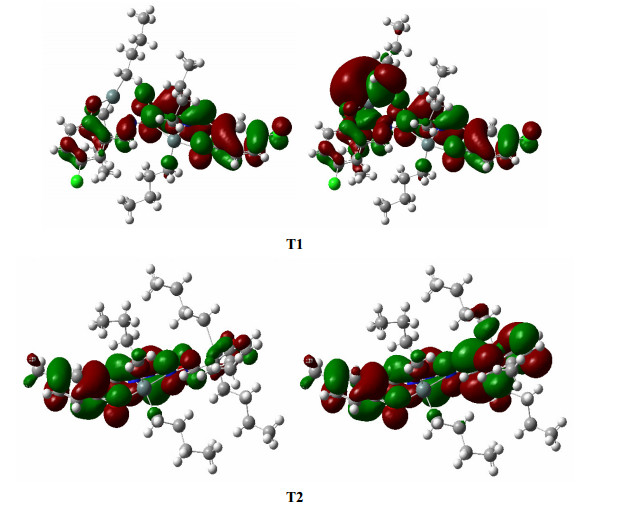
In order to understand the luminescence, according to the atomic coordinates of the crystal structure, the molecular orbital and electron cloud distribution of the complex unit structure are calculated by using the G03W program at the DFT/b3lyp/lanl2dz base group[21]. The distribution of electron clouds on these atoms is expressed by the sum of squares of the atomic orbital coefficients involved in the combined molecular orbitals. The atoms of the complexes are divided into 10 parts: tin atom Sn, hydroxyl atom and carbonyl oxygen atom O, nitrogen atom N, chlorine atom (T1) or methyl atom carbon (T2), Schiff base carbon atom C (C=N), benzene Cy (1), benzene Cy (2), carbonyl carbon atom C (C=O), butyl carbon atom C (Bu) and atomic H. The results are shown in Table 3 and Fig. 4. The energy gap between the front unoccupied orbital and the front occupied orbital is 0.1047 a.u. ΔE(T1) and 0.1084 a.u. ΔE(T2), respectively, indicating the two complexes are stable. The carbon of carbonyl, oxygen, hydrogen and the chlorine (T1) or methyl carbon (T2) atoms on the benzene ring have similar distributions in the HOMO and LUMO orbitals, and the contribution of other atoms to the frontier orbital composition is obviously different. In complex T1, the lowest unoccupied molecular orbital (LUMO) is mainly concentrated on the tin atom (46.73%) and butyl carbon atom connected with the tin atom, followed by benzene ring carbon atom (6.28%). The total distribution of other atoms to LUMO orbital is only 9.03%. However, the highest occupied molecular orbital (HOMO) of T1 is mainly distributed on the nitrogen atom (37.29%) and another benzene ring carbon atom (20.01%), followed by oxygen atom (15.19%) and imine carbon atom (10.57%), and other atoms are less distributed. Therefore, the conjugated delocalization system formed by the ligand with tin becomes a rigid structure, and the free rotation within the molecule is hindered to form a luminescent substance. When the excited LUMO electrons transfer to HOMO, the electron cloud of tin atom and butyl carbon atom connected with the tin atom is transferred to the nitrogen, oxygen, imino group and benzene ring atoms in the conjugated chain of ligand, so the complex produces luminescence phenomenon[22].
DownLoad:
CSV
| Comp. | FMO | E/a.u. | Sn | O | N | R | C(CN) | Cy(1) | Cy(2) | C(CO) | C(Bu) | H |
| T1 | HOMO | –0.1876 | 0.33 | 15.19 | 37.29 | 2.51 | 10.57 | 20.01 | 4.91 | 1.61 | 6.36 | 1.20 |
| LUMO | –0.0829 | 46.73 | 0.88 | 3.20 | 0.01 | 2.98 | 0.16 | 6.28 | 0.78 | 37.95 | 1.02 | |
| T2 | HOMO | –0.1760 | 0.76 | 14.97 | 38.53 | 0.24 | 10.28 | 3.64 | 22.77 | 2.03 | 5.45 | 1.30 |
| LUMO | –0.0676 | 14.12 | 2.19 | 19.66 | 0.39 | 16.09 | 29.79 | 1.56 | 1.65 | 13.00 | 1.55 |
Estimation of root (R) and stem (S) growth of five target plants of Portulaca oleracea L., Amaranthus spinosus L., Cassia tora L., Brassica campestris L. ssp. chinensis var. utilis Tsen et Lee and Amaranthus tricolor L. by ligand (L) and its butyltin complex (T) was investigated. Fig. 5 shows that both ligands and their complexes have inhibitory effects on the growth of target plants, but the effects of two ligands (L) on the growth of five target plants are significantly smaller than that of their complexes (T). In addition to the 100% inhibitory effect of L1 on the growth of Amaranthus spinosus L. at the test concentration of 100 mg·L-1, the inhibitory effect of L1 on the growth of the other four target plants is small. When L1 was combined with butyltin to form T1, except that T1 inhibited the growth of Amaranthus spinosus L. similar to L1, the inhibitory effects of T1 on Portulaca oleracea L., Cassia tora L., Brassica campestris L. ssp. chinensis var. utilis Tsen et Lee and Amaranthus tricolor L. were significantly higher than those of L1. When the test concentration of T1 was above 50 mg·L-1, the inhibitory effects on growth reached 100%, which had high-efficiency and broad inhibition on the target plant growth.
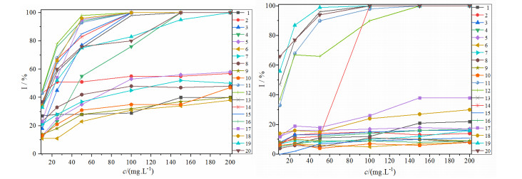
The growth inhibition rate of L2 on five target plants was 5~20%, and the growth inhibition of Cassia tora L. and Brassica campestris L. ssp. chinensis var. utilis Tsen et Lee was not obvious by butyltin complex T2, but that of T2 on Portulaca oleracea L., Amaranthus spinosus L. and Amaranthus tricolor L. increased significantly with the increase of test concentration. When the concentration was above 50 mg·L-1, the growth of three target plants was inhibited up to 100%, which could selectively inhibit the growth of Portulaca oleracea L., Amaranthus spinosus L. and Amaranthus tricolor L.. Thus it could be used as a candidate herbicide for Portulaca oleracea L., Amaranthus spinosus L. and Amaranthus tricolor L.
Two ligands, 1,5-bis(5-chlorosalicylaldehyde) carbazide (L1) and 1,5-bis(5-methylsalicylaldehyde) carbazide (L2), were prepared by the reaction of carbazide with 5-chlorosalicylaldehyde and 5-methylsalicylaldehyde, respectively. Furthermore, bis(5-chlorosalicylaldehyde) carbazide dibutyltin complex (T1) and bis(5-methylsalicylaldehyde) dibutyltin complex (T2) with hexanuclear triangular structure were synthesized by ligand reactions with dibutyltin oxide, respectively. The complexes exhibit fluorescence emission and good aggregation fluorescence enhancement properties in dimethylformamide and its aqueous solution of 0~10% volume fractions of water. Fluorescence quenching occurs when the aqueous volume fractions of water is greater than 10%. T1 can inhibit the growth of Portulaca oleracea L., Cassia tora L., Brassica campestris L. ssp. chinensis var. utilis Tsen et Lee and Amaranthus tricolor L. effectively and broadly. T2 can selectively inhibit the growth of Portulaca oleracea L., Amaranthus spinosus L., Amaranthus tricolor L., and can be used as a candidate herbicide for further study.
Gordon, J. A.; Jencks, W. P. The relationship of structure to the effectiveness of denaturing agents for proteins. Biochemistry 1963, 2, 47–57. doi: 10.1021/bi00901a011
Abdel-Aziz, A. H.; Ghabbour, H. A.; Eldehna, W. M.; Qabeel, M. M.; Fun, H. K. Synthesis, crystal structure, and biological activity of cis/trans amide rotomers of (Z)-N-(2-oxoindolin-3-ylidene)formohydrazide. Eur. J. Med. Chem. 2013, 70, 358–363. doi: 10.1016/j.ejmech.2013.09.060
Dragancea, D.; Addison, A. W.; Zeller, M.; Thompson, L. K.; Hoole, D.; Revenco, M. D.; Hunter, A. D. Dinuclear copper(Ⅱ) complexes with bis-thiocarbohydrazone ligands. Eur. J. Inorg Chem. 2008, 2530–2536.
Angelova, V. T.; Vassilev, N. G.; Nikolova-Mladenova, B.; Vitas, J.; Malbaša, R.; Momekov, G.; Djukic, M.; Saso, L. Antiproliferative and antioxidative effects of novel hydrazone derivatives bearing coumarin and chromene moiety. Med. Chem. Res. 2016b, 25, 2082–2092. doi: 10.1007/s00044-016-1661-4
Kuthubtheen, A. J.; Wickneswari, R.; Das, V. G. K. Structure-activity studies on the fungitoxicity of some triorganotin(Ⅳ) compounds. Appl. Organomet. Chem. 1989, 3, 231–242. doi: 10.1002/aoc.590030306
Wang, H.; Hu, L.; Du, W.; Tian, X. H.; Zhang, Q.; Hu, Z. J.; Luo, L.; Zhou, H. P.; Wu, J. Y.; Tian, Y. P. Two-photon active organotin(Ⅳ) carboxylate complexes for visualization of anticancer action. ACS Biomater. Sci. Eng. 2017, 3, 836–842. doi: 10.1021/acsbiomaterials.6b00786
Salam, M. A.; Affan, M. A.; Arafat, M. A.; Saha, R.; Nasrin, R. Synthesis, characterization, and antibacterial activities of organotin(Ⅳ) complexes with 2-acetylpyridine-N(4)-cyclohexylthiosemicarbazone (HAPCT). Heteroat Chem. 2012, 24, 43–52.
Sadiq, R.; Khadija, S.; Saqib, A.; Moazzam, H.; Bhatti, M. P. Organotin esterification of (E)-3-(3-fluorio-phenyl)-2-(4-chlorophenyl)-2-propenoic acid: synthesis, spectroscopic characterization and in vitro biological activities. Crystal structure of [Ph3Sn(OC(O)C(4-ClC6H4)=CH(3-FC6H4))]. J Organomet. Chem. 2005, 690, 1396–1408. doi: 10.1016/j.jorganchem.2004.12.004
Fang, X. N.; Dai, M. Z.; Li, X. H.; Kuang, R. Y. Synthesis and antimicrobial activities of dibutyltin (Ⅳ) complex of 1,5-bis(2-hydroxybenzylidene)thiocarbohydrazide. Chin. J. Chem. Res. Appl. 2009, 21, 680–684.
Salam, M. A.; Haque, R. A. Diorganotin(Ⅳ) complexes of 3,5-dichloro-2-hydroxybenzaldehyde-N(4)-ethylthiosemicarbazone: synthesis, spectral characterization and crystal structure. Inorg. Chim. Acta 2015, 435, 103–108. doi: 10.1016/j.ica.2015.06.015
Yang, C. L.; Feng, Y. L.; Zhang, F. X.; Yu, J. X.; Jiang, W. J.; Kuang, D. Z.; Yang, N. F. Microwave-solvent thermal syntheses, crystal struture and herbicidal activity of bis(3,5-di-t-butylsalicylaldehyde) carbohydrazide dibutyltin complexes. Chin J. Inorg. Chem. 2017, 33, 1397–1402.
Sheldrick, G. M. SHELXL-2014/7, University of Göttingen, Germany 2014.
Zhu, J. H.; Zheng, X. D.; Guo, G. Z.; Zhang, Y. Q.; Wu, B. W. Microwave-assisted synthesis of asymmetrical 1,5-disubstituted carbonohydrazide and crystal structure. Chin. J. Org. Chem. 2015, 35, 1975–1980. doi: 10.6023/cjoc201503004
Feng, Y. L.; Zhang, F. X.; Kuang, D. Z. Syntheses, structures and herbicidal activity of bis(5-R-2-hydroxy-benzylidene) thiocarbohydrazide monobutyltin complexes [R: H(T1), Me(T2)]. Chin. J. Struct. Chem. 2019, 38, 719–726.
Ruan, B. F.; Tian, Y.; Zhou, H. P.; Wu, J. Y.; Zhu, C. H.; Yang, J. X.; Zhu, H. L. Synthesis, characterization and in vitro antitumor activity of three organotin(Ⅳ) complexes with carbazole ligand. Inorg. Chim. Acta 2011, 365, 302–308. doi: 10.1016/j.ica.2010.09.024
Xiao, X.; Du, D. F.; Tian, M, Han, X.; Liang, J. W.; Zhu, D. S.; Xu, L. Organotin(Ⅳ) carboxylates based on benzenedicarboxylic acid derivatives: syntheses, crystal structures and characterizations. J. Organomet. Chem. 2012, 715, 54–63 doi: 10.1016/j.jorganchem.2012.05.038
Mirta, R.; Nives, G.; Ivan, H.; Tomislav, J.; Nenad, J.; Janez, P.; Primoz, S.; Predrag, N. Multiple solid forms of 1,5-bis(salicylidene)carbohydrazide: polymorph-modulated thermal reactivity. Cryst. Growth Des. 2014, 14, 2900–2912. doi: 10.1021/cg500203k
Frisch, M. J.; Trucks, G. W.; Schlegel, H. B. Gaussian 03, Revision B. 03, Gaussian, Inc.: Pittsburgh, PA 2003.
Feng, Y. L.; Kuang, D. Z.; Zhang, F. X.; Yu, J. X.; Jiang, W. J.; Zhu, X. M. Syntheses and crystal structures of bis(4-(diethylamino)salicylaldehyde) azodicarbonhydrazide dibutyltin complex with aggregation induced emission properties. Chin. J. Inorg. Chem. 2019, 35, 307–313.
Tang, W. X.; Xiang, Y.; Tong, A. J. Salicylaldehyde azines as fluorophores of aggregation-induced emission enhancement characteristics. J. Org. Chem. 2009, 74, 2163–2166. doi: 10.1021/jo802631m
Pan, L. X.; Luo, W. W.; Chen, M.; Liu, J. K.; Xu, L.; Hu, R. R.; Zhao, Z. J.; Qin, A. J.; Tang, B. Z. Tetraphenylpyrazine-based luminogens with aggregation-enhanced emission characteristics: preparation and property. Chin. J. Org. Chem. 2016, 36, 1316–1324. doi: 10.6023/cjoc201602020
Chen, W. T. Fluorescent, theoretical and structural investigations of a 2, 2΄-biimidazole cadmium compound. Chin. J. Inorg. Chem. 2013, 29, 2455–2459.

Scheme 1 Ketone-enol and its dehydrogenation to anion interconversion of 1,5-disubstituted carbohydrazide and metal coordination; R = Cl(T1), Me(T2); Butyl was omitted for clarity

Figure 1 Molecular structure of complexes with 15% probability ellipsoids: T1 (left) and T2 (right)

Figure 2 Triangular framework formed by hexanuclear of complexes.
The geometric data of Sn···Sn are shown in Table 1

Figure 3 Fluorescence spectra of complexes in DMF-H2O with different volume fractions of water (The left picture is T1 (0.3906 μM·L-1)-DMF-H2O system, and the right is T2 (0.4229 μM·L-1)-DMF-H2O system; λex = 414 nm (T1), 288 nm (T2); Inset shows a plot of maximum fluorescence intensity of complexes in DMF-H2O mixtures with different volume fractions of water)

Figure 4 Electron-density distribution of HOMO (left) and LUMO (right) calculated for T by the DFT/b3lyp/lanl2dz method

Figure 5 Herbicidal activity of ligand with its complexes (L1 and T1 are on the left, and L2 and T2 are on the right) Target plant: Portulaca oleracea L., Amaranthus spinosus L., Cassia tora L., Brassica campestris L.ssp. var.utilis Tsen et Lee and Amaranthus tricolor L.. Effects of ligand on plants: (1)~(10), chinensis effects of complex on plants: (11)~(20); where odd number represents root (R), and even number represents stalk (S)
Table 1. Selected Bond Lengths (Å) and Bond Angles (°) for Complexes T1 and T2
| T1 | T2 | T1 | T2 | |||
| Bond | Dist. | Bond | Bond | Dist. | Dist. | |
| O(1)–Sn(1) | 2.089(8) | 2.101(12) | N(11)–Sn(6) | 2.423(7) | 2.449(11) | |
| O(2)–Sn(2) | 2.104(6) | 2.087(9) | N(1)–N(2) | 1.389(9) | 1.383(16) | |
| O(3)–Sn(2) | 2.365(6) | 2.339(10) | N(3)–N(4) | 1.398(9) | 1.402(16) | |
| O(4)–Sn(3) | 2.098(7) | 2.061(11) | N(5)–N(6) | 1.391(9) | 1.388(16) | |
| O(5)–Sn(4) | 2.098(7) | 2.062(11) | N(7)–N(8) | 1.393(9) | 1.385(19) | |
| O(6)–Sn(4) | 2.341(6) | 2.337(10) | N(9)–N(10) | 1.397(10) | 1.370(15) | |
| O(7)–Sn(5) | 2.063(7) | 2.080(10) | N(11)–N(12) | 1.400(9) | 1.399(15) | |
| O(8)–Sn(6) | 2.093(6) | 2.082(10) | Sn(1)…Sn(3) | 10.490 | 10.933 | |
| O(9)–Sn(6) | 2.393(5) | 2.352(11) | Sn(1)…Sn(5) | 10.846 | 10.892 | |
| N(2)–Sn(1) | 2.238(6) | 2.221(12) | Sn(3)…Sn(5) | 10.858 | 10.921 | |
| N(4)–Sn(1) | 2.229(7) | 2.163(13) | Sn(1)…Sn(2) | 5.503 | 5.504 | |
| N(3)–Sn(2) | 2.423(6) | 2.419(12) | Sn(2)…Sn(3) | 5.481 | 5.432 | |
| N(5)–Sn(2) | 2.416(7) | 2.375(13) | Sn(3)…Sn(4) | 5.552 | 5.490 | |
| N(6)–Sn(3) | 2.224(7) | 2.199(13) | Sn(4)…Sn(5) | 5.577 | 5.439 | |
| N(8)–Sn(3) | 2.190(7) | 2.188(11) | Sn(5)…Sn(6) | 5.437 | 5.486 | |
| N(7)–Sn(4) | 2.467(7) | 2.409(15) | Sn(1)…Sn(6) | 5.423 | 5.414 | |
| N(9)–Sn(4) | 2.353(7) | 2.352(10) | Sn(2)…Sn(6) | 5.059 | 5.174 | |
| N(10)–Sn(5) | 2.247(7) | 2.220(11) | Sn(2)…Sn(4) | 5.129 | 5.138 | |
| N(12)–Sn(5) | 2.179(7) | 2.157(11) | Sn(4)…Sn(6) | 5.079 | 5.112 | |
| N(1)–Sn(6) | 2.360(7) | 2.354(12) | ||||
| Angle | (°) | (°) | Angle | (°) | (°) | |
| O(1)–Sn(1)–N(4) | 83.6(3) | 82.5(5) | O(5)–Sn(4)–N(9) | 79.4(2) | 80.6(4) | |
| O(1)–Sn(1)–N(2) | 154.8(3) | 151.3(5) | O(6)–Sn(4)–N(7) | 132.1(2) | 132.4(4) | |
| N(4)–Sn(1)–N(2) | 71.3(2) | 71.3(4) | O(6)–Sn(4)–N(9) | 68.6(2) | 67.8(3) | |
| O(2)–Sn(2)–O(3) | 144.2(2) | 145.9(4) | N(9)–Sn(4)–N(7) | 159.2(2) | 159.5(4) | |
| O(2)–Sn(2)–N(3) | 81.9(2) | 82.9(4) | O(7)–Sn(5)–N(10) | 152.4(3) | 154.9(4) | |
| O(2)–Sn(2)–N(5) | 77.4(2) | 79.5(4) | O(7)–Sn(5)–N(12) | 83.2(3) | 83.5(4) | |
| O(3)–Sn(2)–N(3) | 133.6(2) | 130.8(4) | N(12)–Sn(5)–N(10) | 71.2(3) | 71.9(4) | |
| O(3)–Sn(2)–N(5) | 67.0(2) | 67.7(4) | O(8)–Sn(6)–O(9) | 147.5(2) | 145.9(4) | |
| N(5)–Sn(2)–N(3) | 159.3(2) | 160.7(4) | O(8)–Sn(6)–N(1) | 79.4(2) | 78.9(4) | |
| O(4)–Sn(3)–N(6) | 151.4(3) | 153.3(4) | O(8)–Sn(6)–N(11) | 81.5(2) | 82.1(4) | |
| O(4)–Sn(3)–N(8) | 82.3(3) | 83.3(4) | O(9)–Sn(6)–N(1) | 68.1(2) | 67.2(4) | |
| N(8)–Sn(3)–N(6) | 70.8(3) | 70.7(4) | O(9)–Sn(6)–N(11) | 131.0(2) | 132.0(4) | |
| O(5)–Sn(4)–O(6) | 147.7(2) | 148.2(4) | N(1)–Sn(6)–N(11) | 160.2(2) | 160.8(4) | |
| O(5)–Sn(4)–N(7) | 80.2(2) | 79.3(5) |
 下载: 导出CSV
Table 2. Intermolecular Hydrogen Bond Geometry (Å, °) of Complexes T1 and T2
| Comp. | D–H…A | D–A | H…A | D…A | ∠DHA | Symmetry code |
| T1 | C(44)–H(44)…O(10ⅰ) | 0.931 | 2.380 | 3.264 | 158.44 | ⅰ1+x, y, z |
| C(60)–H(60B)…Cl(1ⅱ) | 0.970 | 2.914 | 3.426 | 114.09 | ⅱ1–x, 1–y, 1–z | |
| C(82)–H(82A)…Cl(4 iii) | 0.971 | 2.819 | 3.563 | 134.02 | ⅲ–1+x, y, z | |
| C(84)–H(84B)…Cl(4ⅲ) | 0.969 | 2.926 | 3.661 | 132.52 | ⅲ–1+x, y, z | |
| C(70)–H(70B)…Cl(6ⅳ) | 0.970 | 2.859 | 3.815 | 168.52 | ⅳ1–x, –y, 2–z | |
| T2 | C(13)–H(13)…O(7ⅰ) | 0.930 | 2.782 | 3.208 | 113.66 | ⅰx, 1+y, z |
下载: 导出CSV
Table 3. Frontier Molecular Orbital Composition of Complexes (%) (DFT/b3lyp/lanl2dz)
| Comp. | FMO | E/a.u. | Sn | O | N | R | C(CN) | Cy(1) | Cy(2) | C(CO) | C(Bu) | H |
| T1 | HOMO | –0.1876 | 0.33 | 15.19 | 37.29 | 2.51 | 10.57 | 20.01 | 4.91 | 1.61 | 6.36 | 1.20 |
| LUMO | –0.0829 | 46.73 | 0.88 | 3.20 | 0.01 | 2.98 | 0.16 | 6.28 | 0.78 | 37.95 | 1.02 | |
| T2 | HOMO | –0.1760 | 0.76 | 14.97 | 38.53 | 0.24 | 10.28 | 3.64 | 22.77 | 2.03 | 5.45 | 1.30 |
| LUMO | –0.0676 | 14.12 | 2.19 | 19.66 | 0.39 | 16.09 | 29.79 | 1.56 | 1.65 | 13.00 | 1.55 |
下载: 导出CSV

 扫一扫看文章

扫一扫关注我们