Table 1.
Crystal and Structure Refinement Data
Citation:
WU Xiang-Wen, YIN Shi, MA Jian-Ping. Syntheses and Crystal Structures of Two New Complexes Generated from 2-((Pyridin-3-ylmethyl)thio)-5-(6-quinolinyl)-1,3,4-oxadiazole and AgI Salts[J]. Chinese Journal of Structural Chemistry,
2016, 35(12): 1894-1901.
doi:
10.14102/j.cnki.0254-5861.2011-1148

Syntheses and Crystal Structures of Two New Complexes Generated from 2-((Pyridin-3-ylmethyl)thio)-5-(6-quinolinyl)-1,3,4-oxadiazole and AgI Salts
English
Syntheses and Crystal Structures of Two New Complexes Generated from 2-((Pyridin-3-ylmethyl)thio)-5-(6-quinolinyl)-1,3,4-oxadiazole and AgI Salts
Abstract:
Two new AgI-complexes have been synthesized based on the semi-rigid ligand 2-((pyridin-3-ylmethyl)thio)-5-(6-quinolinyl)-1,3,4-oxadiazole (L), obtained by the condensation reaction of 5-(6-quinolinyl)-1,3,4-oxadiazole-2-thiol and 3-chloromethyl pyridine hydrochloride. Crystallization of L with AgOTf and AgPF6 in a CH2Cl2/MeOH mixed solvent system at room temperature affords a novel supramolecular[Ag2L2(CF3SO3)2] (I) and a coordination polymer[AgL(PF6)]n (II), respectively. Two complexes were characterized by TGA, X-ray powder and single-crystal diffraction.
-
Key words:
- crystal structure
- / coordination polymer
- / 1,3,4-oxadiazole
- / quinolinyl
-
1. INTRODUCTION
Metal-organic frameworks (MOFs) are currently of significant interest and importance, not only because of their fascinating compositions and topologies but also of their potential applications as functional solid materials[1-3], particularly in the areas of magnetism, sensors, catalysis, gas sorption, and ion exchange[4-7]. Due to the introduction of the bridging five-membered heterocycle, the semi-rigid organic ligands containing 1, 3, 4-oxadiazole have special curving geometry. The specific geometry of this type of ligands may result in coordination polymers with novel network patterns not achievable by other rigid linking ligands, such as the rigid linear bidentate ligands mentioned earlier[8, 9]. What's more, the introduction of thioether group can not only increase the length but also add the plasticity and flexibility of these ligands. In addition, quinoline derivatives could generate hydrogen bonding and π-π stacking interactions because of the quinoline ring and conjugate π-system[10, 11]. It is well known that AgI is a favorable and fashionable connecting node for the construction of coordination polymers due to its coordination diversity and flexibility, and it shows positive coordination tendency with sulfur or nitrogen donors[12-14]. We report herein a novel supermolecular [Ag2L2(CF3SO3)2] (I) and a coordination polymer [AgL (PF6)]n (II), based on the same semi-rigid ligand namely 2-((pyridin-3-ylmethyl) thio)-5-(6-quinolinyl)-1, 3, 4-oxadiazole (L). The ligand L exhibits different coordination modes in these complexes. In addition, X-ray crystal structures, IR, thermal stability and Powder XRD of two complexes were measured and discussed.
2. EXPERIMENTAL
2.1. Reagents and instruments
All reagents and solvents for synthesis and analysis were purchased and used without further purification. Elemental analyses were performed on a PerkinElmer model 2400 analyzer. Infrared (IR) samples were prepared as KBr pellets, and spectra were obtained in the 400~4000 cm-1 range using a Perkin- Elmer1600 FTIR spectrometer. 1H-NMR data were col- lected using an AM-300 spectrometer. Chemical shifts are reported in δ relative to TMS.
2.2. Synthesis of L
A mixture of 5-(6-quinolinyl)-1, 3, 4-oxadiazole- 2-thiol (1.145 g, 5 mmol), K2CO3 (6.9 g, 50 mmol), 3-chloromethyl pyridine hydrochloride (0.98 g, 6 mmol) and KI (0.282 g, 1.7 mmol) in DMF (30 mL) was stirred at room temperature for 24 h. The mixture was poured into 100 mL of water, forming brown solid which was isolated by filtration after drying in air. Then the residue was purified on silica gel by a column using CH2Cl2/CH3OH = 50:1 as the eluent to afford L as a yellow crystalline solid (yield 1.18 g, 74%). H NMR (300 MHz, DMSO, 298 K, TMS): δ 9.02~9.01(d, 1H, -C5H3N), 8.72(s, 1H, -C5H4N), 8.64(s, 1H, -C6H4), 8.63~8.58(d, 1H, -C5H4N), 8.50~8.48(d, 1H, -C5H3N), 8.27~8.25(d, 1H, -C6H4), 8.24~8.19(d, 1H, -C6H4), 7.95~8.16(d, 1H, -C5H4N), 7.93~7.63(t, 1H, -C5H4N), 7.41~ 7.37(t, 1H, -C5H3N), 4.89 (s, 2H, -CH2-); IR (KBr, pellet, cm-1): 3034(w), 2924 (w), 1573 (w), 1462 (s), 1423(s), 1301(m), 1166 (s), 1114 (s), 1018 (w), 882(m), 801 (w), 707 (s), 630(w), 476 (m). Elemental analysis calcd. (%) for C17H12N4OS: C, 63.75; H, 3.75; N, 17.5. Found (%): C, 63.08; H, 3.56; N, 17.11.
2.3. Syntheses of I and II
For the preparation of I, a solution of AgOTf (5.14 mg, 0.02 mmol) in methyl alcohol (2 mL) was layered onto a solution of L (3.2 mg, 0.01 mmol) in DCM (2 mL). The solutions were left for about a week at room temperature, then colourless crystals of I were obtained (4.30 mg, yield 74%). IR (KBr, pellet, cm-1): 3079(w), 2366(w), 1800(w), 1599(m), 1554(w), 1507(w), 1462(s), 1431(w), 1225(s), 1160(s), 1056(m), 1021(s), 906(m), 825(s), 694(s), 629(s), 568(m), 511(s). Elemental analysis calcd. (%) for C18H12AgF3N4O4S2: C, 37.41; H, 2.08; N, 9.7. Found (%): C, 36.13; H, 1.91; N, 8.96.
For the preparation of II, a solution of AgPF6 (5.06 mg, 0.02 mmol) in methyl alcohol (2 mL) was layered onto a solution of L (3.2 mg, 0.01 mmol) in DCM (2 mL). The solutions were left for about a week at room temperature, forming colourless crystals of II (3.96 mg, yield 69%). IR (KBr, pellet, cm-1): 3007(w), 1628(w), 1599(w), 1551(m), 1466(s), 1434(w), 1371(w), 1312(w), 1166(m), 1126(w), 1062(m), 971(w), 826(s), 699(m), 551(s), 480(m). Elemental analysis calcd. (%) for C17H12AgF6N4OPS: C, 35.59; H, 2.09; N, 9.77. Found (%): C, 34.77; H, 1. 99; N, 9.15.
2.4. Crystal structure determination
The suitable single crystals of I and II were selected and mounted in air onto thin glass fibers. X-ray intensity data of I and II were measured at 100 K on an Agilent supernova CCD-based diffractometer (CuKα radiation, λ = 1.54184 Å). The raw frame data for I and II were integrated into SHELX-format reflection files and corrected for Lorentz and polariz ation effects using CrysAlisPro[15, 16]. Corrections for incident and diffracted beam absorption effects were applied using SCALE3ABSPACK. The crystals sho wed no evidence of crystal decay during data collection. The structures were solved by a combination of direct methods and difference Fourier syntheses and refined against F2 by the full-matrix least-squares tec hnique. Crystal data and experimental details for I and II are contained in Table 1, and the selected bond distances/angles in Tables 2 and 3.
Table 1

 DownLoad:
CSV
DownLoad:
CSV
Temperature(K) 298 100 Crystal system Triclinic Monoclinic Space group P1 P21/c a(Å) 8.2946(12) 15.1940(5) b(Å) 9.9266(14) 8.1723(6) c(Å) 14.280(3) 15.5374(11) α(°) 88.455(14) 90 β(°) 74.996(15) 91.360(3) γ(°) 65.813(14) 90 Volume(Å3) 1031.8(3) 1928.7(2) Crystal size 0.15×0.09×0.02 0.20×0.07×0.04 Z 2 4 Dc(Mg/m3) 1.856 1.974 μ(mm-1) 10.306 1.312 F(000) 572 1128 θ range(°) 3.22~67.07 3.11~25.68 Reflections collected 6560 10200 Independent reflections 3651 3648 Data/restraints/parameters 3651/0/289 3648/0/280 Goodness-of-fit 1.081 1.062 Final R indices(italic>2σ(I)) R=0.0499,wR=0.1316 R=0.0252,wR=0.0568 R indices(all data) R=0.0619,wR=0.1425 R=0.0305,wR=0.0600 (Δρ)max,(Δρ)min(e·Å–3) 1.353,–0.911 0.511,–0.405 Reflections collected 6560 10200 Table 2
Table 2. Selected Bond Lengths (Å) and Bond Angles (°) for Complex IDownLoad:
CSV
Bond Dist. Bond Dist. Bond Dist. O(4)–Ag(1) 2.751(5) Ag(1)–N(4) 2.0339(18) Ag(1)–N(1)i 2.153(4) Angle (°) Angle (°) Angle (°) N(4)–Ag(1)–N(1)i 165.37(18) N(4)–Ag(1)–O(4) 104.52(16) N(1)i–Ag(1)–O(4) 89.88(16) Symmetry transformation: (i) –x+1, –y+1, –z Table 3
Table 3. Selected Bond Lengths (Å) and Bond Angles (°) for Complex IIDownLoad:
CSV
Bond Dist. Bond Dist. Bond Dist. Ag(1)–N(4) 2.187(2) Ag(1)–N(1)i 2.234(2) Ag(1)–N(3)ii 2.405(2) Angle (°) Angle (°) Angle (°) N(4)–Ag(1)–N(1)i 143.36(8) N(4)–Ag(1)–N(3)ii 116.48(7) N(1)i–Ag(1)–N(3)ii 96.92(7) Symmetry transformation: (i) –x+1, y+1/2, –z+1/2; (ii) x, y+1, z; (iii)–x+1, y–1/2, –z+1/2; (iiii) x, y–1, z 3. RESULTS AND DISCUSSION
3.1. Crystal structures of I and II
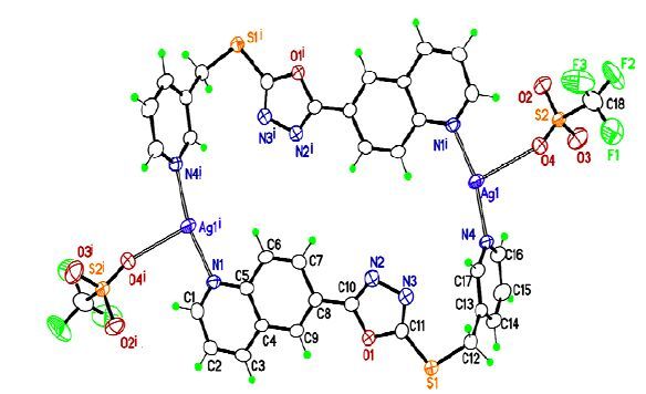
The single-crystal X-ray structural analysis reveals that complex I crystallizes in the triclinic space group P1, and the asymmetric unit contains one AgI centre, one L ligand, and one SO3CF3 - anion. As shown in Fig. 1, each Ag (I) center is located in a distorted trigonal coordination sphere consisting of one triflate O-donor , one quinoline N-donor and one pyridine N-donor from two L ligands. The corresponding Ag (I)-Otriflate, Ag (I)-Nquinoline, and Ag (I)-Npyridine bond distances are 2.751(1), 2.153(8), and 2.133(1) Å, respectively. All of the Ag-N bond distances found in I are within the normal range for N-containing heterocyclic Ag (I) complexes[17, 18]. Two L ligands are arranged in an end-to-end fashion to bind two silver atoms into a distorted rectangular bimetallic ring with the Ag⋅⋅⋅Ag separation of 11.510(1) Å. A novel bimetallic ring has been reported in [Cu (L2)(CH3CN)- (NO3)]2 [19]. In the rectangular-like ring, the quinoline and oxadiazole heterocycles are almost coplanar with a small dihedral angle of 7.7(1)º and the dihedral angle between oxadiazole and pyridine rings is 75.684(2)º. The bond angles of S (1)- C (12)-C (13)) and (N (1)-Ag (1)-N (4) are 114.3(3)º and 165.4(1)º , respectively. Moreover, two pyridine rings and two oxadiazole rings are parallel completely. Two coordinated SO3CF3 - anions point outside the ring, and no guest solvent molecules are found among these bimetallic rings.
Figure 1
 Figure 1. View of the asymmetric unit showing the coordination environment of Ag (I) in I. Symmetry code: (i) -x+1, -y+1, -z
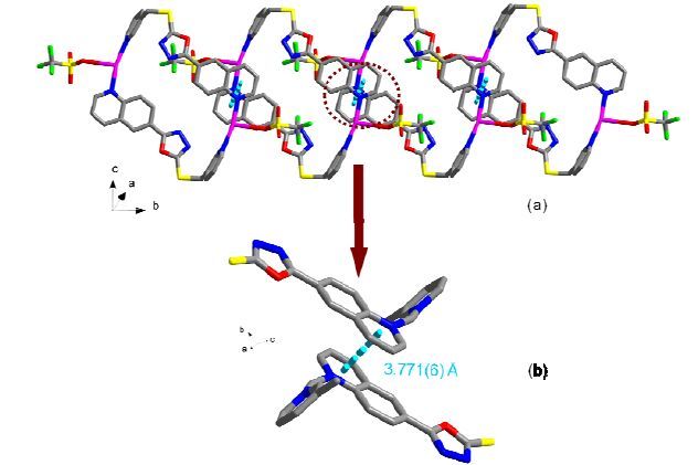
In the solid state, bimetallic silver (I) macrocycles are stacked together along the b axis by interquinoline rings π-π (3.771(6) Å) (Fig. 2b) interactions to generate a one-dimensional chain (Fig. 2a). Also, extensive π-π stacking interactions are found between the pyridine groups of L ligands with the centroid-to-centroid distance of 3.784(6) Å (Fig. 3b). These weak interactions connect the one-dimension chains into a two-dimensional network in the bc plane (Fig. 3a). As shown in Fig. 3(c), the adjacent layers are completely parallel with each other, eventually forming a three-dimensional structure via parallel stacking.
Figure 2
 Figure 2. (a) One-dimensional chain of I, and (b) π-π interactions (bright-blue dashed lines) between the bimetallic rings
Figure 3
 Figure 3. (a) Two-dimensional network of I, (b) Interchain π-π interactions (purple dashed line) between two adjacent chains, and (c) Three-dimensional framework of I
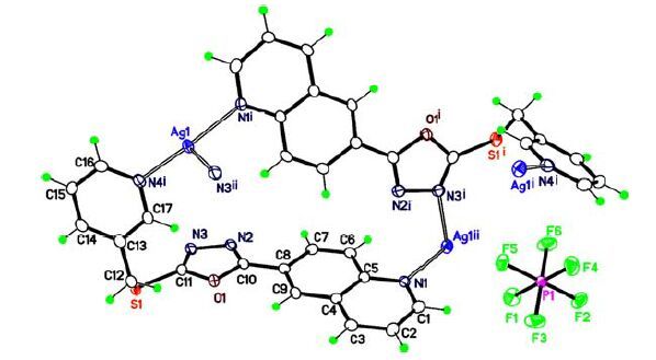
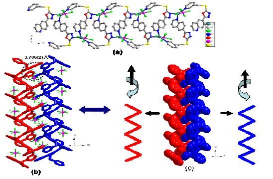
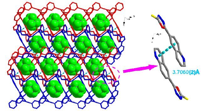
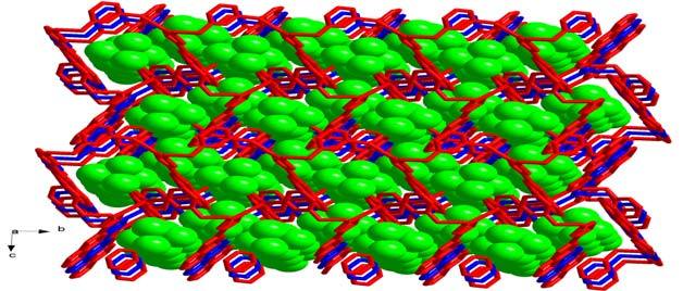
In order to study the coordination chemistry of the ligand L and Ag (I) salts, the AgPF6 was used instead of AgSO3CF3. The result of X-ray diffraction analysis reveals that complex II crystallizes in the monoclinic space group P21/c with Z = 4. As shown in Fig. 4, an irregular cyclic structure is formed by two L ligands and Ag-N bonds. Compared to I, the coordination sphere of the Ag (I) atom changed from [AgN2O] to [AgN3], which is clearly caused by the difference of SO3CF3 - and PF6 - anions. There is only one kind of crystallographic Ag (I) centers in II and every Ag (I) adopts a distorted trigonal coordination environment which consists of three N-donors from one pyridyl (N (4)), one quinoline (N (1)) and one oxadiazole (N (3)) ring. The Ag-N bond lengths lie in the range of 2.187 ~ 2.405 Å. The Ag (I)- Nquinoline and Ag (I)-Npyridine bond distances (Ag- N (1) and Ag-N (4)) are slightly longer than those corresponding in I. While the quinoline and oxadiazole rings of L in II are almost coplanar with a dihedral angle of 5.7(6)º, the dihedral angle between the pyridyl and oxadiazole rings is 75.8(8)º and the bond angle of the thioether group (C (11)-S (1)-C (12)) is 100.9(1)º, which are similar to I. In the extended structure of II, the irregular rings are connected to each other by Ag-N bonds into an infinite one-dimensional helical chain along the b axis (Fig. 5a). As indicated in Fig. 5(c), two adjacent one-dimensional chains show opposite helicity and intertwine together to generate a double-stranded helix through a kind of interchain π-π interactions between the two quinoline rings. A similar helix structure has been reported by Dong[20]. The corresponding face-to-face separation is 3.706(2) Å. The PF6 - counterions were located in the gap in the interior of the double helix (Fig. 5b). Moreover, these double-helical chains interact via the same interchain π-π interactions (quinoline…quinoline rings) (Fig. 6b) into a two-dimensional network with big griddings parallel to the bc plane. It is worth noting that the PF6 - counterions were located inside the griddings (Fig. 6a). When viewed down the a axis, two-dimensional layers are found (Fig. 7), which interact via π-π stacking to develop a three-dimensional framework.
Figure 4
 Figure 4. View of the asymmetric unit showing the coordination environment of Ag (I) in II. Symmetry codes: (i) -x+1, y+1/2, -z+1/2; (ii) x, y+1, z; (iii) -x+1, y-1/2, -z+1/2; (iiii) x, y-1, z
Figure 5
 Figure 5. (a) One-dimensional helical chain of II. (b) Stick representation of the double-stranded helix generated by the intertwining of two single helical chains through a kind of interchain π-π interactions. The PF6 - anions are located in the gap in the interior of the double helix. (c) Space-filling representation of the doublestranded chain and the direction of the spiral are showed
Figure 6
 Figure 6. (a) Two-dimensional network of II with the PF6 - anions located inside the griddings. (b) Interchain π-π interactions (blue dashed line) between two adjacent helical chains
Figure 7
 Figure 7. Three-dimensional framework of II
3.2. IR spectrum
The IR spectra show several characteristic bands: In compound I, the medium intensity bands at 1599 and 1507 cm-1 can be ascribed to the C=C and C=N stretching modes. The very strong peak at 1462 cm-1 in the complexes can be assigned to the characteristic band of SO3CF3 - ion. In compound II, the very strong absorption at about 1608 cm-1 is attributed to characteristic band of PF6 - ion, and the vibration modes of the monosubstituted pyridine ring are observed at about 826 and 699 cm-1. The bands at about 1434 and 11266 cm-1 can be ascribed to the C-N and C-O stretching modes of the pyridine and quinoline rings, respectively.
3.3. Thermogravimetric analysis and powder XRD
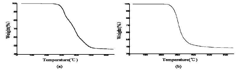
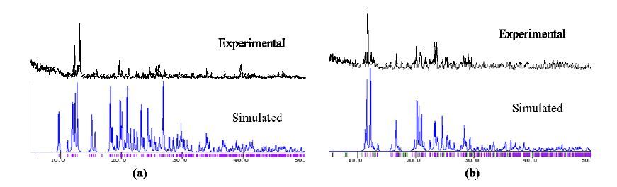
To study the thermal stability of two complexes, thermogravimetric analyses (TGA) are performed under N2 atmosphere from 25 to 650 ℃ at a heating rate of 10 ℃/min, as shown in Fig. 8. TGA of complex I shows it is stable up to 254 ℃. On heating, a large weight loss occurs in the range of 254~615 ℃, which can be attributed to the decomposition of the framework and the loss of organic component. Similar to that in I, TGA of complex II is stable up to 236 ℃, and the decomposition of the framework structure occurs in the range of 236~566 ℃. Powder XRD experiment was carried out to confirm the phase purity of two complexes, as shown in Fig. 9, the main peaks displayed in measured patterns closely match those in the simulated patterns.
Figure 8
 Figure 8. TG curves of I (a) and II (b)
Figure 9
 Figure 9. (a) Experimental and simulated XRD spectra of I, (b) Experimental and simulated XRD spectra of II
4. CONCLUSION
In summary, a new semi-rigid organic ligand, namely 2-((pyridin-3-ylmethyl) thio)-5-(quinoline-6- yl)-1, 3, 4-oxadiazole, is prepared and can be used as a polydentate ligand to coordinate to the transition metal ions. A novel supermolecular [Ag2L2- (CF3SO3)2] (I) and a coordination polymer [AgL (PF6)]n (II) have been synthesized and structurally characterized. Although, in the two complexes, the Ag (I) metal centers are both trigonally coordinated, we get different conformations of them. Thus, we can get completely different structures of the compounds by using different anions. By changing the anions, we anticipate that this new type of organic ligand will result in a variety of new coordination polymers or supramolecular complexes with novel polymeric patterns.
-
-
[1]
Lin W. B, Evans O. R, Xiong R. G, Wang Z. Y. Supramolecular engineering of chiral and acentric 2D networks. Synthesis, structures, and second-order nonlinear optical properties of bis(nicotinato)zinc and bis{3-[2-(4-pyridyl)ethenyl]benzoato} cadmium[J]. J. Am. Chem. Soc, 1998, 120: 13272-13273. doi: 10.1021/ja983415h
-
[2]
Gardner G. B, Kiang Y. H, Lee S, Asgaonkar A, Venkataraman D. Exchange properties of the three-dimensional coordination compound 1,3,5-tris(4-ethynylbenzonitrile) benzene AgO3SCF3[J]. J. Am. Chem. Soc, 1996, 118: 6946-6953. doi: 10.1021/ja960595r
-
[3]
Inoue K, Hayamizu T, Iwamura H, Hashizume D, Ohashi Y. Assemblage and alignment of the spins of the organic trinitroxide radical with a quartet ground state by means of complexation with magnetic metal ions. A molecule-based magnet with three-dimensional structure and high TC of 46 K[J]. J. Am. Chem. Soc, 1996, 118: 1803-1804. doi: 10.1021/ja952301s
-
[4]
Li M, Yao Y, Ding J, Liu L, Qin J. H, Zhao Y. P, Hou H. W, Fan Y. T. Spectroscopic and crystallographic investigations of novel BODIPY-derived metal-organic frameworks[J]. Inorg. Chem, 2015, 54: 1346-1353. doi: 10.1021/ic502219y
-
[5]
Wang K. C, Feng D. W, Liu T. F, Su J, Yuan S, Chen Y. P, Bosch M, Zou X. D, Zhou H. C. A series of highly stable mesoporous metalloporphyrin Fe-MOFs[J]. J. Am. Chem. Soc, 2014, 136: 13983-13986. doi: 10.1021/ja507269n
-
[6]
Kitagawa S, Kitaura R, Noro S. Functional porous coordination polymers[J]. Angew. Chem. Int. Ed, 2004, 43: 2334-2375. doi: 10.1002/(ISSN)1521-3773
-
[7]
Pachfule P, Das R, Poddar P, Banerjec R. Structural, magnetic, and gas adsorption study of a series of partially fluorinated metal-organic frameworks (HF-MOFs)[J]. Inorg. Chem, 2011, 50: 3855-3865. doi: 10.1021/ic1017246
-
[8]
Dong Y. B, Zhang Q, Wang L, Ma J. P, Huang R. Q, Shen D. Z, Chen D. Z. Organometallic coordination polymers generated from bent bis(acetylenylphenyl)oxadiazole ligands and Ag(I) salts[J]. Inorg. Chem, 2005, 44: 6591-6608. doi: 10.1021/ic0508223
-
[9]
Dong Y. B, Xu H. X, Ma J. P, Huang R. Q. Silver(I) coordination polymers based on a nano-sized bent bis(3-acetylenylphenyl-(4-cyanophenyl))oxadiazole ligand: the role of ligand isomerism and the templating effect of polyatomic anions and solvent intermediates[J]. Inorg. Chem, 2006, 45: 3325-3343. doi: 10.1021/ic052158w
-
[10]
Wu X. W, Zhang D, Ma J. P. Two different one-dimensional supramolecular chains formed from the reaction of 2-[1-(pyridin-4-ylmethyl)-1H-benzimidazol-2-yl] quinoline with two different precursors, Co(NO3)2 and CoCl2[J]. Acta Cryst, 2014, C70: 522-527.
-
[11]
Wu X. W, Wu W. F, Yin S, Ma J. P. Syntheses and structures of one CuI-containing coordination polymer and two macrocyclic supramolecular complexes based on flexible1,3,4-oxadiazole-containing ligands[J]. Acta Cryst, 2015, C71: 683-689.
-
[12]
Zheng Y, Li J. R, Du M, Zou R. Q, Bu X. H. Novel silver(I) coordination polymers with a series of bis(arylthio)ether ligands bearing a trans-2-butene backbone[J]. Crystal Growth & Design, 2005, 5: 215-222.
-
[13]
Jing X. M, Zhou X. Y, Zhao T. T, Huo Q. S, Liu Y. L. Construction of lanthanide-organic frameworks from 2-(pyridine-3-yl)-1H-4,5-imidazole dicarboxyl-ate and oxalate[J]. Crystal Growth & Design, 2012, 12: 4225-4229.
-
[14]
Wu X. W, Wu W. F, Yin S, Ma J. P. A double helix coordination polymer generated from 2-((pyridin-4-ylmethyl)thio)- 5-(quinoline-2-yl)-1,3,4-oxadiazole and AgI salts[J]. Chin. J. Struct. Chem, 2015, 34: 1496-1502.
-
[15]
Sheldrick, G. M. SHELXS 97, Program for Crystal Structure Solution. University of Gottingen, Germany, 1997.
-
[16]
Sheldrick, G. M. SHELXL-97, Program for X-ray Crystal Structure Refinement. University of Gottingen, Germany, 1997.
-
[17]
Dong Y. B, Cheng J. Y, Ma J. P, Huang R. Q, Smith M. D. Self-assembly of {Ag2N4}-core-containing coordination polymers from AgX (X = NO3 -, ClO4 -, and PF6 -) and oxadiazole-bridged 4,4'- and 3,3'-biphenylamine ligands[J]. Crystal Growth & Design, 2005, 5: 585-591.
-
[18]
Dong Y. B, Cheng J. Y, Huang R. Q, Smith M. D, zur Loye H. C. Self-assembly of coordination polymers from AgX (X = SbF6 -, PF6 -, and CF3SO3 -) and oxadiazole-containing ligands[J]. Inorg. Chem, 2003, 42: 5699-5706. doi: 10.1021/ic034306t
-
[19]
He L, Ma D. X, Duan L, Wei Y. G, Qiao J, Zhang D. Q, Dong G. F, Wang L. D, Qiu Y. Control of intramolecular π-π stacking interaction in cationic iridium complexes via fluorination of pendant phenyl rings[J]. Inorg. Chem, 2012, 51: 4502-4510. doi: 10.1021/ic2021325
-
[20]
Dong Y. B, Sun T, Ma J. P, Zhao X. X, Huang R. Q. Ag(I) and Cu(II) discrete and polymeric complexes based on single and double-armed oxadiazole-bridging organic clips[J]. Inorg. Chem, 2006, 45: 10613-10628. doi: 10.1021/ic0612552
-
[1]
-
Figure 1 View of the asymmetric unit showing the coordination environment of Ag (I) in I. Symmetry code: (i) -x+1, -y+1, -z
Figure 2 (a) One-dimensional chain of I, and (b) π-π interactions (bright-blue dashed lines) between the bimetallic rings
Figure 3 (a) Two-dimensional network of I, (b) Interchain π-π interactions (purple dashed line) between two adjacent chains, and (c) Three-dimensional framework of I
Figure 4 View of the asymmetric unit showing the coordination environment of Ag (I) in II. Symmetry codes: (i) -x+1, y+1/2, -z+1/2; (ii) x, y+1, z; (iii) -x+1, y-1/2, -z+1/2; (iiii) x, y-1, z
Figure 5 (a) One-dimensional helical chain of II. (b) Stick representation of the double-stranded helix generated by the intertwining of two single helical chains through a kind of interchain π-π interactions. The PF6 - anions are located in the gap in the interior of the double helix. (c) Space-filling representation of the doublestranded chain and the direction of the spiral are showed
Figure 6 (a) Two-dimensional network of II with the PF6 - anions located inside the griddings. (b) Interchain π-π interactions (blue dashed line) between two adjacent helical chains
Figure 9 (a) Experimental and simulated XRD spectra of I, (b) Experimental and simulated XRD spectra of II
Table 1. Crystal and Structure Refinement Data
Temperature(K) 298 100 Crystal system Triclinic Monoclinic Space group P1 P21/c a(Å) 8.2946(12) 15.1940(5) b(Å) 9.9266(14) 8.1723(6) c(Å) 14.280(3) 15.5374(11) α(°) 88.455(14) 90 β(°) 74.996(15) 91.360(3) γ(°) 65.813(14) 90 Volume(Å3) 1031.8(3) 1928.7(2) Crystal size 0.15×0.09×0.02 0.20×0.07×0.04 Z 2 4 Dc(Mg/m3) 1.856 1.974 μ(mm-1) 10.306 1.312 F(000) 572 1128 θ range(°) 3.22~67.07 3.11~25.68 Reflections collected 6560 10200 Independent reflections 3651 3648 Data/restraints/parameters 3651/0/289 3648/0/280 Goodness-of-fit 1.081 1.062 Final R indices(italic>2σ(I)) R=0.0499,wR=0.1316 R=0.0252,wR=0.0568 R indices(all data) R=0.0619,wR=0.1425 R=0.0305,wR=0.0600 (Δρ)max,(Δρ)min(e·Å–3) 1.353,–0.911 0.511,–0.405 Reflections collected 6560 10200  下载: 导出CSV
Table 2. Selected Bond Lengths (Å) and Bond Angles (°) for Complex I
Bond Dist. Bond Dist. Bond Dist. O(4)–Ag(1) 2.751(5) Ag(1)–N(4) 2.0339(18) Ag(1)–N(1)i 2.153(4) Angle (°) Angle (°) Angle (°) N(4)–Ag(1)–N(1)i 165.37(18) N(4)–Ag(1)–O(4) 104.52(16) N(1)i–Ag(1)–O(4) 89.88(16) Symmetry transformation: (i) –x+1, –y+1, –z
下载: 导出CSV
Table 3. Selected Bond Lengths (Å) and Bond Angles (°) for Complex II
Bond Dist. Bond Dist. Bond Dist. Ag(1)–N(4) 2.187(2) Ag(1)–N(1)i 2.234(2) Ag(1)–N(3)ii 2.405(2) Angle (°) Angle (°) Angle (°) N(4)–Ag(1)–N(1)i 143.36(8) N(4)–Ag(1)–N(3)ii 116.48(7) N(1)i–Ag(1)–N(3)ii 96.92(7) Symmetry transformation: (i) –x+1, y+1/2, –z+1/2; (ii) x, y+1, z; (iii)–x+1, y–1/2, –z+1/2; (iiii) x, y–1, z
下载: 导出CSV
-


 扫一扫看文章
计量
- PDF下载量: 0
- 文章访问数: 5201
- HTML全文浏览量: 158
