 Figure 1.
Chemical structure of compound 1
Figure 1.
Chemical structure of compound 1
Citation:
ZHANG Fan, PENG Hao, SHANG Zheng-Hui, LIANG Jie, RU Neng. A Novel 3-D Zn(II) Coordination Polymer for Inhibiting the Human Osteosarcoma Cells Growth[J]. Chinese Journal of Structural Chemistry,
2016, 35(7): 1019-1023.
doi:
10.14102/j.cnki.0254-5861.2011-1059

A Novel 3-D Zn(II) Coordination Polymer for Inhibiting the Human Osteosarcoma Cells Growth
English
A Novel 3-D Zn(II) Coordination Polymer for Inhibiting the Human Osteosarcoma Cells Growth
Abstract:
A new three-dimensional Zn(II) coordination polymer, namely[Zn4(bpydb)3(tz)2(H2O)2]n (1), has been synthesized by the self-assembly reactions of Zn(NO3)2·6H2O, bpydbH2, Htz and DMF. Single-crystal X-ray structural analysis reveals that compound 1 features a three-dimensional framework structure and is the first example of Zn-containing coordination polymers based on two kinds of ligands bpydbH2 and Htz. It crystallizes in triclinic, space group P1 with a=14.9953(12), b=17.5335(17), c=20.2381(11)Å, α=115.225(7), β=92.329(5), γ=105.606(8)°, V=4561.9(7)Å3, Z=2, F(000)=1644, Dc=1.177 Mg/m3, Mr=1616.76 and μ=1.098 mm-1. The antitumor activities of compound 1 and its corresponding organic ligands (bpydbH2 and Htz) were investigated for inhibiting human osteosarcoma cells (MG-63 and U-2 OS) growth by MTT assay. It was found that compared with the two ligands, compound 1 exerted rather potent activities against all of these cell lines.
-
Key words:
- coordination
- / X-ray
- / three-dimensional
-
1 INTRODUCTION
Cancer is a major health problem worldwide. Improvements in treatment and prevention have led to a decrease in cancer deaths, but the number of new diagnoses continues to rise. Chemotherapy is one of the most commonly used treatment options, especially for unresectable patients[1, 2]. However, the use of conventional cytotoxic drugs, including doxorubicin, cisplatin and fluorouracil, has not shown any improvement in survival, and severe adverse effects have been frequently observed in treated patients. Thus, it is urgent to develop novel chemotherapeutic agents for the treatment of cancer[3].
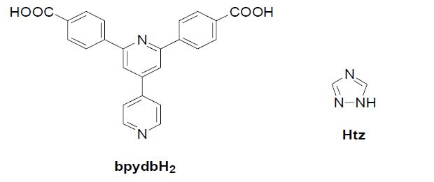
Medicinal inorganic chemistry is a field of increasing prominence as metal-based compounds, offering possibilities for the design of therapeutic agents not readily available to organic compounds[4, 5]. The wide range of coordination numbers and geometries, accessible redox states, thermodynamic and kinetic characteristics, and the intrinsic properties of cationic metal ion and ligand itself offer the medicinal chemist a wide spectrum of reactivities that can be exploited[6, 7]. In this work, we prepared a new Zn(II) coordination polymer, namely [Zn4(bpydb)3(tz)2(H2O)2]n (bpydbH2 = 4, 4΄-(4, 4΄- bipyridine-2, 6-diyl)dibenzoic acid, Htz = 1, 2, 4-1Htriazole) (Fig. 1), and then evaluated their antitumor activities.
Figure 1.
Chemical structure of compound 1
2 EXPERIMENTAL
2.1 Apparatus and materials
All the starting materials and reagents used in this work were obtained commercially and used without further purification. Element analyses (C, H and N) were determined with an elemental Vairo EL III analyzer. Single-crystal X-ray diffraction data for compound 1 were recorded on a Mercury CCD diffractometer. The melting points were taken on a XT-4 micro melting apparatus, and the thermometer was uncorrected.
2.2 Synthesis and characterization of Zn4(bpydb)3(tz)2(H2O)2 (1)
A mixture of Zn(NO3)2·6H2O (0.060 g, 0.2 mmol), bpydbH2 (0.020 g, 0.05 mmol), Htz (0.070 g, 0.1 mmol), DMF (2 mL) and H2O (0.5 mL) was sealed in a 5 mL glass bottle at 70 ℃ for 72 h under autogenous pressure. After cooling to room temperature, yellow block crystals suitable for X-ray diffraction analysis were obtained. The yield was 36% for 1 (based on bpydbH2). Analytical analysis found for compound 1 (C76H50N12O14Zn4): C, 56.75; H, 3.29; N, 9.94%. Calcd.: C, 56.41; H, 3.09; N, 10.39%.
2.3 Crystal structure determination
A yellow crystal of compound 1 with approximate dimensions of 0.20mm × 0.22mm × 0.20mm was selected and mounted on a glass fiber. The intensity data were collected on a Bruker Smart APEX CCDbased diffractometer equipped with a graphite-monochromator equipped with a MoKα radiation (λ = 0.71073 Å) by using a φ-ω scan mode at 293(2) K. The empirical absorption was applied to the intensity data. A total of 38946 reflections were collected in the range of 2.939<θ<24.999° (-17≤h≤17, -20≤k ≤20, -24≤l≤24), of which 4783 were independent (Rint = 0.0697) and 3434 were observed with I > 2σ(I). The intensity data were corrected for Lorentz and polarization effects as well as for empirical absorption based on the multi-scan technique. The structure was solved by direct methods and refined by full-matrix least-squares techniques on F2 with SHELX-97[8]. All non-hydrogen atoms were refined anisotropically and hydrogen atoms isotropically by full-matrix least-squares refinement. The organic hydrogen atoms were generated geometrically. The final R = 0.0777, wR = 0.2261 (w = 1/[σ2(Fo 2) + (0.1000P)2 + 0.0000P], where P = (Fo 2 + 2Fc 2)/3), (Δ/σ)max = 0.001, S = 1.058, (Δρ)max = 1.549 and (Δρ)min = -0.863 e/Å3.
2.4 Antitumor activity
Viability of human osteosarcoma cells (MG-63 and U-2 OS) was determined by using the MTT assay. Cells reaching 70~80% confluency were treated with various concentrations of the synthesized compounds with 1% dimethyl sulfoxide (DMSO) as a negative control. After 48 h incubation, 20 μL of MTT solution (5 mg/mL in PBS) was added and incubated for an additional 4 h. Subsequently, the medium was aspirated carefully, and 150 μL of DMSO was added. After incubation for 15 min, the optical density was measured at 490 nm using FlexStation 3 benchtop multi-mode microplate reader (Molecular Devices, USA). Data were recorded and analyzed for the assessment of the effects of the test substances on cell viability and growth inhibition.
3 RESULTS AND DISCUSSION
3.1 Molecular structure
Single-crystal X-ray diffraction analysis indicates that compound [Zn4(bpydb)3(tz)2(H2O)2]n (1) crystallizes in the triclinic space group P1. Compound 1 could be viewed as a three-dimensional framework structure and featured the first example of Zncontaining coordination polymers based on two kinds of ligands bpydbH2 and Htz. Compound 1 enriched the family of the Zn-containing coordination polymers with three-dimensional structures. The molecular structure of 1 contains four Zn2+ cations, three bpydbH2 ligands, two Htz ligands and two coordination water molecules. Fig. 2 shows the molecular structural unit of 1. Within the structure, it is worth noting that the coordination modes of Zn2+ cations can be divided into two types: four-coordination mode (Zn(1), Zn(2) and Zn(3)) and fivecoordination mode (Zn(4)). The Zn(1) and Zn(2) centers are four-coordinated by one nitrogen atom from the bpydbH2 ligand, one nitrogen atom from the Htz ligand and two oxygen atoms from two monodentate carboxylate groups belonging to two different bpydbH2 ligands. The Zn(3) center adopts one oxygen atom from the bpydbH2 ligand, two nitrogen atoms from two different Htz ligands and one N atom from the bpydbH2 ligand, resulting in the formation of a tetrahedral configuration. It is noting that the Zn(4) center is five-coordinated by two nitrogen atoms from two different Htz ligands, one O atom from the bpydbH2 ligand and two coordination water molecules. Bond lengths of Zn-O and Zn-N are in the ranges 1.923(5)~2.300(6) and 2.015(4) ~ 2.036(5) Å, respectively. Bond angles of O-Zn-N, O-Zn-O and N-Zn-N are in the ranges of 104.6(2) ~ 131.26(19)°, 84.3(2) ~ 173.77(18)° and 97.44(18)~ 121.76(18)°, respectively. These bond distances of Zn-O and Zn-N are similar to those in other Zn-containing coordination polymers[9]. On the basis of the bond strength calculations, the bond valence sums (BVS) for all Zn sites are close to their normal valences of +2.
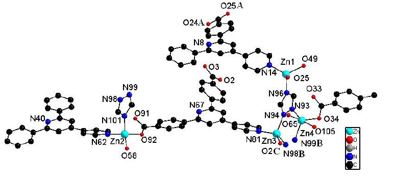 Figure 2.
View of the molecular structural unit of 1
Figure 2.
View of the molecular structural unit of 1
These molecular structural units of 1 are connected with each other, extending into a three-dimensional framework structure. In the packing structure of 1, each bpydbH2 ligand is linked to three Zn2+ cations through one N and two O atoms from its carboxylate groups. Actually, each Htz ligand is connected to three Zn2+ cations via its three N atoms, leading to a three-dimensional framework structure (Fig. 3). It is interesting that the packing structure contains 4- and 6-membered ring channels consi-dering each Zn2+ cation as a connected node along the [100] direction. The sizes of 4- and 6-membered ring channels in the packing structure are 17.050Å × 16.435Å and 22.344Å × 12.785Å, respectively. In addition, there is a void presented in the structure with the size of 150 Å3, and single-crystal X-ray diffraction analysis indicates no other fragment in this crystal structure, which means that compound 1 may own the potential gap (such as: N2 and CO2) adsorption properties.
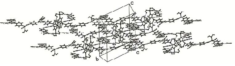 Figure 3.
Three-dimensional framework structure for compound 1
Figure 3.
Three-dimensional framework structure for compound 1
In addition, there are O-H···O hydrogen bonding interactions between the coordinate water molecules and carboxylate groups belonging to the bpydbH2 ligands (O(65)-H(65A)···O(33) (-x+1, -y-1, -z+1), 153.00º, 2.738(6) Å). Moreover, the π-π stacking between the pyridine rings from the bpydbH2 ligands is also observed in the packing structure. Such π-π stacking can be divided into four types, with their centroid-to-centroid distances of 3.561(3), 3.519(3), 3.529(4) and 3.561(3) Å, respectively. The hydrogen bonds and π-π stacking further stabilize the threedimensional framework structure.
3.2 Antitumor activity
Two human osteosarcoma cells (MG-63 and U-2 OS) representing two different tumor types were used in the systematic analysis of the antitumor activities of the newly synthesized compound 1 and its corresponding organic ligands (bpydbH2 and Htz) in vitro. For comparison purpose, the cytotoxicity of doxorubicin, a standard antitumor drug, was evaluated under the same conditions.
The results showed that the tested compounds possess a certain degree of antitumor activities against the two tumor cell lines and their inhibitory action gets stronger with the corresponding higher concentration. The related half maximal inhibitory concentration (IC50) and IC90 values (dose of the compound which causes 50% and 90% reduction of the survival values, respectively) are shown in Table 1.
 Table 1.
IC50 and IC90 Values of Compound 1, Organic Ligands (bpydbH2 and Htz) and Doxorubicin agains t Two Tumor Cell Lines (μg/mL)
Table 1.
IC50 and IC90 Values of Compound 1, Organic Ligands (bpydbH2 and Htz) and Doxorubicin agains t Two Tumor Cell Lines (μg/mL)
bpydbH2 Htz 133.45 267.89 70.34 178.34 Compound 1 25.67 52.45 26.33 45.89 Table 1. IC50 and IC90 Values of Compound 1, Organic Ligands (bpydbH2 and Htz) and Doxorubicin agains t Two Tumor Cell Lines (μg/mL)As can be seen in Table 1, there is great difference in the antitumor activity among the three compounds. Compound 1 showed more potent antitumor activity against the two tested tumor cells with IC50 and IC90 values of 25.67~26.33 and 45.89~52.45 μg/mL, respectively, which is much lower than the IC50 and IC90 values (54.12 ~ 67.78 and 118.12 ~ 159.33 μg/mL) of the standard antitumor drug doxorubicin. However, organic ligands (bpydbH2 and Htz) demonstrated lower antitumor activity with relatively higher IC50 and IC90 values.
4 CONCLUSION
In summary, a new three-dimensional Zn(II) coordination polymer, namely [Zn4(bpydb)3(tz)2(H2O)2]n (1), has been synthesized by the self-assembly reactions of Zn(NO3)2·6H2O, bpydbH2, Htz and DMF and structurally characterized by single-crystal Xray diffraction. The in vitro antitumor activity experiment showed that when the organic compound bpydbH2 and Htz coordinated with Zn2+, the antitumor activity of the title Zn(II) complex 1 has been much improved. However, the exact target is still unknown, whether it has an exact target or is just a cytotoxic agent, the detailed mechanisms of the inhibitory effects need to be further investigated.
-
-
[1]
Dedhar S., Argraves W. S., Suzuki S., Ruoslahti E., Pierschbacher M. D.. Human osteosarcoma cells resistant to detachment by an Arg-Gly-Asp-containing peptide overproduce the fibronectin receptor[J]. J. Cell Biol., 1987, 105: 1175-1182. doi: 10.1083/jcb.105.3.1175
-
[2]
Heino J., Massagué J.. Transforming growth factor-beta switches the pattern of integrins expressed in MG-63 human osteosarcoma cells and causes a selective loss of cell adhesion to laminin[J]. J. Biol. Chem., 1989, 264: 21806-21811.
-
[3]
Franceschi, R. T.; James, W. M.; Zerlauth, G. 1α,25-Dihydroxyvitamin D 3 specific regulation of growth, morphology, and fibronectin in a human osteosarcoma cell line. J. Cell. Physiol. 1985, 123, 401-409.
-
[4]
Xu X., Park J., Hong Y. K., Lane A. M.. Synthesis and characterization of hollow mesoporous BaFe12O19 spheres[J]. J. Solid. State. Chem., 2015, 222: 84-89. doi: 10.1016/j.jssc.2014.11.008
-
[5]
Xu X., Park J., Hong Y. K., Lane A. M.. Magnetically self-assembled SrFe12O19/FeCo core/shell nanoparticles[J]. Mater. Chem. Phys., 2015, 152: 9-12. doi: 10.1016/j.matchemphys.2014.11.061
-
[6]
Xu X., Park J., Hong Y. K., Lane A. M.. Ethylene glycol assisted spray pyrolysis for the synthesis of hollow BaFe12O19 spheres[J]. Mater. Lett., 2015, 144: 119-122. doi: 10.1016/j.matlet.2015.01.034
-
[7]
Xu X., Hong Y. K., Park J., Lee W., Lane A. M.. Magnetic self-assembly for the synthesis of magnetic exchange coupled MnBi/FeCo composites[J]. J. Solid. State Chem., 2015, 231: 108-113. doi: 10.1016/j.jssc.2015.08.019
-
[8]
Sheldrick, G. M. (1997) SHELXL 97, Program for the Solution of Crystal Structure, University of Gottingen, Germany.
-
[9]
Teo T. L., Vetrichelvan M., Lai Y. H.. Infinite three-dimensional polymeric metalloporphyrin network via six-coordinate Zn(II) and two axial oxygen donors[J]. Org. lett., 2003, 5: 4207-4210. doi: 10.1021/ol035665v
-
[1]
-

Figure 2 View of the molecular structural unit of 1
(Symmetric codes: A: -1+x, y, z; B: x, 1+y, z; C: 1+x, y, z)
Table 1. IC50 and IC90 Values of Compound 1, Organic Ligands (bpydbH2 and Htz) and Doxorubicin agains t Two Tumor Cell Lines (μg/mL)
bpydbH2 Htz 133.45 267.89 70.34 178.34 Compound 1 25.67 52.45 26.33 45.89  下载: 导出CSV
-


 扫一扫看文章
计量
- PDF下载量: 40
- 文章访问数: 1891
- HTML全文浏览量: 269
 下载:
下载:
