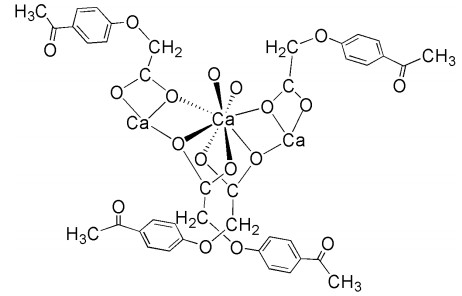
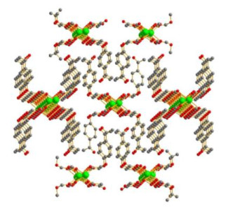
Figure 1.
Coordination mode of Ca(II) ion

The design and synthesis of metal coordination polymers have received considerable attention during the past decades because they show not only structural diversity[1], but also potential widespread applications as functional materials, such as molecular recognition[2], gas storage[3], magnetocaloric effect[4], luminescence[5-7], catalysis[8], magnetic property[9], bioactivity[10], magnetorefrigerant[11], and so on. To our knowledge, many studies on coordination polymers have focused on transition metals in the past years[12-16]. However, few investigations have been done on calcium coordination polymers. In our previous work, some calcium coordination polymers have been synthesized and structurally characterized[17-20]. In order to extend the investigation of novel structures and properties of calcium coordination polymer, in this work, we successfully designed and synthesized a novel 1D Ca(II) coordination polymer, [CaL2(H2O)2]n (HL = 4-acetylphenoxyacetic acid), by 4-acetylphenoxyacetic acid, Ca(ClO4)2·4H2O and NaOH as raw materials. And its antitumor activity against human hepatoma SMMC-7721 cells, human colon carcinoma WiDr cells and human lung adenocarcinoma A549 cells has been investigated. The coordination mode of Ca(II) ion is shown in Fig. 1.
4-Acetylphenoxyacetic acid, NaOH, Ca(ClO4)2·4H2O and ethanol solvent were of analytical grade and used directly without further purification. Carbon, hydrogen and nitrogen were determined using an Elementar Vario III EL elemental analyzer. IR spectra were recorded using KBr discs on a Nicolet AVATAR 360 FTIR spectrophotometer (Nicolet Instrument Inc., Madison, WI, USA) (range 4, 000~400 cm–1). Single-crystal X-ray diffraction data of [CaL2(H2O)2]n were collected on a Bruker Smart CCD diffractometer (Bruker, Billerica, MA, USA).
4-Acetylphenoxyacetic acid (0.1941 g, 1.0 mmol) and sodium hydrate (0.040 g, 1.0 mmol) were dissolved into 10 mL ethanol solution at room temperature. Then 5 mL of aqueous solution containing 0.1195 g Ca(ClO4)2·4H2O (0.5 mmol) was dropped into the above solution. The mixture was heated at 70 ℃ for 6.5 h with stirring, cooled to room temperature and filtered. Colourless crystals of [CaL2(H2O)2]n were obtained in 25 days by slowly volatilizing the filtrate at room temperature. Anal. Calcd. for C20H22O10Ca: C, 51.90; H, 4.76%. Found: C, 51.65; H, 5.09. IR vmax (cm–1): v (COO–): 1675 cm−1.
The culture of tumor cells (Human hepatoma SMMC-7721 cells, human colon carcinoma WiDr cells and human lung adenocarcinoma A549 cells) and the test procedure for the antitumor activity are consistent with the literature[21, 22].
A single crystal of 1 (0.12mm × 0.11mm × 0.10mm) was placed on a Bruker SMART APEX CCD X-ray diffractometer equipped with graphite-monochromated MoKα radiation (λ = 0.71073 Å) at 99.99(10) K. 4441 reflections (Rint = 0.0245) were independent for 1. The structure was solved by direct methods using SHELXL-2014/7 program[23]. The OLEX2[24] program was used to refine the structure. The non-hydrogen atoms were refined anisotropically and the hydrogen atoms were generated by theoretical calculations. Crystal data for complex 1: monoclinic system, space group C2/c with a = 24.4894(11), b = 11.3416(5), c = 7.7164(4) Å, β = 94.750(5)º, V = 2135.88(17) Å3, Z = 4, C20H22O10Ca, Mr = 462.45, Dc = 1.438 Mg/m3, F(000) = 968 and μ(MoKα) = 0.348 mm–1. The final R = 0.0319, wR = 0.0753 (w = 1/[σ2(Fo2) + (0.0342P)2 + 1.4584P], where P = (Fo + 2Fc2)/3), S = 1.067. The maximum and minimum peaks are 0.229 and –0.333 e/Å3, respectively.
Single-crystal X-ray structural analyses show that complex 1 crystallizes in monoclinic space group C2/c. Selected bond distances and bond angles of complex 1 are shown in Table 1. The average distance of the Ca–O bonds is 2.465 Å (ranging from 2.3384(12) to 2.6308(12) Å), which are consistent with those reported[16-21]. And the bond angles of O–Ca–O vary from 50.82(4) to 157.67(4)°.
 DownLoad:
CSV
DownLoad:
CSV
| Bond | Dist. | Bond | Dist. | |
| Ca(1)–O(4) | 2.3384(12) | Ca(1)–O(5) | 2.3828(12) | |
| Ca(1)–O(4A) | 2.6308(12) | Ca(1)–O(5A) | 2.3827(12) | |
| Ca(1)–O(4B) | 2.6308(12) | C(8)–O(4) | 1.259(2) | |
| Ca(1)–O(4C) | 2.3384(12) | C(4)–O(2) | 1.363(2) | |
| Ca(1)–O(3B) | 2.5069(12) | C(7)–O(2) | 1.427(2) | |
| Ca(1)–O(3C) | 2.5069(12) | C(8)–O(3) | 1.252(2) | |
| C(9)–O(1) | 1.224(2) | |||
| Angle | (°) | Angle | (°) | |
| O(4)–Ca(1)–O(4C) | 122.51(4) | O(4)–Ca(1)–O(3B) | 122.51(4) | |
| O(4)–Ca(1)–O(4B) | 72.94(4) | O(4A)–Ca(1)–O(3B) | 77.99(4) | |
| O(4A)–Ca(1)–O(4C) | 72.94(4) | O(4A)–Ca(1)–O(3C) | 122.51(4) | |
| O(4B)–Ca(1)–O(4C) | 108.23(5) | O(4)–Ca(1)–O(3C) | 77.99(4) | |
| O(4A)–Ca(1)–O(4B) | 122.51(4) | O(4A)–Ca(1)–O(5A) | 78.78(4) | |
| O(4)–Ca(1)–O(4A) | 155.97(6) | O(4)–Ca(1)–O(5A) | 84.74(4) | |
| O(4)–Ca(1)–O(5) | 78.78(4) | O(4B)–Ca(1)–O(3C) | 72.78(4) | |
| O(4A)–Ca(1)–O(5) | 84.74(4) | O(4B)–Ca(1)–O(3B) | 50.82(4) | |
| O(4C)–Ca(1)–O(3B) | 72.78(4) | O(3B)–Ca(1)–O(3C) | 75.37(5) | |
| O(4C)–Ca(1)–O(3C) | 50.82(4) | O(4C)–Ca(1)–O(5A) | 83.18(4) | |
| O(4B)–Ca(1)–O(5) | 83.18(4) | O(3C)–Ca(1)–O(5A) | 102.61(4) | |
| O(4C)–Ca(1)–O(5) | 157.67(4) | O(3B)–Ca(1)–O(5) | 102.61(4) | |
| O(4B)–Ca(1)–O(5A) | 157.67(4) | O(3C)–Ca(1)–O(5) | 150.49(4) | |
| O(3B)–Ca(1)–O(5A) | 150.49(4) | O(5)–Ca(1)–O(5A) | 93.09(6) | |
| Symmetry transformation: A: 1 – x, y, 1/2 – z; B: 1 – x, 1 – y, 1 – z; C: x, 1 – y, –1/2 + z | ||||
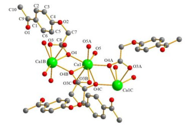
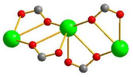
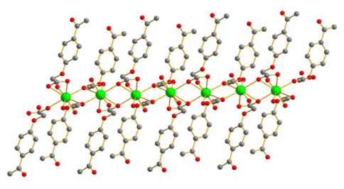
The asymmetric unit of complex 1 is shown in Fig. 2. The fundamental unit of 1 contains one Ca(II) ion, two 4-acetylphenoxyacetate ligands and two coordinated water molecules. Each Ca(II) ion is eight-coordination with six O atoms (O(3B), O(3C), O(4), O(4A), O(4B) and O(4C)) from four 4-acetylphenoxyacetate ligands and two O atoms (O(5) and O(5A)) from two coordinated water molecules, resulting in a distorted triangular dodecahedral geometric configuration. In complex 1, the carboxylate O atoms of 4-acetylphenoxyacetate ligand adopt different coordination modes with Ca(II) ion (Fig. 3): one O atom adopts a bidentate chelating mode to coordinate to different Ca(II) ions, and the other O atom adopts a monodentate chelating mode to coordinate to the Ca(II) ion. The complex forms a one-dimensional (1D) chain structure by the bridging effect of carboxylate O atoms with adjacent Ca(II) ions (Fig. 4). The 3D network structure is also formed by the interactions of 1D chain (Fig. 5). In addition, four uncoordinated water molecules also exist in the crystal structure.
The antitumor activity of 4-acetylphenoxyacetic acid and [CaL2(H2O)2]n was tested against human hepatoma SMMC-7721 cells, human colon carcinoma WiDr cells and human lung adenocarcinoma A549 cells. The data of antitumor activity are given in Table 2. The results show that both 4-acetylphenoxyacetic acid and [CaL2(H2O)2]n exhibit considerable cytotoxicity, but the antitumor effect of [CaL2(H2O)2]n is better than that of 4-acetylphenoxyacetic acid. The antitumor effect of [CaL2(H2O)2]n against WiDr cell is better than that reported[22], while the antitumor effect of [CaL2(H2O)2]n against SMMC-7721 and A549 cells is weaker than that in document[22].
DownLoad:
CSV
| Compound | IC50 (μg/mL) | ||
| SMMC-7721 | WiDr | A549 | |
| [CaL2(H2O)2]n | 12.5 ± 0.2 | 15.3 ± 0.2 | 19.2 ± 0.2 |
| 4-Acetylphenoxyacetic acid | 15.3 ± 0.2 | 22.6 ± 0.2 | 29.1 ± 0.2 |
In summary, we have synthesized and structurally characterized a new eight-coordinated Ca(II) coordination polymer, [CaL2(H2O)2]n (1). The antitumor activities of 4-acetylphenoxyacetic acid and the Ca(II) coordination polymer have also been tested. The above results provide a good idea for the synthesis of Ca(II) complexes with antitumor activity in the future.
Lee, E.; Seo, S.; Lee, S. S.; Lindoy, L. F. Assembling latter d-block heterometal coordination polymers: synthetic strategies and structural outcomes. Coord. Chem. Rev. 2017, 348, 121–170. doi: 10.1016/j.ccr.2017.08.005
Yao, S. L.; Liu, S. J.; Tian, X. M.; Zheng, T. F.; Cao, C.; Niu, C. Y.; Chen, Y. Q.; Chen, J. L.; Huang, H. P.; Wen, H. R. A ZnII-based metal-organic framework with a rare tcj topology as a turn-on fluorescent sensor for acetylacetone. Inorg. Chem. 2019, 58, 3578–3581. doi: 10.1021/acs.inorgchem.8b03316
Ma, S. Q.; Zhou, H. C. Gas storage in porous metal-organic frameworks for clean energy applications. Inorg. Chem. Commun. 2010, 46, 44–53. doi: 10.1039/B916295J
Liu, S. J.; Cao, C.; Xie, C. C.; Zheng, T. F.; Tong, X. L.; Liao, J. S.; Chen, J. L.; Wen, H. R.; Chang, Z.; Bu, X. H. Tricarboxylate-based GdIII coordination polymers exhibiting large magnetocaloric effects. Dalton Trans. 2016, 45, 9209–9215. doi: 10.1039/C6DT01349J
Song, J.; Duan, B. F.; Lu, J. F.; Ge, H. G. Three new lanthanide coordination polymers constructed from 2, 6-bis(pyrazin-2-yl)pyridine-4-carboxylate: syntheses, structures and luminescence. Chin. J. Struct. Chem. 2020, 39, 793–800.
Wang, Y. S.; Zhou. Z. M. Utilization of mixed ligands to construct two new coordination polymers: syntheses, structures and properties. J. Solid State Chem. 2015, 228, 117–123. doi: 10.1016/j.jssc.2015.04.026
Tian, X. M.; Yao, S. L.; Qiu, C. Q.; Zheng, T. F.; Chen, Y. Q.; Huang, H.; Chen, J. L.; Liu, S. J.; Wen, H. R. Turn-on luminescent sensor toward Fe3+, Cr3+ and Al3+ based on a Co(II) metal-organic framework with open functional sites. Inorg. Chem. 2020, 59, 2803-2810. doi: 10.1021/acs.inorgchem.9b03152
Yang, Y. J.; Wang, F. S.; Dong, G. Y. In situ synthesis, structural characterization and catalytic properties of a 2D Cu(I)-cyanide coordination polymer. Chin. J. Struct. Chem. 2019, 38, 811–818.
Zeng, Y.; Liu, S. J.; Liu, C. M.; Xie, Y. R.; Du, Z. Y. Diversified magnetic behaviors in new nickel(II) and copper(II) azido coordination polymers templated by diethyl or triethyl amines. New J. Chem. 2017, 41, 1212–1218. doi: 10.1039/C6NJ03268K
Tai, X. S.; Zhao, W. H. Synthesis, crystal structure, and antibacterial activity of magnesium(II) coordination polymers formed by hydrogen bonding. Res. Chem. Intermed. 2015, 41, 3471–3478. doi: 10.1007/s11164-013-1463-y
Liu, S. J.; Han, S. D.; Zhao, J. P.; Xu, J.; Bu, X. H. In-situ synthesis of molecular magnetorefrigerant materials. Coord. Chem. Rev. 2019, 394, 39–52. doi: 10.1016/j.ccr.2019.05.009
Mostafa, M. A.; Ezzatollah, N.; Ali, D.; Seik, W. N. Structure and optical properties of new lead(II) coordination polymers and PbO nanoparticles core of polymer. J. Mol. Struct. 2015, 1083, 221–228. doi: 10.1016/j.molstruc.2014.12.019
Li, G. L.; Liu, G. Z.; Xin, L. Y.; Li, X. L.; Ma, L. F.; Wang, L. Y. Syntheses, structures and fluorescence properties of four Zn/Cd(II) coordination polymers with 3-nitrobenzene-1, 2-dicarboxylate and dipyridyl-typed coligands. J. Inorg. Organomet. Polym. 2015, 25, 694–701. doi: 10.1007/s10904-014-0147-4
Liu, Y. Y.; Liu, J.; Song, S. Y.; Wu, H.; Ma, J. F. Two new tetrazamacrocycle based Cu(II) complexes with 2D → 0D single-crystal to single-crystal transformation. Inorg. Chem. Commun. 2015, 57, 26–28. doi: 10.1016/j.inoche.2015.04.017
Cai, Z. W.; Sun, J.; Pan, Y. T.; Jiang, T. T.; Li, Q.; Cui, P. P.; Zhang, J. Synthesis of a rare doubly-interpenetrating zinc(II) coordination polymer for applications in photocatalysis. Chin. J. Struct. Chem. 2020, 39, 718–726.
Zhou, X. J.; Guo, X. L.; Liu, L. L.; Shi, Z.; Pang, Y.; Tai, X. S. Synthesis, crystal structures, and magnetic properties of three cobalt(II) coordination polymers constructed from 3, 5-pyridinedicarboxylic acid or 3, 4-pyridinedicarboxylic acid ligands. Crystals 2019, 9, 166, doi: 10.3390/cryst9030166.
Tai, X. S.; Zhao, W. H. Synthesis, crystal structure and antitumor activity of Ca(II) coordination polymer based on 1, 5-naphthalenedisulfonate. J. Inorg. Organomet. Polym. 2013, 23, 1354–1357. doi: 10.1007/s10904-013-9936-4
Tai, X. S.; Zhao, W. H. Synthesis, structural characterization, and antitumor activity of a Ca(II) coordination polymer based on 1, 6-naphthalenedisulfonate and 4, 4′-bipyridyl. Materials 2013, 6, 3547–3555. doi: 10.3390/ma6083547
Tai, X. S.; Wang, X. Synthesis and crystal structure of a 1D chained coordination polymer constructed from Ca2+ and 2-[(E)-(2-furoylhydrazono)methyl]benzenesulfonate. Crystals 2015, 5, 458–465. doi: 10.3390/cryst5040458
Tai, X. S.; Guo, Q. Q.; Li, P. F.; Liu, L. L. A Ca(II) coordination polymer of 2-carboxybenzaldehyde: synthesis, crystal structure, and catalytic activity in oxidation of benzyl alcohol. Crystals 2018, 8, 150, doi: 10.3390/cryst8040150.
Tai, X. S.; Zhou, X. J.; Liu, L. L. Synthesis, crystal structure and antitumor activity of a Na(I) coordination polymer based on 2-propyl-4, 5-imidazoledicarboxylic acid and 1, 10-phenanthroline ligands. Chin. J. Struct. Chem. 2019, 38, 1079–1085.
Tai, X. S.; Wang, X. Synthesis, structural characterization and antitumor activity of a Ca(II) coordination polymer based on 4-formyl-1, 3-benzenedisulfonate-2-furoic acid hydrazide ligands. Crystallogr. Rep. 2017, 62, 242–245. doi: 10.1134/S1063774517020286
Sheldrick, G. M. SHELXT-Integrated space-group and crystal-structure determination. Acta Crystallogr. 2015, A71, 3–8.
Dolomanov, O. V.; Bourhis, L. J.; Gildea, R. J.; Howard, J. A. K.; Puschmann, H. OLEX2: a complete structure solution, refinement and analysis program. J. Appl. Crystallogr. 2009, 42, 339–341. doi: 10.1107/S0021889808042726
Table 1. Selected Bond Lengths (Å) and Bond Angles (°)
| Bond | Dist. | Bond | Dist. | |
| Ca(1)–O(4) | 2.3384(12) | Ca(1)–O(5) | 2.3828(12) | |
| Ca(1)–O(4A) | 2.6308(12) | Ca(1)–O(5A) | 2.3827(12) | |
| Ca(1)–O(4B) | 2.6308(12) | C(8)–O(4) | 1.259(2) | |
| Ca(1)–O(4C) | 2.3384(12) | C(4)–O(2) | 1.363(2) | |
| Ca(1)–O(3B) | 2.5069(12) | C(7)–O(2) | 1.427(2) | |
| Ca(1)–O(3C) | 2.5069(12) | C(8)–O(3) | 1.252(2) | |
| C(9)–O(1) | 1.224(2) | |||
| Angle | (°) | Angle | (°) | |
| O(4)–Ca(1)–O(4C) | 122.51(4) | O(4)–Ca(1)–O(3B) | 122.51(4) | |
| O(4)–Ca(1)–O(4B) | 72.94(4) | O(4A)–Ca(1)–O(3B) | 77.99(4) | |
| O(4A)–Ca(1)–O(4C) | 72.94(4) | O(4A)–Ca(1)–O(3C) | 122.51(4) | |
| O(4B)–Ca(1)–O(4C) | 108.23(5) | O(4)–Ca(1)–O(3C) | 77.99(4) | |
| O(4A)–Ca(1)–O(4B) | 122.51(4) | O(4A)–Ca(1)–O(5A) | 78.78(4) | |
| O(4)–Ca(1)–O(4A) | 155.97(6) | O(4)–Ca(1)–O(5A) | 84.74(4) | |
| O(4)–Ca(1)–O(5) | 78.78(4) | O(4B)–Ca(1)–O(3C) | 72.78(4) | |
| O(4A)–Ca(1)–O(5) | 84.74(4) | O(4B)–Ca(1)–O(3B) | 50.82(4) | |
| O(4C)–Ca(1)–O(3B) | 72.78(4) | O(3B)–Ca(1)–O(3C) | 75.37(5) | |
| O(4C)–Ca(1)–O(3C) | 50.82(4) | O(4C)–Ca(1)–O(5A) | 83.18(4) | |
| O(4B)–Ca(1)–O(5) | 83.18(4) | O(3C)–Ca(1)–O(5A) | 102.61(4) | |
| O(4C)–Ca(1)–O(5) | 157.67(4) | O(3B)–Ca(1)–O(5) | 102.61(4) | |
| O(4B)–Ca(1)–O(5A) | 157.67(4) | O(3C)–Ca(1)–O(5) | 150.49(4) | |
| O(3B)–Ca(1)–O(5A) | 150.49(4) | O(5)–Ca(1)–O(5A) | 93.09(6) | |
| Symmetry transformation: A: 1 – x, y, 1/2 – z; B: 1 – x, 1 – y, 1 – z; C: x, 1 – y, –1/2 + z | ||||
 下载: 导出CSV
Table 3. Antitumor Activity of 4-Acetylphenoxyacetic Acid and [CaL2(H2O)2]n
| Compound | IC50 (μg/mL) | ||
| SMMC-7721 | WiDr | A549 | |
| [CaL2(H2O)2]n | 12.5 ± 0.2 | 15.3 ± 0.2 | 19.2 ± 0.2 |
| 4-Acetylphenoxyacetic acid | 15.3 ± 0.2 | 22.6 ± 0.2 | 29.1 ± 0.2 |
下载: 导出CSV

 扫一扫看文章

扫一扫关注我们


