Scheme1.
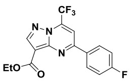
Chemical diagram of ethyl 5-(4-fluorophenyl)-7-(trifluoromethyl)pyrazolo[1, 5-a]pyrimidine-3-carboxylate
Synthesis, Crystal Structure and Biological Activity of Ethyl 5-(4-Fluorophenyl)-7-(trifluoromethyl)-pyrazolo[1, 5-a]pyrimidine-3-carboxylate
Juan SONG , Juan ZHAO , Bo-Feng DUAN , Rui WU , Soumendra ROY

With the increase of morbidity and mortality worldwide, cancer is a life-threatening disease and remains a major health problem around the globe[1]. Therefore, the search for highly efficient and safe antitumor agents has become more urgent than ever before[2, 3]. In recent years, heterocyclic structure has attracted great attention of many scientists because of their important utility in medicine[4, 5]. Among them, pyrazolo-[1, 5-a]pyrimidines are considered privileged scaffolds in drug discovery with a wide array of biological activities[6]. In the literature, pyrazolo[1, 5-a]pyrimidine derivatives have been described as antitumor[7, 8], antifungal[9], antibacterial[10], analgesics[11] and anti-inflammatory[12]. Insomnia agent indiplon[13], anticancer agent dinaciclib[14] and type 2 diabetes mellitus agent anagliptin[15] are all approved drugs containing a pyrazolo[1, 5-a]pyrimidine moiety.
In the course of our search for new anticancer agents, we have recently reported the synthesis and biological activities of a series of novel fused heterocycles. Several compounds showed excellent broad spectrum anticancer activities[16-18]. The title compound (Scheme 1) was synthesized by a published method in our laboratory[16]. In this paper, we present the crystal structural analysis of ethyl 5-(4-fluorophenyl)-7-(trifluoromethyl)pyrazolo[1, 5-a]pyrimidine-3-carb-oxylate and evaluated for its anticancer properties against two human cancer cell lines (MKN45 and H460) using MTT assay.
All melting points were taken on a Beijing Taike X-4 microscopy melting point apparatus and were uncorrected. 1H NMR spectra were recorded on a Bruker Biospin 600 MHz instrument using TMS as the internal standard. All chemical shifts were reported in ppm. IR spectra were recorded as KBr pellets on a PerkinElmer Spectrum one FTIR spectrometer. Mass spectra were obtained on a 6460 QQQ mass spectrometer (Agilent, USA) analysis system (ESI, direct injection). Elemental analysis was carried out on a Carlo Erba 1108 analyser and are found within the range of theoretical value. All reagents used in the synthesis were obtained commercially and used without further purification unless otherwise specified.
A solution of ethyl 5-amino-1H-pyrazole-4-carboxylate (1.00 g, 6.45 mmol) and 4, 4, 4-trifluoro-1-(4-fluorophenyl)-butane-1, 3-dione (1.51 g, 6.45 mmol)in acetic acid (50 mL) was heated at reflux for 6 h. After cooling to room temperature, the formed precipitate was filtered off, washed with water, and dried to give 1.97 g of the title compound in 86.5% yield. m.p.: 167~169 ℃; IR (KBr) ν: 2965 (Et, m), 1697 (C=O, s), 1634, 1594 (Ar, s), 1570 (Ar, s), 1466 (Ar, m), 1397, 1327 (CF3, s), 1198, 1171, 1027, 848, 778; 1HNMR (600MHz, DMSO-d6): δ 8.75 (s, 1H, Ar–H), 8.48 (m, 2H, Ar–H), 8.37(s, 1H, Ar–H), 7.45 (m, 2H, Ar–H), 4.34 (q, J = 7.2 Hz, 2H, CH2), 1.36 (t, J = 7.2 Hz, 3H, CH3); MS (ESI) m/z (%): 354.3 [M+H]+. Anal. Calcd. for C16H11F4N3O2 (%): C, 54.40; H, 3.14; N, 11.89. Found (%): C, 54.46; H, 3.13; N, 11.92.
The powder of the title compound was dissolved in ethyl acetate/tetrahydrofuran mixed solvents = 3:2 (V/V). After slowly evaporating the solvents for several days, some colourless single crystals suitable for X-ray analysis were obtained. A suitable single crystal of the title compound was mounted on glass fibers for data collection performed on a Bruker APEX-II CCD automatic diffractometer equipped with a graphite-monochromatic MoKα radiation (λ = 0.71073 Å) at 293 K. The data collected were integrated using the SAINT program[19]. Multi-scan absorption corrections were applied using the SADABS program[20]. The structures were solved by direct methods and refined against F2 by full-matrix least-squares methods using the SHELXTL package[21]. Location of H atoms was found by geometric calculations, and their positions and thermal parameters were fixed during the structure refinement.
The title compound was evaluated for its in vitro cytotoxic activity against two cancer cell lines (MKN-45 and H460) by the MTT-based assay method under standard conditions using sorafenib tosylate as a positive control (Table 1).
 DownLoad:
CSV
DownLoad:
CSV
| Compound | IC50 (μmol/L) | |
| MKN45 | H460 | |
| The title compound | 57.12 | 88.75 |
| Sorafenib tosylate | 3.45 | 3.74 |
| aTest MTT colourimetric assay in MKN45 and H460 human cancer cell lines | ||
The synthesis route of the title compound by one step with good yield was depicted in Scheme 1. This compound was obtained as stable solid and easily purified by filtration, and its structure was elucidated by IR, 1H NMR, MS and elemental analysis. IR shows the peak at about 1697 cm−1 resulting from the carbonyl group stretching vibration of ester. The 1H NMR spectrum for the title compound exhibits a triple peak at 1.36 ppm and a quadruple peak at 4.34 ppm, corresponding to the ethenyl proton of ester. In the mass spectrum, the peak appeared at m/z 354.3 ([M+H]+, 100%), which is in accordance with its molecular formula. IR, 1H NMR, MS, elemental analyses and single-crystal X-ray of the target compounds confirmed their structural integrity.
The single crystal of the title compound was cultured using acetate/tetrahydrofuran mixed solvents = 3:2 (V/V) at room temperature and determined by X-ray diffraction method to confirm its structure. Selected bond lengths and bond angles are listed in Table 2.
DownLoad:
CSV
| Bond | Dist. | Bond | Dist. | Bond | Dist. | ||
| F(1)–C(1) | 1.352(3) | F(2)–C(10) | 1.335(4) | F(3)–C(10) | 1.321(4) | ||
| F(4)–C(10) | 1.324(4) | O(1)–C(14) | 1.201(4) | O(2)–C(14) | 1.329(4) | ||
| O(2)–C(15) | 1.432(4) | N(1)–C(7) | 1.329(3) | N(1)–C(13) | 1.334(3) | ||
| N(2)–N(3) | 1.354(3) | N(2)–C(9) | 1.366(3) | N(2)–C(13) | 1.387(3) | ||
| N(3)–C(11) | 1.320(4) | C(1)–C(2) | 1.360(4) | C(1)–C(6) | 1.371(4) | ||
| C(2)–C(3) | 1.371(4) | C(3)–C(4) | 1.384(4) | C(4)–C(5) | 1.388(3) | ||
| Angle | (°) | Angle | (°) | Angle | (°) | ||
| C(14)–O(2)–C(15) | 116.6(3) | C(7)–N(1)–C(13) | 118.4(2) | N(3)–N(2)–C(9) | 125.8(2) | ||
| C(9)–N(2)–C(13) | 120.2(2) | C(11)–N(3)–N(2) | 102.3(2) | F(1)–C(1)–C(2) | 118.7(3) | ||
| F(1)–C(1)–C(6) | 119.1(3) | C(2)–C(1)–C(6) | 122.3(3) | C(1)–C(2)–C(3) | 118.6(3) | ||
| N(1)–C(7)–C(4) | 117.6(2) | C(2)–C(3)–C(4) | 121.3(3) | C(3)–C(4)–C(5) | 117.9(3) | ||
| C(3)–C(4)–C(7) | 122.6(2) | C(5)–C(4)–C(7) | 119.5(3) | C(6)–C(5)–C(4) | 121.4(3) | ||
| C(5)–C(6)–C(1) | 118.4(3) | N(1)–C(7)–C(8) | 120.7(3) |
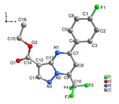
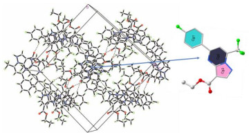
The title compound crystallizes in monoclinic system with one molecule per asymmetric unit cell. Its crystal structure (Fig. 1) shows that the molecule has a pyrazolo[1, 5-a]pyrimidine skeleton, in which all bond lengths and bond angles fall in normal ranges. The dihedral angles between pyrazolo[1, 5-a]pyrimidine and phenyl rings is 2.74°, which indicate that the two planes are almost coplanar. Furthermore, there exists a mass of intermolecular and intramolecular hydrogen bonds (Table 3), which plays a major role in stabilizing the molecule. It is worth noting that the crystal packing is further stabilized by weak π-π stacking interactions between the Cg(1)⋅⋅⋅Cg(3) and Cg(2)⋅⋅⋅Cg(2) rings, as shown in Fig. 2 and Table 4. These interactions together with intermolecular hydrogen bond result in the formation of a three-dimensional framework. In addition, the bioassay results showed the title compound exhibited weak inhibitory activity against MKN45 and H460 cell lines with IC50 values of 57.12 and 88.75 μM, which was fairly less potent than sorafenib.
 DownLoad:
CSV
DownLoad:
CSV
| D–H⋅⋅⋅A | d(D–H) | d(H⋅⋅⋅A) | d(D⋅⋅⋅A) | ∠DHA |
| C(32)–H(32C)⋅⋅⋅F(6)a | 0.96 | 2.63 | 3.561(4) | 162.4 |
| C(31)–H(31B)⋅⋅⋅F(2)b | 0.97 | 2.55 | 3.274(4) | 132 |
| C(27)–H(27)⋅⋅⋅F(5)c | 0.93 | 2.57 | 3.276(4) | 133.5 |
| C(22)–H(22)⋅⋅⋅N(6)d | 0.93 | 2.56 | 3.422(4) | 155 |
| Symmetry transformations used to generate the equivalent atoms: (a) –x + 1, –y, –z + 1; (b) x – 1, –y + 1/2, z + 1/2; (c) –x + 1, y – 1/2, –z + 3/2; (d) –x + 1, y + 1/2, –z + 3/2 | ||||
 DownLoad:
CSV
DownLoad:
CSV
| π-π stacking parameters | Cg(2)⋅⋅⋅Cg(2) | Cg(1)⋅⋅⋅Cg(3) |
| Centroid-centroid distance (Å) | 3.6175 | 3.6135 |
| Perpendicular interplanar distance (Å) | 3.5858 | 3.5229 |
| Slip angle (°) | 0.27 | 2.76 |
| Cg(1): C(1)~C(6); Cg(2): N(1), C(7)~C(9), N(2), C(13); Cg(3): N(2), N(3), C(11)~C(13) | ||
The title compound 5-(4-fluorophenyl)-7-(trifluoromethyl)pyrazolo[1, 5-a]pyrimidine-3-carboxylate was synthesized and characterized by IR, 1H-NMR, MS, elemental analysis and single-crystal X-ray diffraction. The antitumor activities of the title compound were evaluated for cytotoxicity against two human cancer cell lines (MKN-45 and H460), using MTT assay. Cytotoxicity assay results revealed that the title compound showed poor cytotoxicity against MKN-45 and H460 cells.
Kamel, M. M.; Megally Abdo, N. Y. Synthesis of novel 1, 2, 4-triazoles, triazolothiadiazines and triazolothiadiazoles as potential anticancer agents. Eur. J. Med. Chem. 2014, 86, 75–80. doi: 10.1016/j.ejmech.2014.08.047
Ju, L.; Xuechen, H.; Shi, D.; Ye, C.; Yang, W. Synthesis of novel 1-arylpyrazolo[3, 4-b][1, 5]benzodiazepine derivatives. J. Liaoning Univ. Nat. Sci. Ed. 2018, 45, 244–248.
Dörsam, B.; Fahrer, J. The disulfide compound α-lipoic acid and its derivatives: a novel class of anticancer agents targeting mitochondria. Cancer Lett. 2016, 371, 12–19. doi: 10.1016/j.canlet.2015.11.019
Norman, B. H.; Lander, P. A.; Gruber, J. M.; Kroin, J. S.; Cohen, J. D.; Jungheim, L. N. Cyclohexyl-linked tricyclic isoxazoles are potent and selective modulators of the multidrug resistance protein (MRP1). Bioorg. Med. Chem. Lett. 2015, 25, 5526–5530.
You, W. K.; Sennino, B.; Williamson, C. W.; Falcón, B.; Hashizume, H.; Yao, L. C.; Aftab, D. T.; McDonald, D. M. VEGF and c-met blockadeamplify angiogenesis inhibition in pancreatic islet. Cancer Res. 2011, 71, 4758–768.
Cherukupalli, S; Karpoormath, R.; Chandrasekaran, B.; Hampannavar, G. A.; Thapliyal, N.; Palakollu, V. N. An insight on synthetic and medicinal aspects of pyrazolo[1, 5-a]pyrimidine scaffold. Eur. J. Med. Chem. 2017, 126, 298–352. doi: 10.1016/j.ejmech.2016.11.019
Asghar, U.; Witkiewicz, A. K.; Turner, N. C.; Knudeen, E. S. The history and future of targeting cyclin-dependent kinases in cancer therapy. Nat. Rev. Drug Discov. 2015, 14, 130–146. doi: 10.1038/nrd4504
Yakaiah, T.; Kurumurthy, C.; Lingaiah, B. P. V.; Narsaiah, B.; Pamanji, R.; Velatooru, L. R.; Gururaj, S.; Parthasarathy, T.; Sridhar, B. GdCl3 promoted synthesis of novel pyrimidine fused indazole derivatives and their anticancer activity. Med. Chem. Res. 2012, 21, 4261–4273. doi: 10.1007/s00044-011-9962-0
Li, Y.; Gao, W. M.; Li, F.; Wang, J. H.; Zhang, J. X.; Yang, Y. F.; Zhang, S. W.; Yang, L. An in silico exploration of the interaction mechanism of pyrazolo[1, 5-a]pyrimidine type CDK2 inhibitors. Mol. Biosyst. 2013, 9, 2266–2281. doi: 10.1039/c3mb70186g
Gouda, M. A.; Berghot, M. A.; Shoeib, A. I. Synthesis and antimicrobial of new anthraquinone derivatives incorporating pyrazole moiety. J. Med. Chem. 2010, 45, 1843–1848. doi: 10.1016/j.ejmech.2010.01.021
Shaaban, M. R.; Saleh, T. S.; Mayhoub, A. S.; Mansour, A.; Farag, A. M. Synthesis and analgesic/anti-inflammatory evaluation of fused heterocyclic ring systems incorporating phenylsulfonyl moiety. Biorg. Med. Chem. 2008, 16, 6344–6352. doi: 10.1016/j.bmc.2008.05.011
Lu, J. F.; Min, S. T.; Ge, H. G. Synthesis, crystal structures and properties of two-dimensional complexes constructed by dicarboxylate and bis(imidazole) co-ligands. J. Chem. Res. 2014, 38, 726–730. doi: 10.3184/174751914X14176001751990
Rivers, E. C.; Mancera, R. L. New anti-tuberculosis drugs in clinical trials with novel mechanisms of action. Drug Discov. Today 2008, 13, 1090–1098. doi: 10.1016/j.drudis.2008.09.004
Lu, J. F.; Zhao, J.; Zhao, C. B.; Yu, X. H. A Chinese lantern-like 2D Cu(II) coordination polymer constructed by bis-imidazole and dicarboxylate co-ligands: synthesis, crystal structure and photocatalytic activity. Chin. J. Struct. Chem. 2020, 39, 321–328.
Kato, N.; Oka, M.; Murase, T.; Yoshida, M.; Sakairi, M.; Yamashita, S.; Yasuda, Y.; Yoshikawa, A.; Hayashi, Y.; Makino, M.; Takeda, M.; Mirensha, Y.; Kakigami, T. Discovery and pharmacological characterization of N-[2-({2-[(2S)-2-cyanopyrrolidin-1-yl]-2-oxoethyl}amino)-2-methylpropyl]-2-methylpyrazolo[1, 5-a]pyrimidine-6-carboxamide hydrochloride (anagliptin hydrochloride salt) as a potent and selective DPP-IV inhibitor. Bioorg. Med. Chem. 2011, 19, 7221–7227. doi: 10.1016/j.bmc.2011.09.043
Lu, J. F.; Jin, L. X.; Ge, H. G.; Ji, X. H.; Guo, X. H.; Tian, G. H.; Song, J.; Jiang, M. Synthesis, crystal, computational study and biological activity of N-(1-(2, 4-dichlorophenyl)-1H-pyrazolo[3, 4-d]pyrimidin-4-yl)-4-(N, N-dipropylsulfamoyl)benzamide. Chin. J. Struct. Chem. 2017, 36, 1810–1816.
Ji, X. H.; Zhao, J.; Lu, J. F.; Jin, L. X.; Ge, H. G. Synthesis, crystal structure and biological activity of 1-(3-amino-4-morpholino-1H-indazole-1-carbonyl)-N-(4-fluorophenyl)cyclopropane-1-carboxamide. Chin. J. Struct. Chem. 2019, 38, 1889–1894.
Lu, J. F.; Zhou, X. L.; Xu, Y. H.; Yue, S. Y.; Ji, X. H.; Zheng, N.; Jin, L. X. Synthesis, crystal structure, and biological activity of 3-amino-4-morpholino-N-[2-(trifluoromethoxy)phenyl]-1H-indazole-1-carboxamide. J. Chem. Res. 2017, 41, 526–528. doi: 10.3184/174751917X15033157981988
SMART & SAINT. Software Reference Manuals, Version 6.22, Madison (WI, USA): Bruker AXS Analytic X-ray Systems Inc. 2000.
Sheldrick, G M. SADABS, Software for Empirical Absorption Correction. University of Göttingen, Germany 2000.
Sheldrick, G. M. SHELXS-97, Program for the Solution of Crystal Structures. University of Göttingen, Germany 1997.

Scheme1 Chemical diagram of ethyl 5-(4-fluorophenyl)-7-(trifluoromethyl)pyrazolo[1, 5-a]pyrimidine-3-carboxylate
Table 1. In Vitro Anticancer Activity Testa of the Title Compound on MKN45 and H460 Cell Lines
| Compound | IC50 (μmol/L) | |
| MKN45 | H460 | |
| The title compound | 57.12 | 88.75 |
| Sorafenib tosylate | 3.45 | 3.74 |
| aTest MTT colourimetric assay in MKN45 and H460 human cancer cell lines | ||
 下载: 导出CSV
Table 2. Selected Bond Lengths (Å) and Bond Angles (°) for 1
| Bond | Dist. | Bond | Dist. | Bond | Dist. | ||
| F(1)–C(1) | 1.352(3) | F(2)–C(10) | 1.335(4) | F(3)–C(10) | 1.321(4) | ||
| F(4)–C(10) | 1.324(4) | O(1)–C(14) | 1.201(4) | O(2)–C(14) | 1.329(4) | ||
| O(2)–C(15) | 1.432(4) | N(1)–C(7) | 1.329(3) | N(1)–C(13) | 1.334(3) | ||
| N(2)–N(3) | 1.354(3) | N(2)–C(9) | 1.366(3) | N(2)–C(13) | 1.387(3) | ||
| N(3)–C(11) | 1.320(4) | C(1)–C(2) | 1.360(4) | C(1)–C(6) | 1.371(4) | ||
| C(2)–C(3) | 1.371(4) | C(3)–C(4) | 1.384(4) | C(4)–C(5) | 1.388(3) | ||
| Angle | (°) | Angle | (°) | Angle | (°) | ||
| C(14)–O(2)–C(15) | 116.6(3) | C(7)–N(1)–C(13) | 118.4(2) | N(3)–N(2)–C(9) | 125.8(2) | ||
| C(9)–N(2)–C(13) | 120.2(2) | C(11)–N(3)–N(2) | 102.3(2) | F(1)–C(1)–C(2) | 118.7(3) | ||
| F(1)–C(1)–C(6) | 119.1(3) | C(2)–C(1)–C(6) | 122.3(3) | C(1)–C(2)–C(3) | 118.6(3) | ||
| N(1)–C(7)–C(4) | 117.6(2) | C(2)–C(3)–C(4) | 121.3(3) | C(3)–C(4)–C(5) | 117.9(3) | ||
| C(3)–C(4)–C(7) | 122.6(2) | C(5)–C(4)–C(7) | 119.5(3) | C(6)–C(5)–C(4) | 121.4(3) | ||
| C(5)–C(6)–C(1) | 118.4(3) | N(1)–C(7)–C(8) | 120.7(3) |
下载: 导出CSV
Table 3. Hydrogen Bond Lengths (Å) and Bond Angles (°)
| D–H⋅⋅⋅A | d(D–H) | d(H⋅⋅⋅A) | d(D⋅⋅⋅A) | ∠DHA |
| C(32)–H(32C)⋅⋅⋅F(6)a | 0.96 | 2.63 | 3.561(4) | 162.4 |
| C(31)–H(31B)⋅⋅⋅F(2)b | 0.97 | 2.55 | 3.274(4) | 132 |
| C(27)–H(27)⋅⋅⋅F(5)c | 0.93 | 2.57 | 3.276(4) | 133.5 |
| C(22)–H(22)⋅⋅⋅N(6)d | 0.93 | 2.56 | 3.422(4) | 155 |
| Symmetry transformations used to generate the equivalent atoms: (a) –x + 1, –y, –z + 1; (b) x – 1, –y + 1/2, z + 1/2; (c) –x + 1, y – 1/2, –z + 3/2; (d) –x + 1, y + 1/2, –z + 3/2 | ||||
下载: 导出CSV
Table 4. π-π Stacking Geometry for the Title Compound
| π-π stacking parameters | Cg(2)⋅⋅⋅Cg(2) | Cg(1)⋅⋅⋅Cg(3) |
| Centroid-centroid distance (Å) | 3.6175 | 3.6135 |
| Perpendicular interplanar distance (Å) | 3.5858 | 3.5229 |
| Slip angle (°) | 0.27 | 2.76 |
| Cg(1): C(1)~C(6); Cg(2): N(1), C(7)~C(9), N(2), C(13); Cg(3): N(2), N(3), C(11)~C(13) | ||
下载: 导出CSV

 扫一扫看文章

扫一扫关注我们

