Scheme 1.
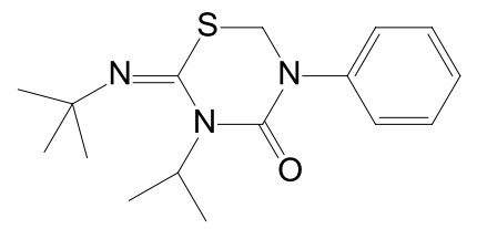
Molecular structure of compound 1
Synthesis, Crystal Structures, Hirshfeld Surfaces, Thermo Characteristics and Solubility of Co-crystal of Buprofezin with Hydrofluoric Acid
Liang MA , Qing-Zhao YAO , Yu-Ming ZHOU , Ming-Liang WANG , Bai-Wang SUN , Yi XUE , Chang GUO

Co-crystal is a common state in the study of small organic molecules and crystallography. Co-crystal can be defined as crystalline solid composed of multiple components in a process where molecular recognition drives assembly[1-4]. The study on co-crystal was first reported in the synthesis of hydroquinone co-crystal in 1893[5], and it is generally believed that co-crystal is a crystal formed by two or more kinds of different molecules in a crystal structure, and it is a synthetic body with certain ordered structure formed by several kinds of molecules through molecular recognition[6, 7]. Cocrystallization is widely used to change the solubility, stability and bioavailability of the active pharmaceutical[8, 9].
Co-crystals have become a hot topic in modern materials chemistry and biochemical pharmacology. Meanwhile, they also have aroused interest of researchers in recent years[10-12]. In the field of pharmacology, a large number of drug molecules have been found, which can use hydrogen bond donors/receptors to compose co-crystal due to the flexibility of molecular conformation and various functions existing in molecules[13, 14]. The hydrogen bonding, hydrophobic forces, van der Waals forces, p-p interactions, and other characteristics will change after the formation of co-crystal, and these investigations have main effect on the areas of materials science and pharmacology[15-17]. The crystallography of pesticides can change the properties of pesticides to some extent and make them play a better role.
Buprofezin (compound 1, Scheme 1) is a selective and highly effective insecticide developed by Japanese pesticide company in 1981, which act to inhibit and restrain the action of insect chitin synthase and interfere with metabolism to block the synthesis of chitin, then prevent insect molting and pupation and other processes and resulting in chronic death[18-21]. High activity, high selectivity, long shelf life and no interaction resistance with conventional pesticides are properties of buprofezin[22]. It is safe for people, livestock, bees and natural enemies, so it is also the most widely used insecticide[23].
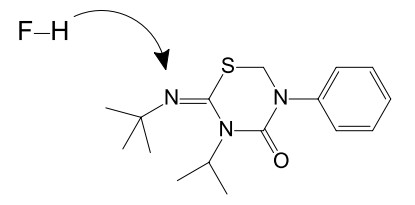
We obtained a co-crystal of buprofezin through many experiments based on previous researches. The co-crystal was obtained through slow evaporation solvent method in this study. Compound 1 has polymorphism[24]. Compound 2 (Scheme 2) was buprofezin co-crystalized with hydrofluoric acid through evaporation solvent method in acetone. Besides, different solvents such as THF, DMSO, dichloromethane, methylbenzene, etc and different inorganic acids like sulfuric acid, nitric acid and so on were also experimented at the same time, intending to investigate the influence factor to co-crystal formation. Compound 2 was a co-crystal, in which the buprofezin was bound with hydrofluoric acid via hydrogen bonds. The result showed that the acetone was the best solvent to crystallize. Then the physical and chemical properties of two compounds were studied by X-ray diffraction, X-ray powder diffraction (XRPD), Fourier-Transform infrared spectra (FT-IR), Hirshfeld surface analysis, differential scanning calorimetry (DSC), thermo gravimetric analysis (TGA) and Raman spectroscopy. Compared with buprofezin, the co-crystal offered stronger thermal stability and water solubility. Therefore, the co-crystal of buprofezin is likely to have longer residual activity than buprofezin, and can increase the spraying concentration and improve the insecticidal efficiency[25].
Buprofezin (purity > 99%, CAS registry number: 69327-76-0), hydrofluoric acid (purity > 99%, CAS registry number: 7664-39-3), solvents such as THF, DMSO, dichloromethane, trichloromethane, acetone and methylbenzene were all purchased by Sinopharm Chemical Reagent Co., Ltd. and used without purification.
Compound 2 was obtained using evaporation solvent method in this solution crystallization experiment. The buprofezin (1.301 g, 4 mmol) and hydrofluoric acid (0.080 g, 4 mmol) were added in acetone (50 mL) solvent. The mixture was stirred at room temperature for 4 h, and both compounds were completely dissolved in acetone. The solution was stood to evaporate the solvent under nitrogen atmosphere at room temperature for 5 days, and colorless transparent crystal was obtained through the evaporation of acetone. The experiment was completed at ambient temperature (~23 ℃).
Crystal structure of compound 2 was measured by single-crystal X-ray diffraction. With graphite-monochromated MoKα radiation (λ = 0.71073 Å), the single-crystal X-ray diffraction data of compound 2 were shown at temperature of 100 K. The SHELXL-2018 program was used for structure solution in direct method. All non-hydrogen atoms were anisotropically extracted and purified with displacement parameters. The MERCURY and DIAMOND programs were used to obtain the molecular graphics of compound 2.
The X-ray powder diffraction(XRPD) was conducted by a Siemens D-5000 diffractometer (Bruker-AXS, Karlsruhe, Germany) at 40 kV and 40 mA with Cu-Ka radiation (λ = 1.5406 Å). The sample was scanned over a 2θ range from 5° to 80° at a step size of 0.02° with a scan rate of 20 °·min-1.
Fourier-transform infrared spectroscopy: Infrared spectra were recorded on a Bruker FT-IR analyzer (VECTOR-22, Bruker Co., Germany) by using ground KBr powder as its measurement background. The sample was determined in the spectral range of 4000~500 cm-1, which shows the form of potassium bromide pellets.
Thermal analysis: Differential scanning calorimetry (DSC) was performed with Mettler-Toledo DSC-822 (Mettler-Toledo, Columbus, OH). Both compounds were heated with 10 K·min-1 rate under the atmosphere of dry nitrogen flowing at 10 cm3·min-1 in the temperature range of 50~200 ℃. All samples were settled in open aluminum oxide crucibles annealed at 1100 ℃. Thermo gravimetric analysis (TGA) was carried out by means of a Shimadzu TGA-50 instrument (Shimadzu, Japan). The sample was measured under an atmosphere of dry N2 flowing at 20 cm3·min−1 at a heating rate of 10 K·min−1 over a range from 30 to 800 ℃.
Raman spectroscopy: Raman spectra were obtained using Renishaw System 1000 micro-Raman spectroscope (Renishaw, U. K.). The sample was measured with a 1000 lines/mm grating in the spectral range 400~3700 cm-1.
The CrystalExplorer17 program was used to perform Molecular Hirshfeld surfaces calculations. When the CIF file of target compound was read into the CrystalExplorer17 program, all bond lengths of hydrogen atoms were automatically converted into typical standard neutron values (C–H = 1.083 and N–H = 1.009 Å). All the Hirshfeld surfaces were generated using a standard (high) surface resolution in this study. Using 0.76(red)~2.4 Å (blue) fixed color label to map the 3-D dnorm surfaces, the shape index was mapped in the range of 0.7~2.5 Å, and the curvature was mapped in the range of 0.72~2.4 Å. The standard 0.6~2.6 Å view in the distance scale of de and di displayed on the axis is used to display the 2-D fingerprint plots.
In order to compare the water solubility of buprofezin and co-crystal at different temperature, the equilibrium solubility of compounds 1 and 2 in pure water was determined over a range from 0 to 40 ℃ to test the solute dissolved in each liter of water.
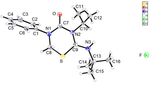
Compound 2 harvested from acetone was colorless and transparent. Crystal structure determination is a very efficient and useful analytical tool for crystal physicochemical property analysis. The structural data of compound 2 can be clearly obtained through X-ray singlecrystal diffraction. The distances and angles of bonds in the crystal structures of compound 2 are depicted in Table 1.
 DownLoad:
CSV
DownLoad:
CSV
| Bond | Dist. | Bond | Dist. | Bond | Dist. | ||
| S(1)–C(9) | 1.738(2) | N(2)–C(10) | 1.505(3) | C(10)–C(12) | 1.515(3) | ||
| O(1)–C(7) | 1.216(3) | N(3)–C(13) | 1.517(3) | C(1)–C(6) | 1.387(4) | ||
| N(2)–C(9) | 1.370(3) | N(1)–C(7) | 1.352(3) | C(2)–C(3) | 1.395(4) | ||
| C(13)–C(15) | 1.526(3) | C(6)–C(5) | 1.384(4) | C(3)– C(4) | 1.384(4) | ||
| Angle | (°) | Angle | (°) | Angle | (°) | ||
| C(9)–S(1)–C(8) | 95.54(11) | O(1)–C(7)–N(2) | 120.0(2) | N(3)–C(13)–C(14) | 110.37(18) | ||
| C(9)–N(2)–C(7) | 122.46(19) | O(1)–C(7)–N(1) | 124.3(2) | N(3)–C(13)–C(15) | 109.93(19) | ||
| C(9)–N(2)–C–(10) | 119.42(18) | N(1)–C(7)–N(2) | 115.76(19) | C(14)–C(13)–C(15) | 112.4(2) | ||
| C(7)–N(2)–C(10) | 116.12(17) | N(2)–C(10)–C(12) | 111.57(19) | C(16)–C(13)–N(3) | 105.29(19) | ||
| C(9)–N(3)–C(13) | 129.4(2) | N(2)–C(10)–C(11) | 111.55(18) | C(16)–C(13)–C(14) | 109.0(2) | ||
| C(7)–N(1)–C(1) | 120.38(19) | C(12)–C(10)–C(11) | 113.2(2) | C(16)–C(13)–C(15) | 109.6(2) | ||
| C(7)–N(1)–C(8) | 119.63(19) | C(2)–C(1)–N(1) | 120.7(2) | C(5)–C(6)–C(1) | 119.5(3) | ||
| C(1)–N(1)–C(8) | 119.73(19) | C(2)–C(1)–C(6) | 120.9(2) | C(4)–C(3)–C(2) | 120.0(3) | ||
| N(2)–C(9)–S(1) | 119.06(17) | C(6)–C(1)–N(1) | 118.4(2) | C(4)–C(5)–C(6) | 120.2(3) | ||
| N(3)–C(9)–S(1) | 120.16(17) | C(1)–C(2)–C(3) | 119.1(3) | C(5)–C(4)–C(3) | 120.3(3) | ||
| N(3)–C(9)–N(2) | 120.8(2) | N(1)–C(8)–S(1) | 108.86(17) |
Compound 2 is of triclinic crystal system and belongs to P
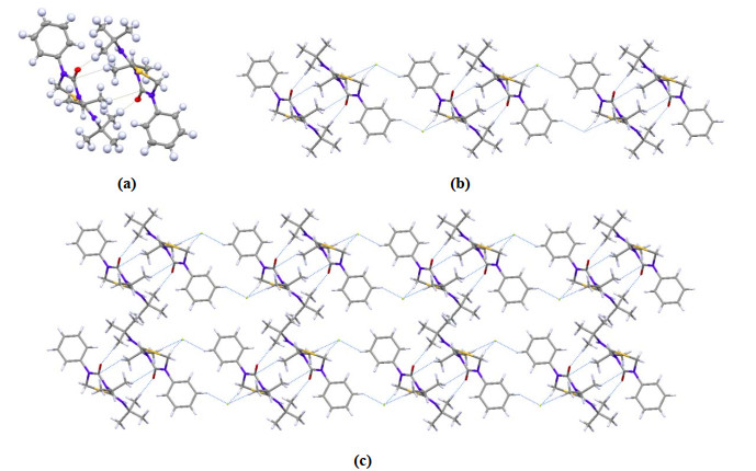
The stacking structure of compound 2 is also characteristic. As shown in Fig. 2a, two molecules are in a unit cell in this co-crystal structure and are connected by C–H and O–H to form a dimer. Then dimers are connected by H–F⋅⋅⋅H bond to form a one-dimensional chain structure, as shown in Fig. 2b. The distance between the two molecules in the dimer is measured to be 1.143 Å, and that between two dimers is 3.76 Å. An infinite number of molecules stack into a two-dimensional structure, as shown in Fig. 2c. Table 2 shows that with more fluorine atoms in the co-crystal system, compound 2 has stronger intermolecular hydrogen bond than 1. Buprofezin is required to have strong stability as an insecticide. Therefore, the progress of co-crystal has a positive significance for the study on the formulation and stability of buprofezin.
DownLoad:
CSV
| Sample | H–H | H–F | C–H | O–H | S–H | S–F | N–H |
| Compound 2 | 61.6 | 16 | 10.2 | 6.2 | 4 | 0.8 | 0.4 |
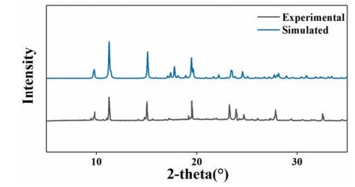
X-ray powder diffraction data provide more chances to confirm the crystal structure though X-ray single-crystal diffraction, which offers more physicochemical properties of compound 2. As shown in Fig. 3, X-ray powder diffraction profiles of compound 2 fall in the 5~245° range, and the experimental pattern has good agreement with the simulated X-ray powder diffraction pattern through refinement in the final Rietveld, indicating that the compound obtained in the experiment showed high thermal stability. Moreover, the crystallization products obtained by solvent evaporation method are fixed in a single form rather than a mixture of multiple forms.
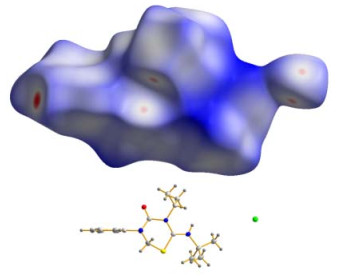
The Hirshfeld surface analysis is an efficient tool for visualizing intermolecular interactions by 3D color-coding scheme which is unique for any crystal structure. The 2D fingerprint plots, ordered by Hirshfeld surface analysis, can be divided by the 3D dnorm surface and could also give a quantitative summary of the nature and type of intermolecular contacts occurring by the molecules in the crystal simultaneously. All interaction types such as hydrogen bonding, close and distant van der Waals contacts, C–H⋅⋅⋅π interactions and π-π stacking are readily obtained. Red-blue-white color scheme served as drawing the 3D dnorm values to the Hirshfeld surface: The red region shows the closer contacts and negative dnorm values, the blue area on behalf of the closer contacts and positive dnorm values, and the white area denotes dnorm value equal to zero and the contacts distance equal to rvdW.
The 3D dnorm surface data of compound 2 are shown in Fig. 4. Large areas of blue and white with several red points could be observed clearly. According to the principle of drawing, the red dots on white areas correspond to the significant longer contacts, and the size of red points indicated the strength of intermolecular force. It is obvious from the 2D fingerprint that a two-dimensional structure was mainly due to the H–H, H–F, C–H, S–H and H–O intermolecular interactions.
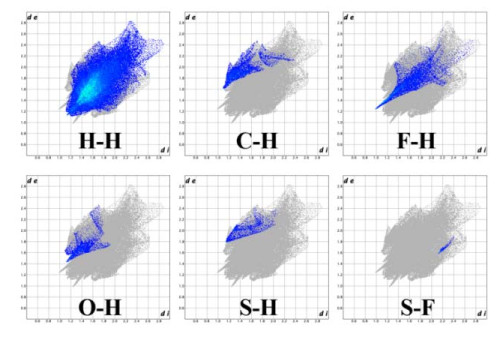
2D Hirshfeld fingerprint plot for compound 2 is shown in Fig. 5. These figures show an important characteristic of the fingerprint plots, which are associated with immediate environment of the compound. 2D fingerprint can be a unique description for the compound with various polymorphic forms, and meantime can be used to differentiate similar compounds. Fig. 5 shows that the dominant interaction in compound 2 is H–H, and the H–H intermolecular interaction of 2 (de + di = 2.32 Å) mainly comprises 61.6% to the total Hirshfeld surface. The O–H intermolecular interaction (de + di = 2.6 Å) mainly comprise 6.2% to the total Hirshfeld surface. Table 2 is listed to sum up various contacts contributions to the Hirshfeld surface in detail.
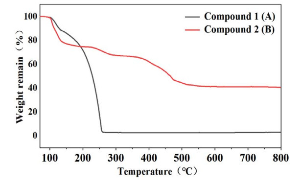
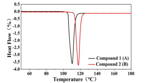
TGA and DSC were used to investigate the thermal properties of compounds 1 (A) and 2 (B). As shown in Fig. 6, obviously compound 1 undergoes two steps of mass loss at 106 and 131 ℃ on TGA, and corresponding mass loss is 11% and 85.5%, respectively. Compound 2 shows three steps in profile, at about 110 ℃ with 24.7% mass loss, 227.2 ℃ with 7.4% mass loss and 334 ℃ with 25.2% mass loss. Fig. 7 shows that compound 1 begins to melt with melting enthalpy of 241 J·g-1 at 105 ℃. Compound 2 begins to melt at 115 ℃ with melting enthalpy of 218 J·g-1. TGA and DSC analysis results combined with the Hirshfeld surface calculations in Table 3 indicate that compound 2 has abundant intermolecular hydrogen bonds which cause its stronger intermolecular force. Thus, comparing the melting point of compounds 2 and 1, the former has a higher melting point, and the structure of co-crystal is more stable than compound 1.
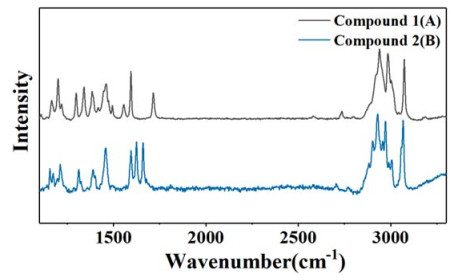
The Raman spectra of compounds 1 (A) and 2 (B) are shown in Fig. 8. From this figure, region ranging from 3200 to 2700 cm-1 and from 1800 to 1100 cm-1 contain the information of proton transfer and hydrogen bonding interactions, so we can obtain important information about the molecular interactions. We can observe the specific differences between A and B clearly. For instance, both compounds have roughly the same peaks, but those at 1341, 1555 and 1717 cm-1 make up with the characteristic of buprofezin molecule. However, compound 1 shows different traits, which shows 1154, 1625 and 1660 cm-1. The lattice vibrations of two polymorphs are mainly concentrates on the high wave-number region, and the primarily chemical structure could be observed from the region from 1800 to 1100 cm-1 in detail.
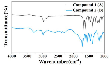
The FT-IR of compounds 1 (A) and 2 (B) are shown in Fig. 9. The individual polymorphs can be distinguished more clearly through FT-IR. Infrared spectra can show almost all molecular vibration modes, which include not only the intermolecular vibration of single bond, double bond and benzene ring, but also hydrogen bonding in strong or weak connection. Besides, the spectra allow conformational changes such as alkyl chains. The absorption peak of compound 2 in the infrared spectrum changed obviously after buprofezin was crystallized with hydrofluoric acid, and the changes mainly focused on the infrared absorption C=O and C=N. Compared with compound 1, the absorption peaks of compound 2 at 1664 and 1619 cm-1 decreased and shifted to 1715 and 1560 cm-1, respectively. This difference can obviously be contributed to a differentiation in the crystal structure. The C=O and C=N in compound 2 formed hydrogen bonds with different hydrogen atoms.
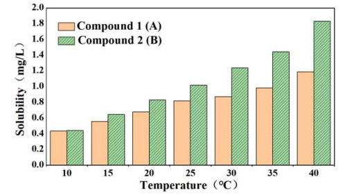
The solubility data of compounds 1 and 2 are depicted in Fig. 10. The water solubility of cocrystal was higher than that of buprofezin over the range of 0~40 ℃ due to the increase of hydrogen bonds. As mentioned above, the molecules in compound 2 are mainly bound by intermolecular hydrogen bonds. Therefore, the molecules of 2 form hydrogen bonds with water molecules when dissolved in water, which makes it more soluble in water than 1. The higher solubility suggests that pesticide concentration can be increased during application to enhance the insecticidal effect. In addition, the water solubility of pesticide is improved by forming co-crystal, so pesticide residues on crops are easier to be washed by water, which improves the safety of pesticides on humans and animals.
The present study is focused on a new kind of co-crystal of buprofezin. A co-crystal with buprofezin and hydrofluoric acid has been discovered through an intensive co-crystal screening, then single-crystal X-ray diffraction method was used to investigate its crystal information. Compared with the single crystal of buprofezin, co-crystal has different thermal behaviors, which indicates that the latter has more stable thermodynamic properties. The Hirshfeld surface analysis, TGA, DSC, Raman, and FT-IR were used to get more co-crystal information, showing that the co-crystal tends to be stable mainly by the interaction of H–H, H–F, C–H, O–H and S–H. Solubility study shows that the water solubility of co-crystal is better than buprofezin in the application.
Dunitz, J. D.; Gavezzotti, A. How molecules stick together in organic crystals: weak intermolecular interactions. Chem. Soc. Rev. 2009, 38, 2622-2633. doi: 10.1039/b822963p
Xiao, M.; Xian, Y.; Shi, F. Precise macroscopic supramolecular assembly by combining spontaneous locomotion driven by the Marangoni effect and molecular recognition. Angew. Chem. Int. Edit. 2015, 54, 8952-8956. doi: 10.1002/anie.201502349
Seth, S. K.; Das, N. K.; Aich, K.; Sen, D.; Fun, H. K.; Goswami, S. Exploring contribution of intermolecular interactions in supramolecular layered assembly of naphthyridine co-crystals: insights from Hirshfeld surface analysis of their crystalline states. J. Mol. Struct. 2013, 1048, 157-165. doi: 10.1016/j.molstruc.2013.05.048
Schneider, H. J. Binding mechanisms in supramolecular complexes. Angew. Chem. Int. Edit. 2009, 48, 3924-3977. doi: 10.1002/anie.200802947
Ling, A. R. LII. Studies on isomeric change. No. Ⅳ. Halogen derivatives of quinone. Part Ⅰ. J. Chem. Soc., Trans. 1892, 61, 558-581. doi: 10.1039/CT8926100558
Parthasarathi, R.; Subramanian, V.; Sathyamurthy, N. Hydrogen bonding without borders: an atoms-in-molecules perspective. J. Phys. Chem. A 2006, 110, 3349-3351. doi: 10.1021/jp060571z
Shimizu, G. K. H.; Vaidhyanathan, R.; Taylor, J. M. Phosphonate and sulfonate metal organic frameworks. Chem. Soc. Rev. 2009, 38, 1430-1449. doi: 10.1039/b802423p
Liu, X. G.; Bao, S. S.; Li, Y. Z.; Zheng, L. M. Polymorphism in homochiral zinc phosphonates. Inorg. Chem. 2008, 47, 5525-5527. doi: 10.1021/ic800663t
Nechipadappu, S. K.; Trivedi, D. R. Cocrystal of nutraceutical sinapic acid with active pharmaceutical ingredients ethenzamide and 2-chloro-4-nitrobenzoic acid: equilibrium solubility and stability study. J. Mol. Struct. 2018, 1171, 898-905. doi: 10.1016/j.molstruc.2018.06.074
Zhou, P.; Liang, Y.; Zhang, H.; Jiang, H.; Feng, K.; Xu, P.; Wang, J.; Wang, X.; Ding, K.; Luo, C.; Liu, M.; Wang, Y. Design, synthesis, biological evaluation and cocrystal structures with tubulin of chiral β-lactam bridged combretastatin A-4 analogues as potent antitumor agents. Eur. J. Med. Chem. 2018, 144, 817-842. doi: 10.1016/j.ejmech.2017.12.004
Qin, X.; Hao, Z.; Tian, Q.; Zhang, Z.; Zhou, C.; Xie, W. Cocrystal structures of glycyl-tRNA synthetase in complex with tRNA suggest multiple conformational states in glycylation. J. Biol. Chem. 2014, 289, 20359-20369. doi: 10.1074/jbc.M114.557249
Ames, B.; Nguyen, C.; Bruegger, J.; Smith, P.; Xu, W.; Ma, S.; Wong, E.; Wong, S.; Xie, X.; Li, W. H.; Vederas, J.; Tang, Y.; Tsai, S. C. Crystal structure and biochemical studies of the trans-acting polyketide enoyl reductase LovC from lovastatin biosynthesis. Proc. Natl. Acad. Sci. 2012, 109, 11144-11149. doi: 10.1073/pnas.1113029109
Shaikh, R.; Singh, R.; Walker, G. M.; Croker, D. M. Pharmaceutical cocrystal drug products: an outlook on product development. Trends Pharmacol. Sci. 2018, 39, 1033-1048. doi: 10.1016/j.tips.2018.10.006
Yan, Y.; Chen, J.; Lu, T. B. Simultaneously enhancing the solubility and permeability of acyclovir by crystal engineering approach. CrystEngComm. 2013, 15, 6457-6460. doi: 10.1039/c3ce41017j
Khan, E.; Shukla, A.; Jadav, N.; Telford, R.; Ayala, A. P.; Tandon, P.; Vangala, V. R. Study of molecular structure, chemical reactivity and H-bonding interactions in the cocrystal of nitrofurantoin with urea. New J. Chem. 2017, 41, 11069-11078. doi: 10.1039/C7NJ01345K
Pandey, J.; Prajapati, P.; Shimpi, M.; Tandon, P.; Velaga, S.; Srivastava, D. A.; Sinha, K. Studies of molecular structure, hydrogen bonding and chemical activity of a nitrofurantoin-L-proline cocrystal: a combined spectroscopic and quantum chemical approach. RSC Adv. 2016, 6, 74135-74154. doi: 10.1039/C6RA13035F
Srivastava, K.; Khan, E.; Shimpi, M.; Tandon, P.; Sinha, K.; Velaga, S. Molecular structure and hydrogen bond interactions of a paracetamol-4, 4'-bipyridine cocrystal studied using vibrational spectroscopic and quantum chemical approach. CrystEngComm. 2017, 20, 213-222.
Cabras, P.; Angioni, A.; Garau, V.; Melis, M.; Pirisi, F.; Cabitza, F.; Dedola, F.; Navickiene, S. Determination of buprofezin, pyridaben, and tebufenpyrad residues by gas chromatography-mass-selective detection in clementine citrus. J. Agric. Food Chem. 1998, 46, 4255-4259. doi: 10.1021/jf9802171
Wang, Z.; Zhou, C.; Long, G. Y.; Yang, H.; Jin, D. C. Sublethal effects of buprofezin on development, reproduction, and chitin synthase 1 gene (SfCHS1) expression in the white-backed planthopper, Sogatella furcifera (Hemiptera: Delphacidae). J. Asia-Pac. Entomol. 2018, 21, 585-591. doi: 10.1016/j.aspen.2018.03.009
Ali, E.; Liao, X.; Yang, P.; Mao, K.; Zhang, X.; Shakeel, M.; Markaz, A.; Wan, H.; Li, J. Sublethal effects of buprofezin on development and reproduction in the white-backed planthopper, Sogatella furcifera (Hemiptera: Delphacidae). Sci. Rep. 2017, 05, 16913-9.
Zia Qureshi, I.; Bibi, A.; Shahid, S.; Ghazanfar, M. Exposure to sub-acute doses of fipronil and buprofezin in combination or alone induces biochemical, hematological, histopathological and genotoxic damage in common carp (Cyprinus carpio L. ). Aquat. Toxicol. 2016, 179, 103-114. doi: 10.1016/j.aquatox.2016.08.012
Ji, X.; Ku, T.; Zhu, N.; Ning, X.; Wei, W.; Li, G.; Sang, N. Potential hepatic toxicity of buprofezin at sublethal concentrations: ros-mediated conversion of energy metabolism. J. Hazard. Mater. 2016, 320, 176-186. doi: 10.1016/j.jhazmat.2016.08.027
Wang, G.; Xu, D.; Xiong, M.; Zhang, H.; Li, F.; Liu, Y. Novel degradation pathway and kinetic analysis for buprofezin removal by newly isolated Bacillus sp. J. Environ. Manage. 2016, 180, 59-67. doi: 10.1016/j.jenvman.2016.04.061
Zhu, Z.; Zhou, Y.; Yao, Q.; Sun, B.; Wang, M.; Zhong, X.; Wang, B.; Xue, Y.; Chen, X. Two polymorphs and a sulfate of buprofezin: crystal structure and Hirshfeld surface analysis. Polyhedron 2018, 155, 85-93. doi: 10.1016/j.poly.2018.08.034
Chen, X.; Zhou, Z.; Chen, J.; Chu, C.; Zheng, J.; Wang, S.; Jia, W.; Zhao, J.; Li, R.; Han, D. Solubility determination and thermodynamic modeling of buprofezin in different solvents and mixing properties of solutions. J. Chem. Eng. Data 2019, 64, 1177-1186. doi: 10.1021/acs.jced.8b01099

Figure 2 (a) Dimer structure of compound 2, (b) One-dimensional chain structure of compound 2, (c) Two-dimensional stacking motif of compound 2
Table 1. Selected Bond Lengths (Å) and Bond Angles (°) of Compound 2
| Bond | Dist. | Bond | Dist. | Bond | Dist. | ||
| S(1)–C(9) | 1.738(2) | N(2)–C(10) | 1.505(3) | C(10)–C(12) | 1.515(3) | ||
| O(1)–C(7) | 1.216(3) | N(3)–C(13) | 1.517(3) | C(1)–C(6) | 1.387(4) | ||
| N(2)–C(9) | 1.370(3) | N(1)–C(7) | 1.352(3) | C(2)–C(3) | 1.395(4) | ||
| C(13)–C(15) | 1.526(3) | C(6)–C(5) | 1.384(4) | C(3)– C(4) | 1.384(4) | ||
| Angle | (°) | Angle | (°) | Angle | (°) | ||
| C(9)–S(1)–C(8) | 95.54(11) | O(1)–C(7)–N(2) | 120.0(2) | N(3)–C(13)–C(14) | 110.37(18) | ||
| C(9)–N(2)–C(7) | 122.46(19) | O(1)–C(7)–N(1) | 124.3(2) | N(3)–C(13)–C(15) | 109.93(19) | ||
| C(9)–N(2)–C–(10) | 119.42(18) | N(1)–C(7)–N(2) | 115.76(19) | C(14)–C(13)–C(15) | 112.4(2) | ||
| C(7)–N(2)–C(10) | 116.12(17) | N(2)–C(10)–C(12) | 111.57(19) | C(16)–C(13)–N(3) | 105.29(19) | ||
| C(9)–N(3)–C(13) | 129.4(2) | N(2)–C(10)–C(11) | 111.55(18) | C(16)–C(13)–C(14) | 109.0(2) | ||
| C(7)–N(1)–C(1) | 120.38(19) | C(12)–C(10)–C(11) | 113.2(2) | C(16)–C(13)–C(15) | 109.6(2) | ||
| C(7)–N(1)–C(8) | 119.63(19) | C(2)–C(1)–N(1) | 120.7(2) | C(5)–C(6)–C(1) | 119.5(3) | ||
| C(1)–N(1)–C(8) | 119.73(19) | C(2)–C(1)–C(6) | 120.9(2) | C(4)–C(3)–C(2) | 120.0(3) | ||
| N(2)–C(9)–S(1) | 119.06(17) | C(6)–C(1)–N(1) | 118.4(2) | C(4)–C(5)–C(6) | 120.2(3) | ||
| N(3)–C(9)–S(1) | 120.16(17) | C(1)–C(2)–C(3) | 119.1(3) | C(5)–C(4)–C(3) | 120.3(3) | ||
| N(3)–C(9)–N(2) | 120.8(2) | N(1)–C(8)–S(1) | 108.86(17) |
 下载: 导出CSV
Table 2. Summing up Different Contacts Contributions to the Hirshfeld Surface in Compound 2
| Sample | H–H | H–F | C–H | O–H | S–H | S–F | N–H |
| Compound 2 | 61.6 | 16 | 10.2 | 6.2 | 4 | 0.8 | 0.4 |
下载: 导出CSV

 扫一扫看文章

扫一扫关注我们


