

Table 1.
Crystal data and structure refinement parameters for compounds 1 and 2


Molecular-based afterglow materials constitute a distinct class of photoluminescent (PL) systems capable of sustained light emission for a finite duration after the cessation of excitation[1-2]. Compared to conventional fluorescent materials[3-6], their significantly extended luminescence lifetimes and pronounced Stokes shifts confer significant potential for diverse applications, including night-vision emergency signage, anti-counterfeiting technologies, optoelectronic devices, and bioimaging[7-8]. Various afterglow materials have been developed to date, including organic compounds, polymers, and carbon dots[1, 7]. Among these, inorganic-organic hybrid (IOH) afterglow materials offer distinct advantages: (ⅰ) readily tunable structural diversity, (ⅱ) a wealth of accessible excited-state energy levels, (ⅲ) the abundance of heavy atoms enhancing spin-orbit coupling (SOC), and (ⅳ) rigid framework environments enforced by coordination bonds that effectively suppress non-radiative decay pathways (e.g., molecular rotation/vibration), thereby stabilizing triplet excitons[9]. Although some IOH afterglow systems have been reported[10], the development of materials exhibiting efficient afterglow while being low-cost and synthetically straightforward remains a considerable challenge.
Open-framework materials (OFMs) represent an important class of IOH compounds, often constructed from building units featuring TO4 tetrahedra (T=Si, P, Se, Ge, etc.)[11]. These materials are typically characterized by convenient synthesis protocols and robust stability. Their inherent porosity underpins widespread applications in areas such as separation science and catalysis[11]. PO43- and SiO44- units have been extensively utilized in the preparation of such OFMs. In contrast, research on sulfate-based OFMs commenced relatively recently. It was not until 2001 that the first series of cadmium sulfate OFMs, templated by piperazine, was reported[12]. In these and subsequent sulfate OFMs, organic molecules typically function solely as structure-directing agents (SDAs) or templates, facilitating the formation of specific framework architectures and/or balancing framework charge[11-12]. More recently, our research group has discovered and demonstrated the critical role played by non-aromatic organic SDAs in governing the room-temperature phosphorescence (RTP) properties of sulfate-based OFMs[13]. However, the influence of aromatic organic SDAs on the afterglow performance of sulfate-based OFMs remains largely unexplored, and systematic investigations in this area are therefore warranted.
To address these issues, we report two new zero-dimensional (0D) ZnSO4 IOHs: (NMe3CH2Ph)2[Zn(H2O)6] (SO4)2 (1) and (NMe3CH2Ph)4[Zn2Cl2(SO4)3] (2). Notably, 1, obtained via direct synthesis, can be converted to 2 upon soaking in the mother liquor for 3 d. In both structures, the organic layers are formed by cations linked through aromatic stacking interactions. The inorganic layers are constructed from a {[Zn(H2O)6](SO4)2}2- hydrogen-bonded network in 1 and binuclear [Zn2Cl2(SO4)3]2- anionic clusters in 2. Both compounds demonstrate RTP, albeit with distinct characteristics. Compound 1 exhibited a lifetime of 14.00 ms with emission invisible to the naked eye, whereas 2 displayed a significantly prolonged lifetime of 290.00 ms and a visible green afterglow. By employing 1 as the host matrix, a series of host-guest doped afterglow materials was developed. Through variation of the guest molecules, the afterglow color could be continuously tuned from cyan to orange-yellow, and the afterglow lifetimes were extended by up to approximately 65 times compared to those of the pure host material. Details of structures, afterglow properties, and anti-counterfeiting applications are presented and discussed.
All chemicals used in this study were commercially available reagents of analytical grade and were utilized as received. Luminescence spectra were obtained using a Hitachi F-7000 FL spectrophotometer. Powder X-ray diffraction (PXRD) patterns were collected on a D/MAX-2400 X-ray diffractometer employing Cu Kα radiation (λ=0.154 060 nm) at a scan rate of 10 (°)·min-1 (voltage: 40 kV, current: 25 mA, scan range: 5°-50°). Infrared spectra were recorded in a range of 400-4 000 cm-1 using a Nicolet-iS50 spectrometer via the KBr pellet pressing method (Instrumental Analysis Center of Dalian University of Technology). Thermogravimetric analyses (TGA) were performed under nitrogen atmosphere with a heating rate of 10 ℃·min-1 using a TA-Q50 thermogravimetric analyzer.
A mixture of (NMe3CH2Ph)Cl (1.0 mmol, 0.151 7 g), ZnSO4·7H2O (1.0 mmol, 0.287 6 g) in 2.0 mL DMF was placed in a 20 mL scintillation vial. The vial was sealed and heated at 85 ℃ for 24 h, then cooled to room temperature. Colorless square-plate crystals were collected, washed with DMF, and dried in air (63.2% yield based on (NMe3CH2Ph)Cl). Element analysis Calcd. for C20H44N2O14S2Zn(%): C, 36.06; H, 6.66; N, 4.20. Found(%): C, 35.89; H, 6.63; N, 4.12. IR (cm-1): 3 175 (s), 1 644 (m), 1 420 (m), 1 220 (w), 1 106 (vs), 970 (s), 892 (m), 734 (s), 700 (s), 593 (vs).
A mixture of (NMe3CH2Ph)Cl (1.0 mmol, 0.151 7 g), ZnSO4·7H2O (1.0 mmol, 0.287 6 g) in 2.0 mL DMF was placed in a 20 mL scintillation vial. The vial was sealed and heated at 85 ℃ for 24 h, then cooled to room temperature. Subsequently, the vial was kept at room temperature for 3 d. The initial colorless square-plate crystals transformed into rhombic-plate crystals, which were collected, washed with DMF, and dried in air (58.8% yield based on (NMe3CH2Ph)Cl). Element analysis Calcd. for C40H64Cl2N4O12S3Zn2(%): C, 44.04; H, 5.91; N, 5.14. Found(%): C, 43.84; H, 5.63; N, 5.31. IR (cm-1): 3 027 (w), 1 478 (m), 1 200 (s), 1 182 (vs), 1 105 (vs), 977 (m), 890 (m), 648 (s), 606 (s), 520 (w).
Intensity data of single crystals were collected at 296(2) K on a Bruker SMART APEX Ⅱ CCD area detector system. Data were corrected for absorption effects using the multi-scan technique (SADABS). The structure was solved by direct methods using SHELXS-2014 and was refined by full-matrix least squares methods using SHELXL-2014[14]. All non-hydrogen atoms were refined anisotropically. The hydrogen atoms related to the carbon atoms were generated geometrically. Hydrogen atoms attached to oxygen atoms were located from the difference Fourier map and refined with restrained O—H and H…H distances. A summary of the most important crystal and structure refinement data is given in Table 1. Selected bond distances and angles are given in Table S1 and S2 (Supporting information).
 下载:
导出CSV
下载:
导出CSV
| Parameter | 1 | 2 |
| Empirical formula | C20H44N2O14S2Zn | C40H64Cl2N4O12S3Zn2 |
| Formula weight | 666.06 | 1 090.77 |
| Crystal system | Monoclinic | Orthorhombic |
| Space group | P21/c | Pbca |
| a / nm | 1.582 57(7) | 2.017 01(11) |
| b / nm | 0.806 87(4) | 1.934 22(10) |
| c / nm | 1.229 18(5) | 2.619 38(12) |
| β / (°) | 110.016(2) | |
| V / nm3 | 1.474 77(12) | 10.219 1(9) |
| Z | 2 | 8 |
| Dc / (g·cm-3) | 1.500 | 1.418 |
| μ / mm-1 | 1.041 | 1.224 |
| F(000) | 704 | 4 560 |
| θ range / (°) | 2.740-24.998 | 2.462-25.000 |
| Unique reflection collected, observed | 21 477, 2 581 | 202 197, 8 986 |
| Rint | 0.040 2 | 0.178 9 |
| GOF on F 2 | 1.074 | 1.034 |
| R1a, wR2b [I>2σ(I)] | 0.035 6, 0.090 1 | 0.062 7, 0.155 0 |
| R1, wR2 (all) | 0.042 4, 0.093 7 | 0.108 8, 0.176 9 |
| Max, mean shift in final cycle | 0.000, 0.000 | 0.001, 0.000 |
| a R=∑(||Fo|-|Fc||)/∑|Fo|, b wR={∑w[(Fo2-Fc2)2]/∑w[(Fo2)2]}1/2, w=1/[σ2(Fo2)+(aP)2+bP], P=(Fo2+2Fc2)/3], a=0.050 8, b=1.034 7 for 1, a=0.107 0, b=0.243 52 for 2. | ||
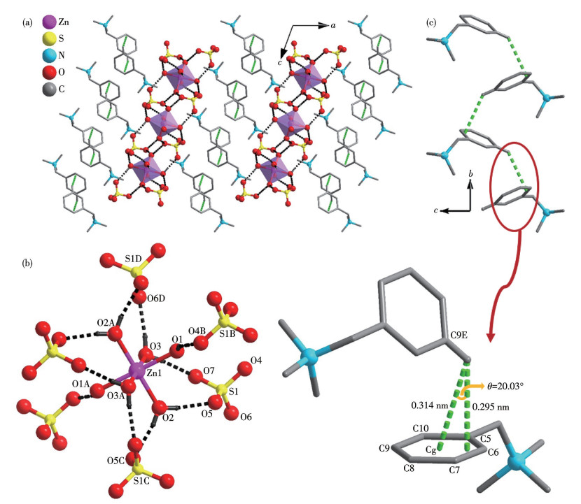
Structural analysis reveals that 1 belongs to the monoclinic space group P21/c. One-half of an independent Zn(Ⅱ) ion, one SO42- anion, one (NMe3CH2Ph)+ cation, and three water molecules are included in the asymmetric unit (Fig.1). The Zn(Ⅱ) ion is octahedrally coordinated by six terminal coordinated H2O molecules to form a [Zn(H2O)6]2+ cation. The Zn—O bond lengths and ∠O—Zn—O bond angles fall in the range of 0.208 2(2)-0.213 3(2) nm and 86.43(9)°-180°, respectively.
Symmetry codes: A:-x, 2-y, 2-z; B: 2-x, -y, 1-z; C: 2-x, 1/2+y, 1/2-z; D: x, 1/2-y, 1/2+z; E: 1-x, -1/2+y, 1/2-z.
Each [Zn(H2O)6]2+ cation is connected to six neighboring SO42- anions, and each SO42- anion is connected to three [Zn(H2O)6]2+ cations through hydrogen bonds. The related parameters, including O(H2O)…O(SO42-) distances, H…O(SO42-) distances, and O(H2O)—H… O(SO42-) angles fall in the ranges of 0.267 9(3)-0.276 1(3) nm, 0.185 0(17)-0.20 0(2) nm, and 157(3)°-173(2)°, respectively (Table S3). The thickness of the resulting inorganic layer reaches 0.614 nm and extends along the b- and c-axes (Fig.S1). The (NMe3CH2Ph)+ cations are linked by edge-to-face C—H…π interaction to form a 1D zigzag chain running along the b-axis. The C—H…π interaction involves the C9E—H bond (Symmetry code: E: 1-x, -1/2+y, 1/2-z) and the π-system of the benzene ring formed by atoms C5, C6, C7, C8, C9, and C10. The centroid of the benzene ring is denoted as Cg. The measured H…π and H…Cg distances are 0.295 and 0.314 nm, respectively, and the corresponding angle between the H…Cg vector and the normal to the ring plane is 20.03°. The accumulation of these chains forms an organic layer. The alternating stacking of organic and inorganic layers forms the final structure.
Single-crystal X-ray diffraction analysis indicates that compound 2 crystallizes in the orthorhombic space group Pbca and possesses a discrete dinuclear structure. The asymmetric unit contains two crystallographically independent Zn2+ ions, two independent Cl- ions, three independent SO42- ions, and four independent (NMe3CH2Ph)+ cations. As illustrated in Fig.2, both Zn2+ centers display a distorted tetrahedral {ClO3} coordination geometry, each coordinated by one terminal Cl- ion and three oxygen atoms derived from three distinct SO42- ligands. The Zn—Cl and Zn—O bond distances range from 0.220 62(15) to 0.222 50(15) nm and from 0.190 4(4) to 0.193 1(4) nm, respectively. Each of the three SO42- ligands adopts an identical μ2-κO1∶κO1 bridging mode, connecting two metal ions. This connectivity results in the formation of the dinuclear [Zn2Cl2(SO4)3]4- anion, which exhibits a crankshaft-like structure featuring three bridging sulfate groups. Notably, two of these sulfate bridges are bent in a clockwise orientation relative to the Zn…Zn vector, while the third is bent counterclockwise. The intramolecular Zn…Zn separation within the anion is about 0.36 nm.
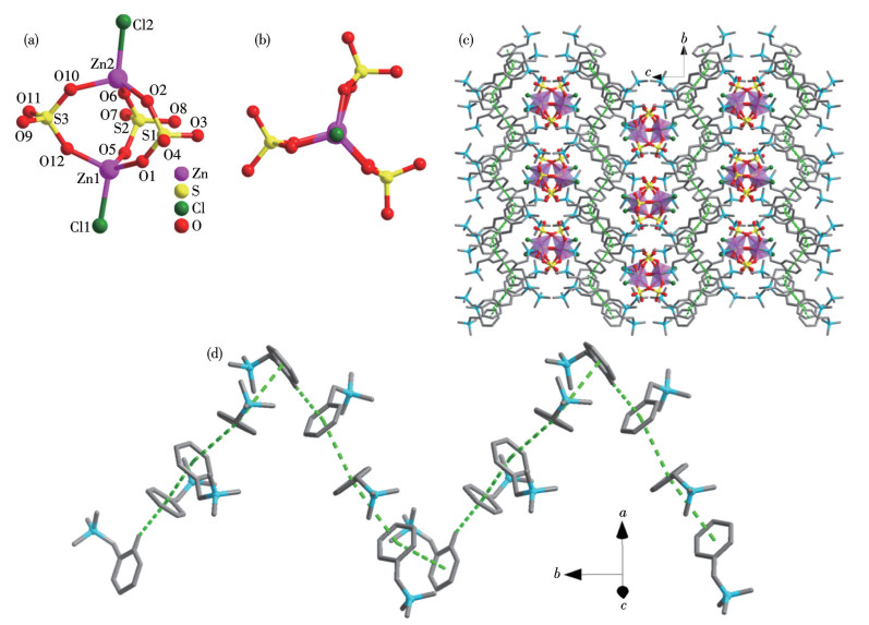
Adjacent (NMe3CH2Ph)+ cations engage in close packing, stabilized by π-π stacking interactions. Fig.2d and S2 illustrate two distinct types of intermolecular contacts: (ⅰ) C—H…π interactions, with H…Ph distances of 0.276 and 0.320 nm, and (ⅱ) offset face-to-face π-π stacking interactions, exhibiting centroid-to-centroid distances of approximately 0.413 and 0.465 nm. These π-interactions link the cations into continuous zigzag chains propagating along the b-axis. Subsequent stacking of these cationic chains, in conjunction with the [Zn2Cl2(SO4)3]4- anions, yields an organic-inorganic layered structure. The cohesion between the cationic layers and anionic moieties is primarily governed by electrostatic interactions.
Using identical starting materials and heating conditions, two structurally distinct compounds were obtained simply by varying the standing time at room temperature after the reaction. Immediate filtration upon heating cessation yielded compound 1 with square-plate morphology. In contrast, allowing the reaction vessel to stand at room temperature for three days before filtration afforded compound 2 with rhombic-plate morphology, indicating a structural transformation occurring during the standing period. Notably, this transformation proceeds only within the mother liquor; immersing crystals of 1 directly in DMF solvent does not induce the conversion.
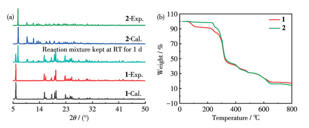
PXRD analyses were performed on both compounds (Fig.3a). The experimental patterns of 1 and 2 matched well with the simulated patterns derived from their respective single-crystal structures, confirming high phase purity and good crystallinity. Additionally, the PXRD pattern of the solid isolated after one day of standing exhibited diffraction peaks characteristic of both 1 and 2, further corroborating the occurrence of a gradual structural transformation.
FTIR spectrum provided additional insights. For compound 1, characteristic bands at 3 175 and 1 644 cm-1 arise from O—H stretching of water molecules, while the intense peak at 1 106 cm-1 corresponds to the bending vibration of free SO42-. In contrast, the spectrum of compound 2 displayed two strong peaks at 1 182 and 1 105 cm-1 attributable to bridging SO42- bending vibrations[15], together with distinctive bands at 648 and 606 cm-1 arising from asymmetric stretching of bridging SO42-. These spectral features are consistent with the crystallographically determined structures.
TGA was performed to evaluate the thermal stability of the two compounds. Compound 1 remained stable up to 65 ℃. In the temperature range of 66-115 ℃, it lost two water molecules (Obsd. 6.53%, Calcd. 5.40%). A plateau region was observed between 116 and 225 ℃, followed by rapid structural decomposition above 225 ℃. In contrast, compound 2 was stable up to 225 ℃ but underwent rapid mass loss above this temperature, indicating immediate framework collapse.
The luminescent behavior of the compounds was systematically evaluated. When exposed to 254 nm UV light, both compounds 1 and 2 emitted blue fluorescence. After the UV source was turned off, 2 exhibited a cyan afterglow visible to the naked eye for about 1.5 s, whereas 1 displayed no observable afterglow (Fig.4a).
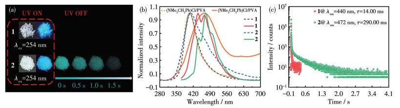
Then the spectra and lifetimes of the two compounds were tested. Under 254 nm excitation, the prompt emission spectrum of compound 1 exhibited a single peak at 405 nm, while its delayed spectrum revealed two distinct peaks at 440 and 466 nm (Fig.4b). Phosphorescence lifetime analysis revealed a decay time of 14.00 ms for the maximum emission peak (Fig.4c). For compound 2, the prompt spectrum peaked at 409 nm, and the delayed spectrum exhibited a main peak at 472 nm accompanied by two shoulders at 443 and 500 nm. The lifetime associated with the maximum delayed peak was determined to be 290.00 ms (Fig.4b and 4c).
To identify the luminescence origin, a PVA film doped with (NMe3CH2Ph)Cl (1% loading) was prepared to simulate the singlet emission of the (NMe3CH2Ph)+ cation (Fig.4b). Its prompt emission peaked at 404 nm, while the delayed spectrum showed a broad band centered around 466 nm. These values closely matched the emission peaks of both compounds, suggesting that their luminescence primarily arises from the (NMe3CH2Ph)+ moiety.
Notably, (NMe3CH2Ph)Cl alone exhibits no afterglow and very weak RTP emission. However, its combination with inorganic components to form compounds 1 and 2 significantly enhances RTP performance—attributed to the rigid crystalline lattice that stabilizes triplet excitons. The superior RTP characteristics of compound 2 over 1 can be explained by two synergistic effects. First, the presence of Cl- in 2 introduces a stronger external heavy-atom effect[13], promoting efficient intersystem crossing from the singlet to triplet state. Second, their molecular packing modes differ: compound 1 is stabilized mainly by hydrogen bonds and edge-to-face C—H…π interactions, whereas compound 2 features partially offset face-to-face π-π stacking. This arrangement increases structural conjugation, stabilizing triplet excited states and suppressing non-radiative relaxation[2]. The red-shifted emission peaks of 2 relative to 1 further support this enhanced conjugation. Together, these factors enable compound 2 to display a brighter and longer-lived afterglow.
Host-guest doping has emerged as an effective strategy for constructing RTP materials. Using compound 1 as the host matrix, we doped a series of phosphorescent guests (G) to prepare G/1 systems with enhanced luminescence (Fig.5a). Taking 6-msa/1 (6-msa=6-methylsalicylic acid) as an example, it was synthesized similarly to 1 but with in situ addition of 6-msa (n6-msa∶
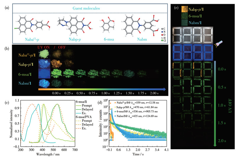
Under 254 nm UV, 6-msa/1 exhibited blue emission and a bright green afterglow lasting about 2.00 s (Fig.5b). Its prompt spectrum peaked at 414 nm, while the delayed spectrum showed a strong peak at 536 nm with shoulders at 412, 455, and 600 nm. The lifetime at 536 nm reached 905.73 ms (Fig.5c, 5d, and Table S4). Compared to pristine 1, both afterglow brightness and duration were significantly enhanced, demonstrating that trace doping boosts RTP performance.
To clarify the RTP origin, a 6-msa/PVA film was prepared. Its emission profile matched that of 6-msa/1 (Fig.5c), confirming that the guest molecule 6-msa contributes decisively to the observed phosphorescence.
By varying the guest, the RTP color of the G/1 system could be tuned widely: from 435 nm (cyan, Nabm/1), 536 nm (green, 6-msa/1), dual peaks at 470/570 nm (yellow, Nabp-p/1), to 559 nm (orange, NabaCl-p/1) (Fig.5 and S4). Attempts to dope guests into matrix 2 were unsuccessful.
The enhanced RTP in doped systems arises from two factors[16]: (ⅰ) the rigid host framework immobilizes the guest, suppressing nonradiative decay; and (ⅱ) competitive absorption between host and guest allows direct excitation of the guest, as evidenced by overlapping excitation spectra (Fig.S5).
Taking advantage of their tunable afterglow colors and lifetimes, selected G/1 materials—Nabm/1 (cyan), 6-msa/1 (green), and Nabp-p/1 (yellow)—were used as inks for information encryption (Fig.5e). Under 254 nm UV, the pattern “8888” appeared uniformly blue. Immediately after turning off the lamp, the digits displayed distinct colors due to different afterglow hues. Over time, the shorter-lived cyan and yellow emissions faded, leaving only the green afterglow of 6-msa/1, which revealed the true information “2026”.
In summary, we have successfully prepared two inorganic-organic hybrid ZnSO4 materials: (NMe3CH2Ph)2[Zn(H2O)6](SO4)2 (1) and (NMe3CH2Ph)4[Zn2Cl2(SO4)3] (2). Weak interactions, including face-to-face π-π and edge-to-face C—H…π interactions, hydrogen bonds, and electrostatic interactions between cations and anions, are utilized to construct their inorganic-organic interlayer structures. The delayed emission lifetime of compound 1 was relatively short. In contrast, compound 2, obtained via structural transformation from 1, exhibited a significantly extended delayed lifetime of 290.00 ms. Its green afterglow was observable to the naked eye for about 1.50 s. By using 1 as the host matrix, a series of host-guest doped materials could be prepared, and their afterglow color could be continuously tuned from cyan to orange-yellow, while the afterglow lifetime was enhanced by up to about 65 times relative to that of the pristine matrix. Benefiting from its excellent luminescence tunability, the doped afterglow materials hold great promise for advanced anti-counterfeiting applications.
SHI H F, YAO W, YE W P, MA H L, HUANG W, AN Z F. Ultralong organic phosphorescence: From material design to applications[J]. Accounts Chem. Res., 2022, 55(23): 3445-3459 doi: 10.1021/acs.accounts.2c00514
王会玉, 刘淑芹, 冯珂新, 张建军. 一个苯并咪唑衍生物的基于晶态-晶态转变的余辉刺激响应行为[J]. 无机化学学报, 2023, 39(8): 1571-1578WANG H Y, LIU S Q, FENG K X, ZHANG J J. Afterglow stimulation response behaviors of a benzimidazole derivative based on crystalline-to-crystalline transition[J]. Chinese J. Inorg. Chem., 2023, 39(8): 1571-1578
QU W B, TIAN R W, YANG B, GUO T Y, WU Z, LI Y, GENG Z R, WANG Z L. Dual-channel/localization single-molecule fluorescence probe for monitoring ATP and HOCl in early diagnosis and therapy of rheumatoid arthritis[J]. Anal. Chem., 2024, 96(14): 5428-5436 doi: 10.1021/acs.analchem.3c05342
陈煜坤, 冯珂新, 张博伦, 宋文涛, 张建军. 两个Mg(Ⅱ)金属有机框架的合成, 晶体结构及截然相反的机械刺激发光响应[J]. 无机化学学报, 2025, 41(6): 1227-1234CHEN Y K, FENG K X, ZHANG B L, SONG W T, ZHANG J J. Syntheses, crystal structures and diametrically opposed mechanically-stimulated luminescence response of two Mg(Ⅱ) metal-organic frameworks[J]. Chinese J. Inorg. Chem., 2025, 41(6): 1227-1234
CHEN X Y, FENG X, ZHANG Z X, DENG X R, DAI F, ZHANG L L, NG S W. Multifunctional lanthanide metal-organic frameworks based on —NH2 modified ligand: Fluorescent ratio probe, CrO42- ions adsorption, and photocatalytic property[J]. Inorg. Chem., 2023, 62(39): 16170-16181 doi: 10.1021/acs.inorgchem.3c02448
CUI S, ZHAO Z Q, WU Z, CHEN J Y, LIU S Y, HU Z P, ZHOU T, LI X L. A 1H-imidazo[4, 5-f][1, 10]phenanthroline based ligand and its binuclear Cu(Ⅰ) complexes: Syntheses, structures and luminescence sensing for Cr2O72- and MnO4-[J]. Polyhedron, 2024, 263: 117210 doi: 10.1016/j.poly.2024.117210
CHEN T H, MA Y J, YAN D P. Single-component 0D metal-organic halides with color-variable long-afterglow toward multi-level information security and white-light LED[J]. Adv. Funct. Mater., 2023, 33(18): 2214962 doi: 10.1002/adfm.202214962
王婷, 张佩佩, 刘淑芹, 王瑞红, 张建军. 一例Bi-CP基固态薄膜传感器的制备及其对生物胺蒸气的发光传感[J]. 无机化学学报, 2024, 40(8): 1615-1621WANG T, ZHANG P P, LIU S Q, WANG R H, ZHANG J J. A Bi-CP-based solid-state thin-film sensor: Preparation and luminescence sensing for bioamine vapors[J]. Chinese J. Inorg. Chem., 2024, 40(8): 1615-1621
FU P Y, LI B N, ZHANG Q S, MO J T, WANG S C, PAN M, SU C Y. Thermally activated fluorescence vs long persistent luminescence in ESIPT-attributed coordination polymer[J]. J. Am. Chem. Soc., 2022, 144(6): 2726-2734 doi: 10.1021/jacs.1c11874
ZHOU B, YAN D P. Long persistent luminescence from metal-organic Compounds: State of the art[J]. Adv. Funct. Mater., 2023, 33(19): 2300735 doi: 10.1002/adfm.202300735
LIN J, GUO D W, TIAN Y Q. Synthesis and structural characterization of open-framework copper(Ⅱ) sulfates[J]. Cryst. Growth Des., 2008, 8(12): 4571-4575 doi: 10.1021/cg8006316
CHOUDHURY A, KRISHNAMOORTHY J, RAO C N R. An approach to the synthesis of organically templated open-framework metal sulfates by the amine-sulfate route[J]. Chem. Commun., 2001, 24: 2610-2611
MIAO L, ZHANG B L, SONG W T, CHEN J, SHI W J, WANG R H, LIU S Q, LI Y J, ZHANG J J. Zinc sulfate open-frameworks with nonconventional room-temperature phosphorescence for selective amine vapor detection[J]. Inorg. Chem., 2025, 64(14): 7214-7223 doi: 10.1021/acs.inorgchem.5c00917
SHELDRICK G M. SHELXT-integrated space-group and crystal-structure determination[J]. Acta Crystallogr. Sect. A, 2015, A71: 3-8
NAKAMOTO K. 无机和配位化合物的红外和拉曼光谱[M]. 黄德如, 汪仁庆, 译. 北京: 化学工业出版社, 1991: 276-279NAKAMOTO K. Infrared and Raman spectra of inorganic and coordination compounds[M]. Translated by HUANG D R, WANG R Q. Beijing: Chemical Industry Press, 1991: 276-279
ZHANG W Q, SONG W T, CHEN J, SHI W J, WANG R H, LI Y J, LIANG J Y, ZHANG J J. Afterglow properties of zinc sulfate open frameworks: Nonconventional chromophore induction and doping modulation[J]. Inorg. Chem., 2026, 65(1): 881-891 doi: 10.1021/acs.inorgchem.5c05229

Figure 1 Structure of compound 1: (a) packing of the structure viewed along the b-axis; (b) coordination environment of the Zn2+ ion, and the interactions between [Zn(H2O)6]2+ and SO42- anions via hydrogen bonds which are presented as black dotted lines; (c) a view of the 1D chain of (NMe3CH2Ph)+ cations based on C—H…π stacking interactions which are presented as green dotted lines
Symmetry codes: A:-x, 2-y, 2-z; B: 2-x, -y, 1-z; C: 2-x, 1/2+y, 1/2-z; D: x, 1/2-y, 1/2+z; E: 1-x, -1/2+y, 1/2-z.

Figure 2 Structure of compound 2: side (a) and top-down (b) views of the dinuclear [Zn2Cl2(SO4)3]4- anion; (c) packing of the structure viewed along the a-axis; (d) 1D chains of the (NMe3CH2Ph)+ cations based on π-π stacking interactions, which are presented as green dotted lines

Figure 3 Basic characterizations of compounds 1 and 2: (a) calculated and experimental PXRD patterns; (b) TGA curves

Figure 4 PL properties of compounds 1 and 2: (a) photographs taken at different time intervals before and after turning off the UV excitation; (b) solid-state emission spectra; (c) delayed emission decay curves (λex=254 nm)

Figure 5 Luminescence properties of G/1: (a) structures of the doped guest molecules and photographs taken under 254 nm UV irradiation and after lamp-off; (b) photos of G/1 under 254 nm UV irradiation and after lamp-off; (c) emission and excitation spectra of 6-msa/1 (solid) and 6-msa/PVA (dashed); (d) time-resolved PL decay curves for the delayed emission; (e) application
Table 1. Crystal data and structure refinement parameters for compounds 1 and 2
| Parameter | 1 | 2 |
| Empirical formula | C20H44N2O14S2Zn | C40H64Cl2N4O12S3Zn2 |
| Formula weight | 666.06 | 1 090.77 |
| Crystal system | Monoclinic | Orthorhombic |
| Space group | P21/c | Pbca |
| a / nm | 1.582 57(7) | 2.017 01(11) |
| b / nm | 0.806 87(4) | 1.934 22(10) |
| c / nm | 1.229 18(5) | 2.619 38(12) |
| β / (°) | 110.016(2) | |
| V / nm3 | 1.474 77(12) | 10.219 1(9) |
| Z | 2 | 8 |
| Dc / (g·cm-3) | 1.500 | 1.418 |
| μ / mm-1 | 1.041 | 1.224 |
| F(000) | 704 | 4 560 |
| θ range / (°) | 2.740-24.998 | 2.462-25.000 |
| Unique reflection collected, observed | 21 477, 2 581 | 202 197, 8 986 |
| Rint | 0.040 2 | 0.178 9 |
| GOF on F 2 | 1.074 | 1.034 |
| R1a, wR2b [I>2σ(I)] | 0.035 6, 0.090 1 | 0.062 7, 0.155 0 |
| R1, wR2 (all) | 0.042 4, 0.093 7 | 0.108 8, 0.176 9 |
| Max, mean shift in final cycle | 0.000, 0.000 | 0.001, 0.000 |
| a R=∑(||Fo|-|Fc||)/∑|Fo|, b wR={∑w[(Fo2-Fc2)2]/∑w[(Fo2)2]}1/2, w=1/[σ2(Fo2)+(aP)2+bP], P=(Fo2+2Fc2)/3], a=0.050 8, b=1.034 7 for 1, a=0.107 0, b=0.243 52 for 2. | ||
 下载: 导出CSV

 扫一扫看文章


扫一扫关注我们
 下载:
下载: