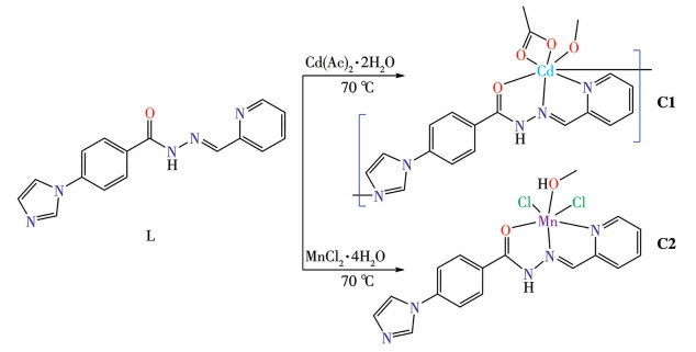
Scheme 1.
Synthetic routes of complexes C1 and C2
Synthesis, crystal structures, and antitumor activity of two metal complexes of imidazolyl acylhydrazones
Zhihui ZONG , Zijie ZHAO , Lei HUANG , Zhicheng PAN , Shan WANG , Lili LIANG , Huaqing LIU , Enli ZHANG

Cancer is one of the most serious diseases endangering human health, posing a substantial challenge to healthcare systems globally[1-3]. Chemotherapy serves as a cornerstone of tumor treatment. However, the systemic toxicity of chemotherapeutic agents and the emergence of multidrug-resistant phenotypes often render initially effective drugs ineffective against tumors[4-6]. Thus, the development of novel anticancer agents has become a pivotal research priority in the fields of chemistry and medicine.
Since the discovery of cisplatin, platinum-based metal complexes such as carboplatin and oxaliplatin have been widely used to treat various types of tumors[7-10]. However, these medications are associated with several disadvantages, including their limited capacity to target tumors, the potential for adverse reactions such as nephrotoxicity and neurotoxicity, and the propensity to induce drug resistance[11-16]. Consequently, this has prompted researchers to focus on the study of non-platinum metal drugs. For the past few years, a considerable number of anti-tumor complexes have been documented, with a subset of these agents undergoing clinical investigation[17-23].
Acylhydrazone represents a distinctive category of Schiff base, exhibiting the capacity to form complexes with metals across diverse forms[24-26]. Acylhydrazone and complexes derived from acylhydrazone have been reported to exhibit a wide range of activities, including anticancer[27-29], antibacterial[30-31], antiviral[32], and antituberculosis properties[33-34]. Similarly, imidazole and its derivatives have demonstrated a broad spectrum of bioactivities, such as anti-HIV, anticancer, anticovid, antifungal, antidiabetic, antidepressant, antioxidant, and antituberculosis[35-36]. Additionally, pyridine and its derivatives have been shown to possess significant biological activities, including antibacterial, antifungal, antiviral, analgesic, antidiabetic, and anticancer[37-38]. These properties have attracted significant attention from researchers in the field of drug development.
To develop new metal-based tumor inhibitors, in this study, we designed and prepared a Schiff base acylhydrazone ligand containing imidazolyl and pyridyl: 4-(1H-imidazol-1-yl)-N′-(pyridin-2-ylmethylene)benzohydrazide (L), and synthesized two novel complexes (Scheme 1). Their structures, antitumor activities, and initial mechanisms were investigated.
The instruments and reagents used in the experiment are listed in the Supporting information.
The synthesis of ligand L was conducted in accordance with a similar method previously reported[30]. 1H NMR (600 MHz, DMSO-d6): δ 12.13 (s, 1H), 8.64 (d, J=5.1 Hz, 1H), 8.51 (s, 1H), 8.44 (s, 1H), 8.10 (d, J=8.5 Hz, 2H), 8.00 (d, J=8.0 Hz, 1H), 7.94-7.84 (m, 4H), 7.43 (t, J=6.0 Hz, 1H), 7.17 (s, 1H) (Fig.S1). 13C NMR (150 MHz, DMSO-d6): δ 162.8, 153.7, 150.0, 148.7, 139.4, 137.4, 136.2, 131.5, 130.8, 130.0, 124.9, 120.4, 120.2, 118.3 (Fig.S2). FTIR (KBr pellet, cm-1): 3 233w, 3 078w, 2 794w, 1 698s, 1 624s, 1 587s, 1 536s, 1 484 w, 1 435s, 1 375s, 1 337s, 1 314w, 1 293s, 1 266w, 1 212 m, 1 184w, 1 115m, 1 065s, 1 016m, 919m, 865w, 837m, 823w, 785m, 745s, 702w, 678m, 638w, 618w, 516w, 490w.
Cd(Ac)2·2H2O (0.0106 g, 0.04 mmol) and L (0.011 7 g, 0.04 mmol) were stirred homogeneously in a mixture of methanol and ethanol (1∶1, V/V). The mixture was then transferred to a Teflon reactor and reacted for three days at 70 ℃ in an oven. Yellow rhombic crystals were obtained after cooling to ambient temperature. A mixture of methanol and ethanol was used to wash the product, which was then dried, and the C1 product was collected with a yield of 48% (based on L). FTIR (KBr, cm-1): 3 353m, 3 124m, 1 606w, 1 591w, 1 557m, 1 523m, 1 505m, 1 465w, 1 421w, 1 358m, 1 340m, 1 300m, 1 257w, 1 185w, 1 153w, 1 062m, 963w, 927m, 850w, 766m, 724w.
MnCl2·4H2O (0.008 g, 0.04 mmol) and L (0.011 7 g, 0.04 mmol) were placed in a mixture of methanol and ethanol (2∶1, V/V) and stirred homogeneously, after which the mixture was transferred to a 25 mL Teflon high-pressure reactor and reacted for three days in an oven at 70 ℃. Yellow rhombic crystals were obtained upon cooling to room temperature. The product was washed with a trace amount of a mixed solution of methanol and ethanol, dried, and product C2 was collected with a yield of 53% (based on L). FTIR (KBr, cm-1): 3 117w, 3 065w, 2 804w, 1 628m, 1 608m, 1 577 w, 1 548w, 1 518m, 1 495m, 1 469w, 1 439w, 1 355w, 1 292m, 1 257w, 1 215w, 1 146w, 1 061m, 1 030w, 962w, 912w, 856m, 762m, 652w。
Single crystal samples of complexes C1 and C2 with regular morphology and appropriate dimensions were selected and mounted on a Bruker APEX-Ⅱ CCD single-crystal diffractometer with Mo Kα radiation (λ=0.154 178 nm) for data collection, and the dimensions of the single crystals were 0.15 mm×0.08 mm×0.05 and 0.12 mm×0.08 mm×0.04 mm, respectively. Diffraction data were collected at 100.0 K in ω-φ scan mode. Data reduction was performed with the SAINT program, and absorption corrections were applied using the SADABS software. The crystal structures were solved by direct methods with SHELXT and refined by full-matrix least-squares techniques on F 2 using SHELXL-2018. Non-hydrogen atomic coordinates were located by direct methods and refined by full-matrix least-squares calculations. Hydrogen atoms were placed in idealized positions and refined using a riding model. Crystal data and structure refinement details for the compounds are presented in Table 1. Selected bond lengths and bond angles are listed in Table 2.
 下载:
导出CSV
下载:
导出CSV
| Parameter | C1 | C2 |
| Empirical formula | C86H113Cd4N23O20 | C17H17Cl2MnN5O2 |
| Formula weight | 2 238.59 | 449.20 |
| Crystal system | Monoclinic | Monoclinic |
| Space group | P21/n | P21/n |
| a/nm | 0.957 22(3) | 0.870 93(3) |
| b/nm | 1.535 85(4) | 1.176 90(4) |
| c/nm | 16.045 3(4) | 18.631 5(7) |
| V/nm3 | 2.287 00(11) | 1.897 17(12) |
| β/(°) | 104.182(3) | 96.574(3) |
| Z | 4 | 4 |
| Dc/(g·cm-3) | 1.625 | 1.573 |
| μ/mm-1 | 1.000 | 1.000 |
| F(000) | 1 142 | 916 |
| θ range/(°) | 2.263-25.347 | 2.051-26.363 |
| Reflection collected | 20 232 | 11 691 |
| Independent reflection | 4 145 (Rint=0.070 2) | 3 787 (Rint=0.029 0) |
| Reflection observed [I>2σ(I)] | 3 372 | 3 200 |
| Data, Nres, Npar* | 4 145, 0, 313 | 3 787, 3, 248 |
| Goodness-of-fit on F 2 | 1.073 | 1.042 |
| R1, wR2 [I>2σ(I)] | 0.052 1, 0.128 2 | 0.030 7, 0.068 7 |
| R1, wR2 (all data) | 0.068 8, 0.146 1 | 0.039 4, 0.072 9 |
| (Δρ)max, (Δρ)min/(e·nm-3) | 2 033, -1 886 | 373, -310 |
| *Nres=number of restraints, Npar=number of parameters. | ||
下载:
导出CSV
| C1 | |||||
| Cd1—O1 | 0.233 1(4) | Cd1—N2 | 0.230 5(4) | Cd1—O2 | 0.248 2(4) |
| Cd1—N5 | 0.232 3(4) | Cd1—O3 | 0.236 4(4) | Cd1—N6 | 0.240 6(4) |
| Cd1—O5 | 0.236 5(4) | ||||
| O3—Cd1—O2 | 82.85(13) | O1—Cd1—O2 | 54.08(12) | N5—Cd1—O2 | 148.25(5) |
| O1—Cd1—O3 | 82.50(13) | N5—Cd1—O3 | 84.03(14) | O1—Cd1—O5 | 88.26(13) |
| N5—Cd1—O5 | 66.69(14) | O1—Cd1—N6 | 135.23(14) | N5—Cd1—N6 | 69.74(15) |
| O3—Cd1—O5 | 91.71(13) | N6—Cd1—O2 | 81.29(13) | O3—Cd1—N6 | 89.25(13) |
| N5—Cd1—O1 | 151.10(13) | O5—Cd1—O2 | 142.32(12) | N2—Cd1—N5 | 103.10(15) |
| O5—Cd1—N6 | 136.10(13) | N2—Cd1—N6 | 98.92(15) | N2—Cd1—O1 | 88.41(14) |
| N2—Cd1—O3 | 170.61(14) | N2—Cd1—O2 | 93.7614) | N2—Cd1—O5 | 85.62(14) |
| C2 | |||||
| Mn1—Cl2 | 0.251 44(6) | Mn1—Cl1 | 0.238 32(6) | Mn1—O2 | 0.227 59(14) |
| Mn1—O1 | 0.219 83(5) | Mn1—N1 | 0.227 28(17) | Mn1—N2 | 0.225 82(7) |
| Cl1—Mn1—Cl2 | 94.49(2) | N2—Mn1—Cl2 | 89.60(4) | O2—Mn1—Cl2 | 91.21(4) |
| N2—Mn1—Cl1 | 170.65(5) | O2—Mn1—Cl1 | 119.18(4) | N2—Mn1—O2 | 69.05(6) |
| O1—Mn1—Cl2 | 173.49(4) | N2—Mn1—N1 | 70.36(6) | O1—Mn1—Cl1 | 91.27(4) |
| N1—Mn1—Cl1 | 101.08(5) | O1—Mn1—O2 | 83.32(6) | N1—Mn1—O2 | 139.30(6) |
| O1—Mn1—N | 90.47(6) | N3—N2—Mn1 | 117.02(2) | O1—Mn1—N2 | 85.16(6) |
| C6—N2—Mn1 | 120.52(4) | N1—Mn1—Cl2 | 91.40(4) | ||
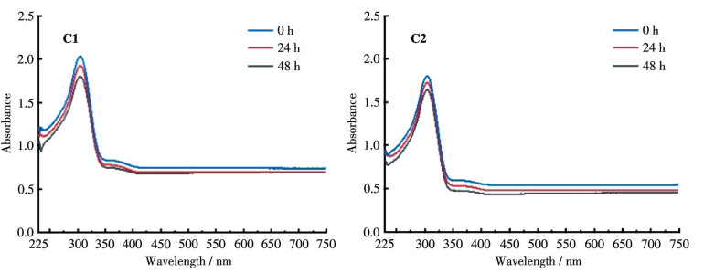
The complexes were dissolved in a small volume of dimethyl sulfoxide (DMSO), then diluted to a final concentration of 128 μmol·L-1 in a Tris-HCl-NaCl buffer. UV-Vis spectra of the resulting solutions were recorded over a wavelength range of 200-750 nm at 0, 24, and 48 h, respectively. The stability of the complexes in the solution was evaluated based on the changes in their characteristic absorption peaks.
Four tumour cells (SMMC-7721 human hepatocellular carcinoma cell lines, MDA-MB-231 human breast adenocarcinoma cell lines, A549 human lung adenocarcinoma cell lines, and A2780 human ovarian carcinoma cell lines) and one normal cell line, HK-2 (human renal cortical proximal tubule epithelial cells) were selected using the CCK-8 assay. The cells were harvested and inoculated into 96-well plates at a density of 4×10³ cells per well (100 μL per well), followed by incubation for 24 h. The supernatant was then discarded, and serial concentrations of the complexes, cisplatinum, and ligand L were added, after which the incubation continued for an additional 24 h. Thereafter, 10 μL of CCK-8 reagent was added to each well, and the incubation was continued for another 0.5 h. Finally, the absorbance value of each well was measured at 450 nm using a microplate reader.
SMMC-7721 and A549 cells were seeded in six-well plates at a density of 2×10⁵ cells per well and cultured under optimal conditions until they reached the predetermined density. The cells were then treated with serial concentrations of C1 for 48 h, followed by trypsin digestion without the addition of EDTA. Subsequently, the cells were rinsed twice with 1 mL of ice-cold PBS (4 ℃), resuspended in the buffer, and stained with Annexin V-FITC/PI staining solution. Finally, C1-induced apoptosis was analyzed via flow cytometry.
SMMC-7721 cells were seeded in six-well plates (1×10⁵ cells per well) and incubated for 24h. Serial concentrations of C1 (6, 12 μmol·L-1) were added, followed by incubation for 48 h. The cells were harvested and fixed in 70% ethanol overnight at 4 ℃. They were then rinsed with ice-cold PBS, followed by resuspension in 200 μL of staining buffer supplemented with 5 μL of propidium iodide (PI). The samples were incubated in the dark at 25 ℃ for 30 min, and the cell cycle distribution was subsequently analyzed via flow cytometry.
Cells in the logarithmic growth phase were rinsed with PBS and then resuspended in serum-free medium containing serial concentrations of C1. A total of 100 μL of the SMMC-7721 cell suspension (1×10⁵ cells·mL-1) was seeded into the upper chamber of the Transwell apparatus, while 500 μL of DMEM (Dulbecco′s modified Eagle medium) supplemented with 10% fetal bovine serum (FBS) was added to the lower chamber. The Transwell system was incubated at 37 ℃ for 48 h. After incubation, non-invaded cells on the upper surface of the membrane were carefully removed with cotton swabs. Cells that had migrated to the lower surface of the membrane were fixed with paraformaldehyde for 25 min, stained with crystal violet, and then rinsed three times with PBS to wash away unbound dye. Finally, the cells were air-dried and counted under an optical microscope.
Cells were seeded into six-well plates and cultured overnight to allow for complete adherence. A sterile 200 μL pipette tip was used to create uniform scratches across the cell monolayers. Detached cells were then rinsed off gently with ice-cold PBS for three times, and the remaining cells were further cultured in medium supplemented with C1 for 24 h. Finally, scratch healing was monitored dynamically, and representative images were captured at designated time points.
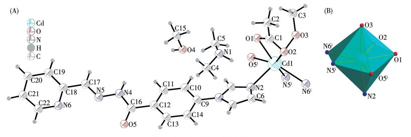
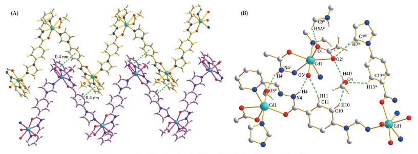
The crystallographic data of complexes C1 and C2 are listed in Table 1. Crystallographic studies show that complex C1 belongs to the monoclinic crystal system with the P21/n space group. The asymmetric unit structure of C1 (Fig.1A) consists of one Cd(Ⅱ) ion, one ligand, one coordinated acetate, one coordinated methoxide, one guest methanol, and one guest dimethylamine. The Cd(Ⅱ) center forms an irregular decahedral configuration (Fig.1B), seven-coordinated by three nitrogen atoms (from imidazole, pyridine, and the carbon-nitrogen double bond of the ligand) and four oxygen atoms (from carbonyl, methoxide, and chelating acetate, respectively). The Cd—O distances are from 0.233 1 to 0.248 2 nm, while the Cd—N distances are from 0.230 5 to 0.274 4 nm. The bond angles are from 54.08(12)° to 136.10(13)°. Each ligand connects two Cd(Ⅱ) ions through acylhydrazone together with 2-pyridyl, forming an NNO chelating group and an imidazole N atom. The dihedral angles between the central benzene ring and the imidazole and pyridine rings at both ends are 15.2(2)° and 20.6(2)°, respectively. Each Cd(Ⅱ) connects two ligands so that the whole structure presents an infinitely extended 1D zigzag chain structure. There are π-π interactions between the benzene ring (C9 to C14) and the pyridine ring (C18 to C22, and N6) of the nearby chains. The dihedral angle is 20.6(2)° and the centroid-to-centroid distance (Cg to Cg) is 0.40 nm (Fig.2A). Besides, there are also a large number of intermolecular hydrogen bonds between coordinated acetate (Table 3), coordinated methoxide, guest methanol and guest dimethylamine (Fig.2B). So complex C1 forms a 3D structure through π-π interactions and abundant intermolecular hydrogen bonds.
Symmetry code: ⅰ 1.5-x, -0.5+y, 1.5-z.
Symmetry codes: ⅰ 1-x, 1-y, 2-z; ⅱ-0.5+x, 0.5-y, 0.5+z; ⅲ 1.5-x, -0.5+y, 1.5-z.
下载:
导出CSV
| Complex | D—H…A | d(D—H)/nm | d(H…A)/nm | d(D…A)/nm | ∠DHA/(°) |
| C1 | N4ⅰ—H4ⅰ…O3ⅲ | 0.088 1 | 0.189 | 0.269 6(6) | 151.3(3) |
| C10—H10…O4 | 0.095 0 | 0.256 8 | 0.336 2(7) | 141.3(4) | |
| C11—H11…O3ⅱ | 0.095 0 | 0.250 | 0.341 6(7) | 162.2(4) | |
| C5ⅱ—H5Aⅱ…O1ⅱ | 0.098 0 | 0.199 6 | 0.282 2(1) | 140.5(6) | |
| C7ⅲ—H7ⅲ…O2ⅱ | 0.095 1 | 0.255 5 | 0.350 2(7) | 174.5(4) | |
| C13ⅲ—H13ⅲ…O4 | 0.095 0 | 0.251 5 | 0.322 0(8) | 131.1(4) | |
| C2 | O1ⅰ—H1ⅰ… N5ⅱ | 0.085 4 | 0.184 0 | 0.269 1(2) | 174.1(1) |
| N3—H3…Cl2ⅰ | 0.088 0 | 0.234 7 | 0.320 4(2) | 164.8(1) | |
| C12—H12…Cl1ⅰ | 0.095 0 | 0.254 7 | 0.345 5(2) | 160.0(1) | |
| Symmetry codes: ⅰ 1-x, 1-y, 2-z; ⅱ -0.5+x, 0.5-y, 0.5+z for C1; ⅰ 0.5-x, -0.5+y, 1.5-z; ⅱ -0.5+x, 0.5-y, 0.5+z; ⅲ 1.5-x, -0.5+y, 1.5-z for C2. | |||||
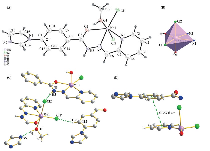
C2 belongs to the monoclinic crystal system with the P21/n space group. The asymmetric unit structure of C2 contains one Mn(Ⅱ) ion, one ligand, two chloride ions, and one coordinated CH3OH (Fig.3A). The Mn(Ⅱ) center forms a twisted octahedral structure (Fig.3B), six-coordinated by two O atoms (one from the carbonyl group of the ligand and one from CH3OH), two N atoms (from the carbon-nitrogen double bond and pyridine), and two Cl atoms. The Mn—O distances are 0.227 59 and 0.219 83 nm, Mn—N distances are 0.225 82 and 0.227 28 nm, and the Mn—Cl distances are 0.238 32(6) and 0.251 44(6) nm. The dihedral angles between the central benzene ring and the imidazole and pyridine rings at both ends are 19.1(8)° and 21.4(7)°, respectively, a little larger than complex C1. Complex C2 exhibits intermolecular hydrogen bonds and π-π interactions. There are intermolecular hydrogen bonds between the coordinated CH3OH, chloride ions and acylhydrazone, imidazole N atoms (Fig.3C). The pyridine (C1 to 5, and N1) and benzene (C8 to C13) rings in complex C2 are approximately parallel, forming a dihedral angle of 20.6(1)° and a centroid-to-centroid distance (Cg to Cg) of 0.3676 nm (Fig.3D), thereby creating a π-π stacking arrangement. In the presence of intermolecular hydrogen bonds and π-π stacking, the complex C2 forms a 3D structure.
Symmetry codes: ⅰ 0.5-x, -0.5+y, 1.5-z; ⅱ-0.5+x, 0.5-y, 0.5+z.
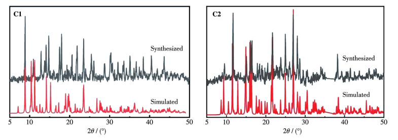
The major peaks in the powder X-ray diffraction (PXRD) patterns of complexes C1 and C2 were consistent with the simulated ones (Fig.4), indicating that the synthesized complexes are of high purity. However, certain peaks of complex C1 showed poor alignment with the simulated pattern, which might be attributable to instrumental parameters.
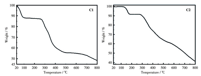
Thermogravimetric analysis (TGA) showed that C1 exhibited an initial weight loss of 11.14% up to 89 ℃, which is attributable to the loss of CH3OH and NH(CH3)2 (Calcd. 13.48%). As the temperature increased further, a rapid weight loss occurred at 261 ℃, indicating the onset of ligand decomposition accompanied by the collapse of the entire framework structure (Fig.5). C2 underwent an initial weight loss starting at 178 ℃, with a weight loss of approximately 9%, which is attributable to the elimination of CH3OH (Calcd. 7%). A subsequent weight loss was then initiated at 267 ℃, accompanied by the collapse of the entire framework structure.
To evaluate the stability of C1 and C2 under physiological conditions, the complexes were dispersed in a Tris-HCl-NaCl buffer and subjected to UV-Vis spectral scanning at 0, 24, and 48 h (Fig.6). The results demonstrated that the spectra showed no significant bathochromic or hypsochromic shifts, nor were any new characteristic peaks detected over the tested time period. These findings confirm that C1 and C2 can maintain structural stability in physiological buffer conditions for up to 48 h, thereby laying a solid foundation for subsequent in vitro experiments.
The antiproliferative activity of the ligand and target complexes against four tumour cells (SMMC-7721, MDA-MB-231, A549, and A2780) and one normal cell (HK-2) was evaluated via the CCK-8 assay, with the experimental results summarized in Table 4. The IC50 values of the ligand against the four tumor cell lines and one normal cell line were significantly higher than those of complexes C1 and C2, indicating that the coordination of metal ions and the formation of novel structures remarkably enhanced the antitumor potency. Complex C1 exhibited superior antiproliferative efficacy to cisplatin in A549 and A2780 cell models, while exerting comparable cytotoxicity against SMMC-7721 cells. In contrast, complex C2 showed weaker inhibitory activity against all four tumor cell lines relative to cisplatin; however, it displayed significantly higher IC50 values toward normal HK-2 cells, suggesting a markedly improved safety profile. C1 exhibited significantly stronger antiproliferative potency than C2 across all tested tumor cell lines. This disparity may be attributable to their distinct metal centers [Cd(Ⅱ) vs Mn(Ⅱ)] and coordination architectures (1D zigzag chain vs mononuclear structure), although further investigations are warranted to elucidate the detailed structure-activity relationships.
下载:
导出CSV
| Compound | IC50/(μmol·L-1) | ||||
| SMMC-7721 | MDA-MB-231 | A549 | A2780 | HK-2 | |
| C1 | 10.23±0.32 | 20.13±0.72 | 10.48±0.24 | 1.27±0.11 | 25.45±0.42 |
| C2 | 19.03±2.24 | 42.27±2.98 | 74.18±0.80 | 68.64±1.02 | 132.9±0.73 |
| Cisplatin | 9.41±0.29 | 16.01±1.27 | 23.56±1.05 | 4.09±0.20 | 3.23±0.07 |
| L | >150 | >150 | >150 | >150 | >150 |
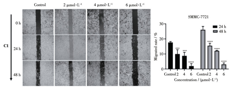
The effect of C1 on the migratory capacity of SMMC-7721 cells was assessed via the scratch wound healing assay (Fig.7). The results demonstrated that the migration rates of SMMC-7721 cells treated with 6 μmol·L-1 of complex C1 were (1.97±1.60)% and (3.04±1.04)% at 24 and 48 h, respectively. These values were significantly lower than those of the control group, which were (17.61±0.68)% at 24 h and (26.01±2.29%) at 48 h, indicating that C1 exerted a highly significant inhibitory effect on the migration of SMMC-7721 cells.
Data are presented as mean±standard deviation (SD) from three independent experiments; * p<0.05, ** p<0.01, *** p<0.001, and **** p<0.000 1 vs the control group.
A Transwell assay was performed to evaluate the effect of C1 on the invasive capacity of SMMC-7721 cells over a 48 h incubation period. The inhibitory effect of the complex on cell invasion was determined by counting the number of cells that had migrated through the membrane to the lower chamber. The results demonstrated that C1 could effectively suppress the invasion of SMMC-7721 cells in a concentration-dependent manner (Fig.8). Specifically, the invasion rates of cells treated with 2, 4, and 6 μmol·L-1 of C1 following 48 h of incubation were 69.4%, 47.6%, and 21.8%, respectively.
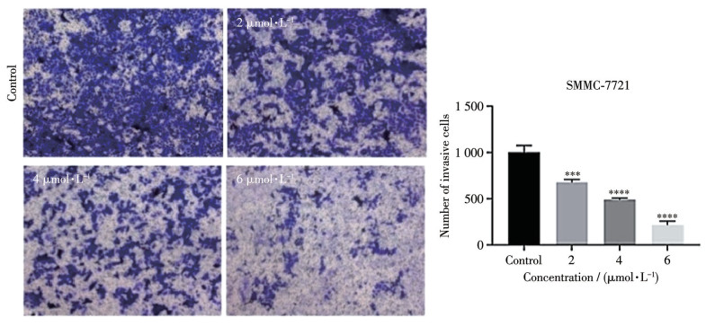
*** p<0.001 and **** p<0.000 1 vs the control group.
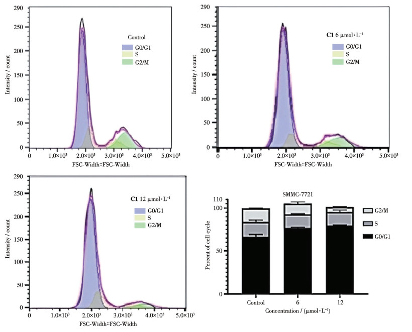
The cell cycle is a critical process governing cell proliferation and division[39-40]. The cell cycle distribution of SMMC-7721 cells treated with C1 was analyzed via flow cytometry using the PI single-staining method (Fig.9). After treatment with 6 and 12 μmol·L-1 of C1 for 48 h, the proportion of SMMC-7721 cells arrested in the G0/G1 phase increased by (10.37±0.87)% and (13.23±0.49)%, respectively, accompanied by a moderate reduction in the S-phase population. Correspondingly, the percentage of cells in the G2/M phase decreased by (3.27±1.81)% and (10.18±0.55)%, respectively. These findings suggest that C1 induces cell cycle arrest at the G0/G1 phase in a concentration-dependent manner.
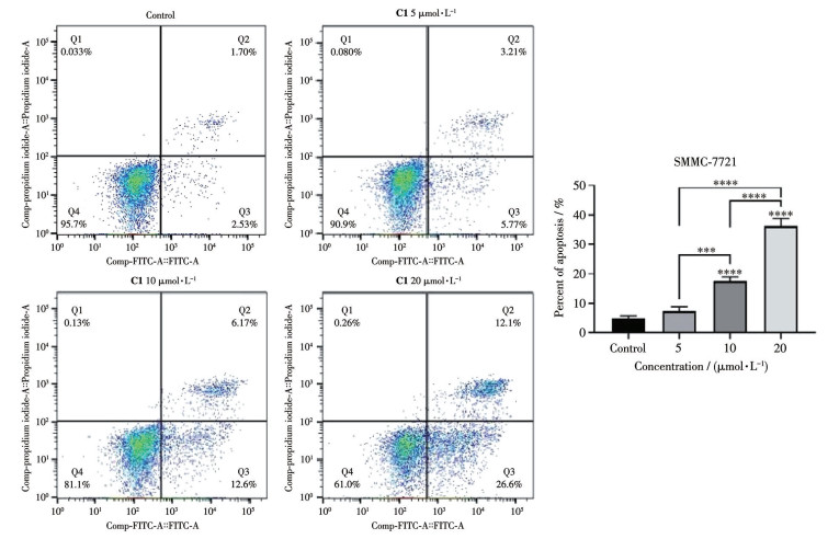
The apoptosis induced by C1 in SMMC-7721 and A549 cells was detected via flow cytometry with the Annexin V-FITC/PI double-staining assay. The results showed that C1 significantly triggered apoptosis in SMMC-7721 cells in a concentration-dependent manner, with a particularly prominent induction of early apoptosis (Fig.10). Specifically, treatment with 5 μmol·L-1 C1 led to early apoptosis in 4.82% of the cells and late apoptosis in 2.50%. When the concentration was increased to 10 μmol·L-1, the proportions of early and late apoptotic cells increased to 11.70% and 5.78%, respectively. At a concentration of 20 μmol·L-1, the percentages of cells undergoing early and late apoptosis further rose to 23.77% and 10.93%, respectively.
*** p<0.001 and **** p<0.000 1 vs the control group.
C1 was also found to induce apoptosis in A549 cells in a concentration-dependent manner, although late apoptosis predominated—similar to the trend observed in SMMC-7721 cells (Fig.11). Specifically, treatment with 5 μmol·L-1 C1 resulted in 2.22% of cells exhibiting early apoptosis and 2.11% demonstrating late apoptosis. When the concentration was increased to 15 μmol·L-1, the percentage of early apoptotic cells rose to 2.95%, while the proportion of late apoptotic cells increased to 9.12%. At a concentration of 20 μmol·L-1, the percentages of early and late apoptotic cells reached 10.93% and 28.33%, respectively. Notably, late apoptosis showed the most pronounced elevation, with an increase of 27.41% relative to the control group.
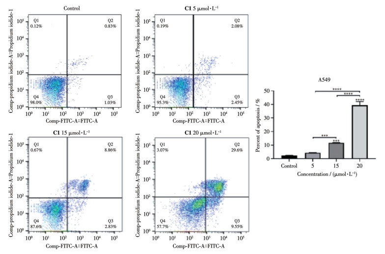
*** p<0.001 and **** p<0.000 1 versus the control group.
Taken together, these data provide compelling evidence that C1 exerts a potent pro-apoptotic effect on both SMMC-7721 and A549 cells in a concentration-dependent manner, yet the apoptotic response is markedly cell-type-specific. The preferential induction of early apoptosis in SMMC-7721 cells implies that C1 may primarily target the initiation phase of the apoptotic pathway in this cell line, whereas the dominant late apoptosis in A549 cells suggests a potential involvement in downstream apoptotic execution processes or a delayed cellular response to C1 stimulation. The 27.41% elevation in late apoptosis of A549 cells at 20 μmol·L-1 further highlights the stronger sensitivity of this cell line to C1-induced late-stage apoptotic events. Collectively, these findings lay a foundation for further investigations into the molecular mechanisms underlying the cell-type-specific apoptotic effects of C1, as well as its potential application as a candidate compound for targeted cancer therapy.
Two complexes, namely [Cd(L)(CH3O)(CH3COO)]·CH3OH·(CH3)2NH (C1) and [Mn(L)Cl2(CH3OH)] (C2), were synthesized using a novel ligand 4-(1H-imidazol-1-yl)-N′-(pyridin-2-ylmethylene)benzohydrazide (L). C1 adopts a zigzag chain structure, while C2 exhibits a mononuclear molecular structure; both complexes further assemble into 3D supramolecular architectures through π-π stacking interactions and intermolecular hydrogen bonds. Both complexes were verified to maintain stability under physiological conditions. Their antitumor activities were evaluated via in vitro assays against four cancer cell lines (SMMC-7721, MDA-MB-231, A549, and A2780) and a normal HK-2 cell line. The results demonstrated that both C1 and C2 exerted significantly stronger inhibitory effects on tumor cell proliferation compared with the free ligand. Notably, C1 exhibited superior antiproliferative activity to cisplatin against A549 and A2780 cells, while displaying comparable cytotoxicity toward SMMC-7721 cells. C1 exhibited significantly stronger antiproliferative potency than C2 across all tested tumor cell lines. Concurrently, both complexes showed reduced toxicity to normal HK-2 cells relative to cisplatin. Subsequent mechanistic investigations revealed that C1 induces apoptosis in SMMC-7721 cells, and also suppresses cell invasion and migration while arresting cell cycle progression at the G0/G1 phase. These findings collectively provide a valuable foundation for further exploring the anticancer potential of this class of metal complexes.
Supporting information is available at
SUNG H, FERLAY J, SIEGEL R L, LAVERSANNE M, SOERJOMATARAM I, JEMAL A, BRAY F. Global cancer statistics 2020: GLOBOCAN estimates of incidence and mortality worldwide for 36 cancers in 185 countries[J]. CA Cancer J. Clin., 2021, 71(3): 209-249
KOCARNIK J M, COMPTON K, DEAN F E, FU W, GAW B L, HARVEY J D, HENRIKSON H J, LU D, PENNINI A, XU R. Cancer incidence, mortality, years of life lost, years lived with disability, and disability-adjusted life years for 29 cancer groups from 2010 to 2019: A systematic analysis for the global burden of disease study 2019[J]. JAMA Oncol., 2022, 8(3): 420-444 doi: 10.1001/jamaoncol.2021.6987
NIERENGARTEN M B. Global cancer statistics 2022: The report offers a view on disparities in the incidence and mortality of cancer by sex and region worldwide and on the areas needing attention[J]. Cancer, 2024, 130(15): 2568 doi: 10.1002/cncr.35444
PIRKER R. Chemotherapy remains a cornerstone in the treatment of nonsmall cell lung cancer[J]. Curr. Opin. Oncol., 2020, 32(1): 63-67 doi: 10.1097/CCO.0000000000000592
KOLODNY G M. New approach to acquired drug resistance and toxicity in cancer chemotherapy[J]. Med. Hypotheses, 2024, 184: 111293 doi: 10.1016/j.mehy.2024.111293
HOLOHAN C, VAN SCHAEYBROECK S, LONGLEY D B, JOHNSTON P G. Cancer drug resistance: An evolving paradigm[J]. Nat. Rev. Cancer, 2013, 13(10): 714-726 doi: 10.1038/nrc3599
DILRUBA S, KALAYDA G V. Platinum-based drugs: Past, present and future[J]. Cancer Chemother. Pharmacol., 2016, 77(6): 1103-1124 doi: 10.1007/s00280-016-2976-z
WANG W K, YANG F Y, ZHANG L P, WANG M S, YIN L, DONG X Y, XIAO H H, XING N Z. Targeting DNA damage and repair machinery via delivering WEE1 inhibitor and platinum(Ⅳ) prodrugs to stimulate STING pathway for maximizing chemo-immunotherapy in bladder cancer[J]. Adv. Mater., 2024, 36(1): 2308762 doi: 10.1002/adma.202308762
BERNAL G, AQUEA G, RAMÍREZ-RIVERA S. Metal-based molecules in the treatment of cancer: From bench to bedside[J]. Oncol. Res., 2025, 33(4): 759 doi: 10.32604/or.2024.057019
WANG X, YANG W F, WANG L L, ZHENG L W, CHOI W S. Platinum-based chemotherapy induces demyelination of Schwann cells in oral squamous cell carcinoma treatment[J]. Toxicol. Appl. Pharmacol., 2023, 481: 116751 doi: 10.1016/j.taap.2023.116751
JIANG X Y, YANG M W, ZHANG W J, SHI D N, LI Y, HE L X, HUANG S M, CHEN B Y, CHEN X W, KONG L Z, PAN Y B, DENG P W, WANG R, OUYANG Y, CHEN X F, LI J, LI Z, ZOU H Q, ZHANG Y N, SONG L B. Targeting the SPC25/RIOK1/MYH9 axis to overcome tumor stemness and platinum resistance in epithelial ovarian cancer[J]. Adv. Sci., 2024, 11(47): 2406688 doi: 10.1002/advs.202406688
MOCHIDA Y, CABRAL H, MIURA Y, OSADA K, NISHIYAMA N, KATAOKA K. Secondary structure-guided assembly of uniform disk-like polymeric micelles incorporating hydrophobic platinum drugs for improved tumor targeting[J]. Chem. Mater., 2025, 37(7): 2457-2473 doi: 10.1021/acs.chemmater.4c02734
GEOHAGEN B C, WEISER D A, LOEB D M, NORDSTROEM L U, LOPACHIN R M. Enolate-forming compounds provide protection from platinum neurotoxicity[J]. Chem. Biol. Interact., 2020, 317: 108961 doi: 10.1016/j.cbi.2020.108961
PANDA T R, M M, VAIDYA S P, CHHATAR S, SINHA S, MEHROTRA M, CHAKRABORTY S, GADRE S, DUARI P, RAY P. The power of kinetic inertness in improving platinum anticancer therapy by circumventing resistance and ameliorating nephrotoxicity[J]. Angew. Chem.‒Int. Edit., 2023, 62(38): e202303958 doi: 10.1002/anie.202303958
LU E Z, GAREEV I, YUAN C, LIANG Y C, SUN J X, CHEN X, BEYLERLI O, SUFIANOV A, ZHAO S G, YANG G. The mechanisms of current platinum anticancer drug resistance in the glioma[J]. Curr. Pharm. Des., 2022, 28(23): 1863-1869 doi: 10.2174/1381612828666220607105746
LI K H, ZHAO Y Y, CHENG H L, YANG J J, CHIEN C Y. Ototoxicity among cisplatin, carboplatin, and oxaliplatin in zebrafish model[J]. Environ. Toxicol., 2024, 39(7): 4058-4065 doi: 10.1002/tox.24285
ZHAO T K, MEI D Y, MA J, LIU N, ZHANG Q, YANG Z D, CORREIA I. Anti-tumor and cellular mechanisms of HfⅣ tetra-(8-hydroxyquinolinato) complexes[J]. J. Inorg. Biochem., 2025: 112945
LIU W K, GUST R. Update on metal N-heterocyclic carbene complexes as potential anti-tumor metallodrugs[J]. Coord. Chem. Rev., 2016, 329: 191-213 doi: 10.1016/j.ccr.2016.09.004
KHAN H Y, PARVEEN S, YOUSUF I, TABASSUM S, ARJMAND F. Metal complexes of NSAIDs as potent anti-tumor chemotherapeutics: Mechanistic insights into cytotoxic activity via multiple pathways primarily by inhibition of COX-1 and COX-2 enzymes[J]. Coord. Chem. Rev., 2022, 453: 214316 doi: 10.1016/j.ccr.2021.214316
PENG K, ZHENG Y, XIA W, MAO Z W. Organometallic anti-tumor agents: Targeting from biomolecules to dynamic bioprocesses[J]. Chem. Soc. Rev., 2023, 52(8): 2790-2832 doi: 10.1039/D2CS00757F
LI X, ZHANG H C, CAO Z, XIAO H H, WENG C, ZHENG Q F. Mitochondria-targeted and ROS-sensitive main-chain ruthenium polymer overcomes cancer drug resistance[J]. J. Control. Release, 2025, 383: 113840 doi: 10.1016/j.jconrel.2025.113840
CAO S Y, XIE Y Q, LU X T, ZHAO Z J, ZHOU F Y, WANG J, LIANG L L. Two multifunctional zero-dimensional Gd(Ⅲ) complexes: Magnetocaloric effect and anticancer mechanisms for lung cancer[J]. J. Inorg. Biochem., 2025, 265: 112832 doi: 10.1016/j.jinorgbio.2025.112832
KARGES J. Clinical development of metal complexes as photosensitizers for photodynamic therapy of cancer[J]. Angew. Chem.‒Int. Edit, 2022, 61(5): e202112236 doi: 10.1002/anie.202112236
ZHU M S Q, JIN H L, SHAO T, LI Y Y, LIU J, GAN L H, LONG M N. Polysaccharide-based fast self-healing ion gel based on acylhydrazone and metal coordination bonds[J]. Mater. Des., 2020, 192: 108723 doi: 10.1016/j.matdes.2020.108723
JIANG T, TIAN L C, HUANG C, ZHU B X, CHEN D M, ZHU C. A new fluorescent chemosensor based on 2, 2′-bipyridyl acylhydrazone Schiff base: Synthesis, sensing properties, and coordination behaviors[J]. Inorg. Chim. Acta, 2023, 547: 121367 doi: 10.1016/j.ica.2022.121367
WANG K, GAO S, LUO Y Z, YAO Z J. Synthesis and catalytic activity of half-sandwich iridium complexes with acylhydrazone ligands for N-alkylation of hydrazides under mild conditions[J]. Appl. Organomet. Chem., 2024, 38(6): e7473 doi: 10.1002/aoc.7473
CHANG Q H, XIE Y Q, LU X T, ZONG Z H, ZHANG E L, CAO S Y, LIANG L L. In vitro and in vivo antiproliferative activity on lung cancer of two acylhydrazone based zinc(Ⅱ) complexes[J]. Bioorg. Chem., 2024, 147: 107422 doi: 10.1016/j.bioorg.2024.107422
YANG W Z, WEN J, LIU X C, LIU J F. Preparation of ferrocene-iridium(Ⅲ) acylhydrazone complexes and their anticancer application against A549 cell line[J]. J. Inorg. Biochem., 2025, 269: 112899 doi: 10.1016/j.jinorgbio.2025.112899
SANTA MARIA DE LA PARRA L, ROMO A I B, RODRIGUEZ-LOPEZ J, NASCIMENTO O R, ECHEVERRIA G A, PIRO O E, LEON I E. Promising dual anticancer and antimetastatic action by a Cu(Ⅱ) complex derived from acylhydrazone on human osteosarcoma models[J]. Inorg. Chem., 2024, 63(11): 4925-4938 doi: 10.1021/acs.inorgchem.3c04085
ZHOU F Y, GAO F X, CHANG Q H, YANG X F, LIANG L L. Three metal complexes with a pyridyl Schiff base: Cytotoxicity, migration and mechanism of apoptosis[J]. Dalton Trans., 2022, 51(39): 14993-15004 doi: 10.1039/D2DT02413F
GAO J Y, ZHANG N, HUANG D S, LIU X R, YANG Z W, ZHAO S S. Synthesis, crystal structures, CT-DNA/BSA binding modes and antibacterial activities of Zn(Ⅱ) and Cr(Ⅲ) with an acylhydrazone ligand[J]. Polyhedron, 2024, 247: 116736 doi: 10.1016/j.poly.2023.116736
CARCELLI M, FISICARO E, COMPARI C, CONTARDI L, ROGOLINO D, SOLINAS C, STEVAERT A, NAESENS L. Antiviral activity and metal ion-binding properties of some 2-hydroxy-3-methoxyphenyl acylhydrazones[J]. Biometals, 2018, 31(1): 81-89 doi: 10.1007/s10534-017-0070-6
SOCEA L I, BARBUCEANU S F, PAHONTU E M, DUMITRU A C, NITULESCU G M, SFETEA R C, APOSTOL T V. Acylhydrazones and their biological activity: A review[J]. Molecules, 2022, 27(24): 8719 doi: 10.3390/molecules27248719
BELYAEVA E R, MYASOEDOVA Y V, ISHMURATOVA N M, ISHMURATOV G Y. Synthesis and biological activity of N-acylhydrazones[J]. Russ. J. Bioorg. Chem., 2022, 48(6): 1123-1150
JAMIL I, NAWAZ F, SHAFIQ M, RASHID M, AKRAM A, SIDDIQUE A, TAIMUR S, ZAHR T. A recent trends on green synthesis and bioactivity of imidazole[J]. Univ. J. Green Chem., 2024: 50-88
GUJJARAPPA R, KABI A K, SRAVANI S, GARG A, VODNALA N, TYAGI U, KALDHI D, VELAYUTHAM R, SINGH V, GUPTA S. Overview on biological activities of imidazole derivatives[M]//SWAIN B P. Nanostructured biomaterials: Basic structures and applications. Singapore: Springer, 2022: 135-227
SAHU D, SREEKANTH P S R, BEHERA P K, PRADHAN M K, PATNAIK A, SALUNKHE S, CEP R. Advances in synthesis, medicinal properties and biomedical applications of pyridine derivatives: A comprehensive review[J]. Eur. J. Med. Chem. Rep., 2024, 12: 100210
ALLAKA T R, KATARI N K. Synthesis of pyridine derivatives for diverse biological activity profiles: A review[M]//SINGH P. Recent developments in the synthesis and applications of pyridines. Amsterdam: Elsevier, 2023: 605-625
GAO Y F, LU W, JIAN L Y, MACHATY Z, LUO H L. Vitamin E promotes ovine Sertoli cell proliferation by regulation of genes associated with cell division and the cell cycle[J]. Anim. Biotechnol., 2022, 33(2): 392-400
LI S, MU R R, GUO X Q. Defensins regulate cell cycle: Insights of defensins on cellular proliferation and division[J]. Life Sci., 2024, 349: 122740

Figure 1 (A) Molecular structure of C1 (50% probability ellipsoids); (B) Coordination mode of Cd(Ⅱ)
Symmetry code: ⅰ 1.5-x, -0.5+y, 1.5-z.

Figure 2 (A) π-π interactions between the benzene ring and pyridine ring of the nearby zigzag chains in C1; (B) Intermolecular hydrogen bonds (green dotted lines)
Symmetry codes: ⅰ 1-x, 1-y, 2-z; ⅱ-0.5+x, 0.5-y, 0.5+z; ⅲ 1.5-x, -0.5+y, 1.5-z.

Figure 3 (A) Molecular structure of C2 (50% probability ellipsoids); (B) Polyhedron picture of the twisted octahedral [MnO2N2Cl2] structure; (C) Three intermolecular hydrogen bonds; (D) π-π interactions (green dotted lines) of C2
Symmetry codes: ⅰ 0.5-x, -0.5+y, 1.5-z; ⅱ-0.5+x, 0.5-y, 0.5+z.

Figure 7 Wound-healing assay of SMMC-7721 cells with treatment of C1 at different concentrations (0, 2, 4, 6 μmol·L-1) at 24 and 48 h
Data are presented as mean±standard deviation (SD) from three independent experiments; * p<0.05, ** p<0.01, *** p<0.001, and **** p<0.000 1 vs the control group.

Figure 8 Invasion of SMMC-7721 in the presence of different concentrations (0, 2, 4, 6 μmol·L-1) of C1 at 48 h in the Transwell assay
*** p<0.001 and **** p<0.000 1 vs the control group.

Figure 9 Cell cycle analysis of SMMC-7721 cells treated with C1 at different concentrations (0, 6, 12 μmol·L-1)

Figure 10 Analysis of apoptosis rate in SMMC-7721 cells following 48 h of C1 treatment
*** p<0.001 and **** p<0.000 1 vs the control group.

Figure 11 Analysis of apoptosis rate in A549 treated with C1 for 48 h
*** p<0.001 and **** p<0.000 1 versus the control group.
Table 1. Crystallographic data and structure refinements for C1 and C2
| Parameter | C1 | C2 |
| Empirical formula | C86H113Cd4N23O20 | C17H17Cl2MnN5O2 |
| Formula weight | 2 238.59 | 449.20 |
| Crystal system | Monoclinic | Monoclinic |
| Space group | P21/n | P21/n |
| a/nm | 0.957 22(3) | 0.870 93(3) |
| b/nm | 1.535 85(4) | 1.176 90(4) |
| c/nm | 16.045 3(4) | 18.631 5(7) |
| V/nm3 | 2.287 00(11) | 1.897 17(12) |
| β/(°) | 104.182(3) | 96.574(3) |
| Z | 4 | 4 |
| Dc/(g·cm-3) | 1.625 | 1.573 |
| μ/mm-1 | 1.000 | 1.000 |
| F(000) | 1 142 | 916 |
| θ range/(°) | 2.263-25.347 | 2.051-26.363 |
| Reflection collected | 20 232 | 11 691 |
| Independent reflection | 4 145 (Rint=0.070 2) | 3 787 (Rint=0.029 0) |
| Reflection observed [I>2σ(I)] | 3 372 | 3 200 |
| Data, Nres, Npar* | 4 145, 0, 313 | 3 787, 3, 248 |
| Goodness-of-fit on F 2 | 1.073 | 1.042 |
| R1, wR2 [I>2σ(I)] | 0.052 1, 0.128 2 | 0.030 7, 0.068 7 |
| R1, wR2 (all data) | 0.068 8, 0.146 1 | 0.039 4, 0.072 9 |
| (Δρ)max, (Δρ)min/(e·nm-3) | 2 033, -1 886 | 373, -310 |
| *Nres=number of restraints, Npar=number of parameters. | ||
 下载: 导出CSV
Table 2. Partial bond lengths (nm) and bond angles (°) of complexes C1 and C2
| C1 | |||||
| Cd1—O1 | 0.233 1(4) | Cd1—N2 | 0.230 5(4) | Cd1—O2 | 0.248 2(4) |
| Cd1—N5 | 0.232 3(4) | Cd1—O3 | 0.236 4(4) | Cd1—N6 | 0.240 6(4) |
| Cd1—O5 | 0.236 5(4) | ||||
| O3—Cd1—O2 | 82.85(13) | O1—Cd1—O2 | 54.08(12) | N5—Cd1—O2 | 148.25(5) |
| O1—Cd1—O3 | 82.50(13) | N5—Cd1—O3 | 84.03(14) | O1—Cd1—O5 | 88.26(13) |
| N5—Cd1—O5 | 66.69(14) | O1—Cd1—N6 | 135.23(14) | N5—Cd1—N6 | 69.74(15) |
| O3—Cd1—O5 | 91.71(13) | N6—Cd1—O2 | 81.29(13) | O3—Cd1—N6 | 89.25(13) |
| N5—Cd1—O1 | 151.10(13) | O5—Cd1—O2 | 142.32(12) | N2—Cd1—N5 | 103.10(15) |
| O5—Cd1—N6 | 136.10(13) | N2—Cd1—N6 | 98.92(15) | N2—Cd1—O1 | 88.41(14) |
| N2—Cd1—O3 | 170.61(14) | N2—Cd1—O2 | 93.7614) | N2—Cd1—O5 | 85.62(14) |
| C2 | |||||
| Mn1—Cl2 | 0.251 44(6) | Mn1—Cl1 | 0.238 32(6) | Mn1—O2 | 0.227 59(14) |
| Mn1—O1 | 0.219 83(5) | Mn1—N1 | 0.227 28(17) | Mn1—N2 | 0.225 82(7) |
| Cl1—Mn1—Cl2 | 94.49(2) | N2—Mn1—Cl2 | 89.60(4) | O2—Mn1—Cl2 | 91.21(4) |
| N2—Mn1—Cl1 | 170.65(5) | O2—Mn1—Cl1 | 119.18(4) | N2—Mn1—O2 | 69.05(6) |
| O1—Mn1—Cl2 | 173.49(4) | N2—Mn1—N1 | 70.36(6) | O1—Mn1—Cl1 | 91.27(4) |
| N1—Mn1—Cl1 | 101.08(5) | O1—Mn1—O2 | 83.32(6) | N1—Mn1—O2 | 139.30(6) |
| O1—Mn1—N | 90.47(6) | N3—N2—Mn1 | 117.02(2) | O1—Mn1—N2 | 85.16(6) |
| C6—N2—Mn1 | 120.52(4) | N1—Mn1—Cl2 | 91.40(4) | ||
下载: 导出CSV
Table 3. Hydrogen bond lengths (nm) and bond angles (°) in complex C1 and C2
| Complex | D—H…A | d(D—H)/nm | d(H…A)/nm | d(D…A)/nm | ∠DHA/(°) |
| C1 | N4ⅰ—H4ⅰ…O3ⅲ | 0.088 1 | 0.189 | 0.269 6(6) | 151.3(3) |
| C10—H10…O4 | 0.095 0 | 0.256 8 | 0.336 2(7) | 141.3(4) | |
| C11—H11…O3ⅱ | 0.095 0 | 0.250 | 0.341 6(7) | 162.2(4) | |
| C5ⅱ—H5Aⅱ…O1ⅱ | 0.098 0 | 0.199 6 | 0.282 2(1) | 140.5(6) | |
| C7ⅲ—H7ⅲ…O2ⅱ | 0.095 1 | 0.255 5 | 0.350 2(7) | 174.5(4) | |
| C13ⅲ—H13ⅲ…O4 | 0.095 0 | 0.251 5 | 0.322 0(8) | 131.1(4) | |
| C2 | O1ⅰ—H1ⅰ… N5ⅱ | 0.085 4 | 0.184 0 | 0.269 1(2) | 174.1(1) |
| N3—H3…Cl2ⅰ | 0.088 0 | 0.234 7 | 0.320 4(2) | 164.8(1) | |
| C12—H12…Cl1ⅰ | 0.095 0 | 0.254 7 | 0.345 5(2) | 160.0(1) | |
| Symmetry codes: ⅰ 1-x, 1-y, 2-z; ⅱ -0.5+x, 0.5-y, 0.5+z for C1; ⅰ 0.5-x, -0.5+y, 1.5-z; ⅱ -0.5+x, 0.5-y, 0.5+z; ⅲ 1.5-x, -0.5+y, 1.5-z for C2. | |||||
下载: 导出CSV
Table 4. IC50 values of ligand L and complexes C1 and C2 against various cell lines
| Compound | IC50/(μmol·L-1) | ||||
| SMMC-7721 | MDA-MB-231 | A549 | A2780 | HK-2 | |
| C1 | 10.23±0.32 | 20.13±0.72 | 10.48±0.24 | 1.27±0.11 | 25.45±0.42 |
| C2 | 19.03±2.24 | 42.27±2.98 | 74.18±0.80 | 68.64±1.02 | 132.9±0.73 |
| Cisplatin | 9.41±0.29 | 16.01±1.27 | 23.56±1.05 | 4.09±0.20 | 3.23±0.07 |
| L | >150 | >150 | >150 | >150 | >150 |
下载: 导出CSV

 扫一扫看文章

扫一扫关注我们