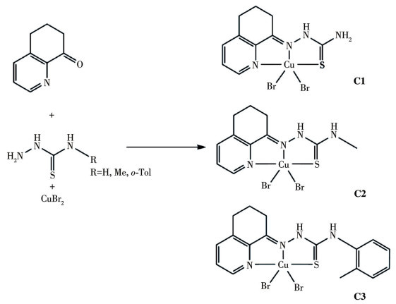
Scheme 1.
Synthesis routes of the Cu(Ⅱ) complexes C1-C3
Endoplasmic reticulum stress and mitochondrial apoptosis in thiosemicarbazone copper(Ⅱ) complex-triggered triple-negative breast cancer cell death
Shuangshuang GAI , Jixiang CAI , Moya YANG , Haijuan WU , Shuzhen FENG , Ming JIANG

Breast cancer represents the most frequently diagnosed malignancy and ranks as the second leading cause of cancer-related mortality in women globally[1]. Approximately 15%-20% of breast cancer cases are classified as triple-negative breast cancer (TNBC), a subtype characterized by heightened invasiveness and an elevated risk of metastasis[2]. Owing to its lack of estrogen receptor, progesterone receptor, and HER2, TNBC is refractory to hormonal therapy and therefore relies predominantly on chemotherapy[3]. Platinum-based agents, such as cisplatin and carboplatin, demonstrate moderate efficacy in suppressing TNBC cell proliferation; however, their clinical utility is often limited by intrinsic or acquired resistance, as well as associated toxicities[4-6]. These limitations have prompted increased research interest in developing non-platinum metal-based chemotherapeutic agents.
Copper complexes are attracting attention as next-generation chemotherapeutics, not only because copper is an essential trace element involved in key physiological processes[7-9], but also because its ions can form complexes with various ligands through diverse coordination modes, creating a synergistic effect that inhibits tumor growth through multiple mechanisms[10-13]. Thiosemicarbazones are a class of compounds known for their broad spectrum of biological activities, including antiviral, antibacterial, and anticancer properties[14-15]. Several thiosemicarbazones, such as Triapine, DpC, and COTI-2, have advanced into clinical trials[16-17]. Furthermore, thiosemicarbazones can function as ligands and exhibit strong chelating ability towards various biologically relevant metal ions[18-19]. Compared to the free ligands, their coordination to metal ions often enhances anticancer efficacy[19-21]. Notably, thiosemicarbazone metal complexes act through diverse mechanisms: they can target DNA[22-24], induce lysosome damage[22, 25], activate immunotherapy[26-27], trigger endoplasmic reticulum (ER) stress[28-29], and suppress protein disulfide isomerase (PDI) activity[30-31], etc. Accordingly, developing a Cu(Ⅱ) complex with multiple targets holds promise as a therapeutic strategy against cancer.
Herein, we report the synthesis and characterization of three copper(Ⅱ) complexes (C1-C3) derived from 6, 7-dihydro-5H-quinoline-8-one thiosemicarbazones (Scheme 1). The in vitro antitumor activities of these Cu(Ⅱ) complexes were investigated against various cancer cell lines. Among them, C3 exhibited the strongest cytotoxicity against MDA-MB-231 cells. Further studies revealed that C3 targeted the ER stress response and induced mitochondrial dysfunction.
Thiosemicarbazide, 4-(2-methylphenyl)-3-thiosemicarbazide, 4-methyl-3-thiosemicarbazide, 6, 7-dihydro-5H-quinoline-8-one, CuBr2, and all other organic solvents were purchased from Bidepharm (Shanghai, China) or Innochem (Beijing, China), and used without further purification.
The Cu(Ⅱ) complexes were synthesized by mixing 6, 7-dihydro-5H-quinoline-8-one (0.1 mmol), the different substituents-thiosemicarbazide (0.1 mmol), CuBr2 (0.1 mmol), and CH3OH (2 mL) in a 10 mL Pyrex glass tube. Then, the glass tubes were heated in an oven at 65 ℃ for 48 h. After cooling the tubes to room temperature slowly, single crystals suitable for single-crystal X-ray diffraction were obtained. The structure of crystals was characterized by X-ray crystallography, elemental analysis, and HRMS.
C1: 80.2% of yield. Anal. Calcd. for C10H12Br2CuN4S·CH3OH(%): C, 27.77; H, 3.39; N, 11.78. Found(%): C, 27.85; H, 3.36; N, 11.71. HRMS(m/z): 282.000 9 [M-CH3OH-2Br-H]+.
C2: 76.5% of yield. Anal. Calcd. for C11H14Br2CuN4S(%): C, 28.87; H, 3.08; N, 12.24. Found(%): C, 28.93; H, 3.07; N, 12.27. HRMS(m/z): 296.017 3 [M-2Br-H]+.
C3: 77.9% of yield. Anal. Calcd. for C17H18Br2CuN4S(%): C, 38.25; H, 3.40; N, 10.50. Found(%): C, 38.31; H, 3.38; N, 10.47. HRMS(m/z): 372.044 9 [M-2Br-H]+.
X-ray diffraction data for single crystals of Cu(Ⅱ) complexes C1-C3 were collected at 296.15 K on a Bruker SMART Apex Ⅱ CCD diffractometer. Structural solution and refinement were achieved using Olex 2. Non-hydrogen atoms were refined for anisotropy and atomic coordinates through the full matrix least squares method F2, while hydrogen atoms were placed in geometrically ideal positions and constrained to ride on their parent atoms[32]. Crystallographic data, refinement parameters, and selected bond angles/lengths for C1-C3 are summarized in Table S1-S3 (Supporting information).
The cytotoxicity of the Cu(Ⅱ) complexes (C1-C3) against various cell lines (MDA-MB-231, HepG2, A549, and WI-38) was examined by MTT assay. In brief, the cell suspension was seeded in 96-well plates at a density of 5 000 cells per well, and then incubated overnight at 37 ℃ under 5% of CO2 volume fraction in medium supplemented with 10% fetal bovine serum and 1% antibiotic-antimycotic solution. Subsequently, the cells were treated with the Cu(Ⅱ) complexes of different concentrations (0, 1, 2, 5, 10, and 20 μmol·L-1), and clinical cisplatin was employed as the control. Following a 48-h incubation, 10 μL of MTT solution (5 mg·mL-1) was added per well, followed by a further 4-h incubation. The medium was then replaced with 100 μL DMSO, and the plate was agitated to solubilize the formazan crystals. Absorbance of the resulting solution was measured at 570 nm using a microplate reader. Finally, the half maximal inhibitory concentration (IC50) values for the Cu(Ⅱ) complexes were determined from the absorbance data using GraphPad Prism software.
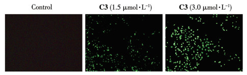
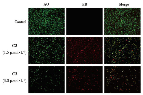
MDA-MB-231 cells were plated in 6-well plates at a density of 5×10⁴ cells per well and incubated overnight. Subsequently, the cancer cells were treated with C3 at concentrations of 0, 1.5, and 3.0 μmol·L-1. After 24 h of incubation, the cells were washed with phosphate-buffered saline (PBS), stained with acridine orange (AO)/ethidium bromide (EB) for 20 min, and then visualized under a fluorescence microscope.
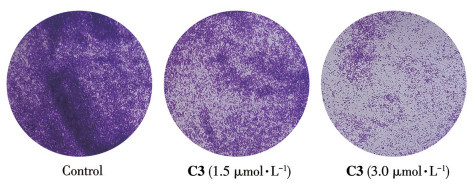
A colony assay was performed to evaluate the inhibitory effect of C3 on colony formation. In brief, MDA-MB-231 cells were seeded into 6-well plates overnight. Then, the cells were treated with C3 at concentrations of 0, 1.5, and 3.0 μmol·L-1. After 3 d of drug exposure, the cells were fixed with 100% cold methanol for 20 min, stained with 0.1% crystal violet for 10 min at room temperature. The samples were washed with water and captured using an inverted microscope.
The effects of Cu(Ⅱ) complex on intracellular levels of reactive oxygen species (ROS) were investigated using 2′,7′-dichlorodihydrofluorescein diacetate (DCFH-DA) fluorescent probe. MDA-MB-231 cells were transplanted into six-well plates and treated with C3 at concentrations of 0, 1.5, and 3.0 μmol·L-1. Following a 24-h incubation, the cells were washed with PBS and incubated with 10 μmol·L-1 DCFH-DA solution for 30 min. The cells were washed with serum-free medium and observed under a fluorescence microscope.
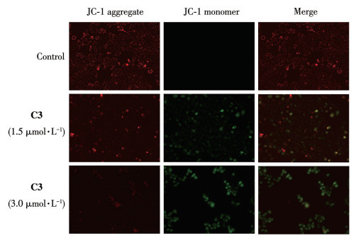
Mitochondrial membrane potential was assessed using the fluorescent probe JC-1. MDA-MB-231 cells were seeded in 24-well plates and incubated for 24 h under standard conditions (37 ℃, 5% of CO2 volume fraction). The medium was then replaced with fresh medium containing C3 (0, 1.5, and 3.0 μmol·L-1), and incubated for another 24 h. Subsequently, the cells were stained with JC-1 in the dark at 37 ℃ for 20 min and immediately imaged under an inverted fluorescence microscope.
MDA-MB-231 cells were seeded in 10 cm dishes and incubated with C3 (0, 1.5, and 3.0 μmol·L-1) for 48 h. Subsequently, total proteins were extracted using radioimmunoprecipitation assay (RIPA) buffer and quantified via bicinchoninic acid (BCA) assay. Proteins were separated by 10% SDS-PAGE, transferred to a polyvinylidene fluoride (PVDF) membrane, and blocked with 5% non-fat milk for 30 min. The membranes were then incubated with primary antibodies overnight at 4 ℃, followed by incubation with secondary antibodies for 1 h at room temperature after thorough washing with TBST (tris-buffered saline with Tween-20).
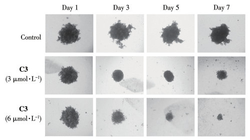
The inhibitory effect of the Cu(Ⅱ) complex on 3D multicellular tumor spheroids was evaluated in vitro. MDA-MB-231 spheroids were established in agarose-coated 96-well plates over 5 d. Subsequently, the 3D spheroids were then exposed to C3 (0, 3, and 6 μmol·L-1). The culture medium containing C3 was refreshed every two days, and spheroid morphology was monitored using an inverted microscope.
The activity of PDI was quantified with the Proteostat PDI assay kit (Enzo Life Sciences, Lausanne, Switzerland), which monitors the insulin-reduction reaction catalyzed by PDI. Assays were performed in strict accordance with the manufacturer′s protocol. The different concentrations of the test Cu(Ⅱ) complexes—or bacitracin as a reference—were incubated with insulin and PDI. The reaction was initiated by the addition of dithiothreitol (DTT) and allowed to proceed for 0.5 h at 37 ℃, then quenched with the provided stop solution. The fluorescence was recorded on a microplate reader. The IC50 values were then calculated.
The interaction mechanism between the Cu(Ⅱ) complex and the protein disulfide isomerase (PDI; PDB ID: 4EKZ) was investigated through molecular docking simulations performed with AutoDock Tool 4.2. The protein structure was prepared by removing crystallographic water molecules. Docking procedures were validated before conducting docking experiments with C3 and PDI. The resulting docking poses were visualized and analyzed using PyMOL software.
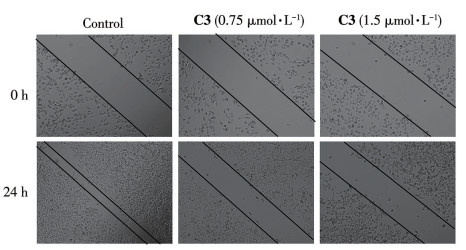
MDA-MB-231 cells were seeded into 6-well plates and incubated overnight. A tip of a sterile toothpick was then used to introduce a vertical scratch through the monolayer. The cells were washed with PBS and exposed to C3 (0.75 or 1.5 μmol·L-1) for 24 h. Subsequently, the medium was aspirated, and the wells were gently rinsed with 1 mL PBS. Migration into the wound area was imaged using an inverted microscope.
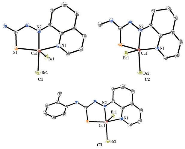
The Cu(Ⅱ) complexes were synthesized by reacting 6, 7-dihydro-5H-quinoline-8-one, thiosemicarbazide, and CuBr2 in a 10 mL Pyrex glass tube at 65 ℃ for 48 h, as illustrated in Scheme 1. The dark green crystals were examined by single-crystal X-ray diffraction, and their structures are shown in Fig. 1. The crystal data for Cu(Ⅱ) complexes are shown in Table S1-S3. Complexes C1-C3 crystallize in the monoclinic crystal system, space group P21/c, P21/n, and P21/c, respectively (Table S1). All these mononuclear Cu(Ⅱ) complexes have similar coordination geometries, which are a five-coordinated distorted trigonal bipyramid geometry with a center Cu2+ ion. C1-C3 consist of mononuclear units comprising one Cu(Ⅱ) ion, one ligand, and two bromide ions. The bond angles of these Cu(Ⅱ) complexes for N1—Cu—N2 are in a range of 79.0°-79.34°, for N1—Cu1—S1 are in a range of 158.89°-159.40°, and for Br2—Cu1—Br1 are in a range of 99.39°-104.34° (Table S2). The Cu—N [0.197 8(5)-0.204 5(5) nm] and Cu—S [0.228 59(12)-0.230 14(17) nm] bond lengths (Table S3) are consistent with those reported for related Cu(Ⅱ) complexes[33-34]. As shown in Fig.S1-S3, the UV-Vis absorption peaks of C1-C3 exhibited no significant change, indicating that all copper complexes maintained good stability in PBS. The purity of complexes C1-C3 was greater than 95% (Fig.S4-S6).
To evaluate the effect of these Cu(Ⅱ) complexes on the activity of cancer cells (MDA-MB-231, HepG2, and A549), the IC50 values were examined using the MTT assay after treatment with various concentrations of C1-C3. As shown in Table 1, the IC50 values of C1-C3 against cancer cells ranged from (1.42±0.22) μmol·L-1 to (7.14±0.35) μmol·L-1. Among the cancer cell lines examined, the Cu(Ⅱ) complexes exhibited the greatest potency toward MDA-MB-231 cells [IC50=(1.42±0.22) μmol·L-1-(4.89±0.21) μmol·L-1]. Interestingly, their cytotoxic efficacy remained markedly higher than that of cisplatin (rank order: C3 > C2 > C1 > cisplatin). Replacing the H atom at the N-4 position with a methyl (C2) or o-Tol (C3) group enhances the anticancer activity compared to the unmodified thiosemicarbazone (C1). This indicates that the N4-substituent plays a pivotal role in determining cytotoxicity, highlighting the potential to develop a diverse library of such Cu(Ⅱ) thiosemicarbazone complexes for systematic anticancer screening. Among all tested complexes, C3 exhibited the highest cytotoxicity against triple-negative breast cancer cells.
 下载:
导出CSV
下载:
导出CSV
| Compound | MDA-MB-231 | HepG2 | A549 | WI-38 |
| C1 | 4.89±0.21 | 4.75±0.25 | 6.91±0.41 | 11.33±0.28 |
| C2 | 2.54±0.19 | 3.65±0.26 | 7.14±0.35 | 8.85±0.51 |
| C3 | 1.42±0.22 | 3.08±0.23 | 4.26±0.29 | 7.44±0.32 |
| CuBr2 | > 100 | > 100 | > 100 | > 100 |
| Cisplatin | 15.22±0.78 | 17.47±0.74 | 15.32±0.80 | 10.03±0.54 |
| *Data are shown as mean±standard deviation (SD, n=3). | ||||
The colony formation assay was employed to assess the capacity of single cells to survive drug treatment and develop into colonies, thereby serving as an indicator of proliferative ability[35]. Following a 24 h treatment with complex C3, MDA-MB-231 cells were cultured for an additional 72 h. The resulting colonies were then stained with crystal violet to visualize and quantify cell viability and proliferation. As shown in Fig.2, the treatment groups exhibited a significant reduction in colony number compared to the control, indicating that C3 effectively suppressed cell proliferation.
Prior research has demonstrated that cancer cells exhibit greater sensitivity to elevated ROS levels than normal cells, and that such excess ROS dysregulates the redox balance, triggering apoptotic cell death[36-37]. Then, the changes in intracellular ROS were determined using the DCHF-DA as a fluorescent probe[38]. As depicted in Fig.3, the blank control group exhibited negligible green fluorescence. In contrast, following C3 treatment, a pronounced increase in green fluorescence intensity was observed, signifying a substantial elevation of intracellular ROS levels in MDA-MB-231 cells relative to the control. In addition, as the concentration of C3 increased, the intensity of the green fluorescence became higher. These results indicated that C3 can increase the ROS level in MDA-MB-231 cells, and this effect is concentration-dependent.
Targeting cancer cells through the apoptotic pathway and inducing their death represents one of the potential therapeutic objectives of antitumor drugs[39]. Dual staining using nucleic acid-binding dyes such as AO and EB[40], based on their fluorescent emission, can indicate changes in cellular morphological characteristics and thereby reveal apoptotic cell death. As shown in Fig.4, the control cells appeared bright and emitted steady green fluorescence, which is attributable to the well-ordered structure of viable cells. After 24 h of treatment with C3, EB (red fluorescence) penetrated the MDA-MB-231 cells, overshadowing the green fluorescence from AO, indicating that C3 induces cancer cells' apoptosis. In addition, the cells exposed to C3 at 3.0 μmol·L-1 exhibited a lower number of viable cells and a higher number of apoptotic cells compared to those treated with C3 at 1.5 μmol·L-1.
A well-maintained mitochondrial membrane potential (ΔΨm) is essential for mitochondrial integrity and bioenergetic function[41]. The change of ΔΨm in cancer cells is a recognized hallmark event, signifying the initiation of the apoptotic cascade[42]. The JC-1 staining was determined to evaluate the effect of the Cu(Ⅱ) complex on ΔΨm in MDA-MB-231 cells. As shown in Fig.5, complex C3 increased bright green fluorescence and diminished red fluorescence, indicating ΔΨm loss. These results suggested that C3 triggers apoptosis by inducing mitochondrial dysfunction.
The ER is responsible for the biosynthesis, folding, maturation, stability, and transport of membrane proteins and secretory proteins, and it constitutes an interconnected intracellular structure[43]. Studies have shown that ROS can disrupt the folding capacity of the ER, thereby leading to the accumulation of unfolded or misfolded proteins within the ER—a phenomenon known as ER stress[44-46]. Persistent and severe ER stress is closely associated with cell death. As shown in Fig.6A, the expression levels of ER stress-related proteins (CHOP, ATF-4, and p-eIF2α) were increased after treatment with C3 (1.5 and 3.0 μmol·L-1) for 48 h, indicating that C3 mediates the ER stress response.
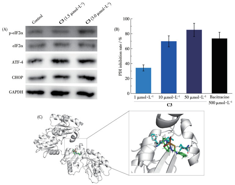
The PDI family, widely recognized as a group of oxidoreductases abundantly present in the endoplasmic reticulum, plays a critical role in disulfide bond formation and is essential for protein folding[47-48]. Subsequently, we used the Proteostat PDI assay kit to detect the reductase activity of PDI in the presence of C3. As shown in Fig.6B, C3 was able to effectively inhibit PDI reductase activity. Moreover, the inhibition was significantly enhanced with increasing concentrations of C3, and its inhibitory effect was much stronger than that of the well-known PDI inhibitor, bacitracin.
Next, to better understand the binding mode of Cu(Ⅱ) complex with PDI (PDB ID: 4EKZ), the molecular docking was performed. As shown in Fig.6C, C3 can directly interact with the active site of PDI and form hydrogen bonds with the surrounding amino acid residues, which may interfere with the function of PDI. These results indicated that the induction of cancer cell death is strongly associated with the activation of ER stress and the unfolded protein response.
After assessing cytotoxicity in 2D cancer cell cultures, the investigation was extended to 3D multicellular tumor spheroids to evaluate therapeutic efficacy[49-50]. Tumor spheroids represent an in vitro model system that recapitulates key histological and pathological features of solid tumors, such as gradients in proliferation and nutrient availability, along with the development of a hypoxic core[51]. To further evaluate the therapeutic effects, the size and morphology of 3D MDA-MB-231 spheroids after treatment with the Cu(Ⅱ) complex were observed. Compared to the untreated control, C3 treatment significantly reduced spheroid size and caused obvious morphological damage (Fig.7). These results demonstrated that the Cu(Ⅱ) complex effectively eradicates multicellular tumor spheroids.
A major factor underlying the therapeutic recalcitrance of triple-negative breast cancer is the exceptionally aggressive biological behavior exhibited by these tumors[2, 52]. Therefore, the suppression of cancer cell metastasis constitutes a critical indicator in drug assessment. To determine whether C3 impedes the migration of MDA-MB-231 cells, an in vitro wound-healing assay was performed. As illustrated in Fig.8, C3 exposure significantly suppressed cell migration in a dose-dependent manner; the anti-migratory potency of 1.5 μmol·L-1 C3 was markedly greater than that of 0.75 μmol·L-1. These results strongly suggested that C3 exhibits potent anti-migratory activity against MDA-MB-231 cells.
In summary, we have designed and synthesized a series of Cu(Ⅱ) complexes (C1-C3) exhibiting pronounced antitumor activity, with C3 emerging as the most potent. C3 displayed potent activity against MDA-MB-231 cells and their 3D spheroids. Mechanistic studies revealed that its potent activity is mediated through a dual-pathway mechanism: C3 induces substantial ROS generation, which concurrently triggers ER stress, inhibits PDI activity, and induces mitochondrial dysfunction. The convergence of these cellular stresses ultimately drives apoptotic cell death. These findings indicate C3 as a promising lead compound for further anticancer development.
Supporting information is available at
DARIE C C, HUKOVIC A, MAYNARD V D, NEAGU A N. Roles of oxygen in the tumorigenesis, progression, and treatment of breast cancer[J]. Med. Gas Res., 2026, 16(1): 41-45 doi: 10.4103/mgr.MEDGASRES-D-25-00023
ZAGAMI P, CAREY L A. Triple negative breast cancer: Pitfalls and progress[J]. npj Breast Cancer, 2022, 8(1): 95 doi: 10.1038/s41523-022-00468-0
NANDHINI S, THIRUPPATHI G, RANJANI M, PUSCHMANN H, RAVI M, SUNDARARAJ P, PRABHAKARAN R. Effect of ruthenium(Ⅱ) complexes on MDA-MB-231 cells and lifespan/tumor growth in gld-1mutant, Daf-16 TF and stress productive genes: A perspective study[J]. J. Inorg. Biochem., 2024, 257: 112580 doi: 10.1016/j.jinorgbio.2024.112580
SONG P Y, LI Y R, DONG Y, LIANG Y Y, QU H N, QI D, LU Y, JIN X S, GUO Y T, JIA Y Y, WANG X Q, XU W H, QUAN C S. Estrogen receptor β inhibits breast cancer cells migration and invasion through CLDN6-mediated autophagy[J]. J. Exp. Clin. Cancer. Res., 2019, 38(1): 354 doi: 10.1186/s13046-019-1359-9
VIDRA R, NEMES A, VIDREAN A, PINTEA S, TINTARI S, DEAC A, CIULEANU T. Pathological complete response following cisplatin or carboplatin-based neoadjuvant chemotherapy for triple-negative breast cancer: A systematic review and meta-analysis[J]. Exp. Ther. Med., 2022, 23(1): 91
LYNCE F, NUNES R. Role of platinums in triple-negative breast cancer[J]. Curr. Oncol. Rep., 2021, 23(5): 50 doi: 10.1007/s11912-021-01041-x
WU Y R, WU D Q, LAN J F, LI A L, HOU L X, XU Y R, GOU Y. Assessment of mononuclear/dinuclear copper acylhydrazone complexes for lung cancer treatment[J]. Bioorg. Chem., 2024, 144: 107122 doi: 10.1016/j.bioorg.2024.107122
SANTINI C, PELLEI M, GANDIN V, PORCHIA M, TISATO F, MARZANO C. Advances in copper complexes as anticancer agents[J]. Chem. Rev., 2014, 114(1): 815-862 doi: 10.1021/cr400135x
JIANG M, SU X L, ZHONG X W, LAN Y H, YANG F, QIN Y M, JIANG C Y. Recent development of Schiff-base metal complexes as therapeutic agents for lung cancer[J]. J. Mol. Struct., 2024, 1318: 139403
MAN X Y, LI W J, ZHU M H, LI S H, XU G, ZHANG Z L, LIANG H, YANG F. Anticancer tetranuclear Cu(Ⅰ) complex catalyzes a click reaction to synthesize a chemotherapeutic agent in situ to achieve targeted dual-agent combination therapy for cancer[J]. Angew. Chem., 2024, 136(51): e202411846 doi: 10.1002/ange.202411846
YANG K, CHEN Q, CHEN J, GENG L F, MA M X, GU Y Q, CHOUDHARY M I, LIANG H, CHEN Z F. Copper(Ⅱ) complexes of pyrazolopyrimidine derivatives as anticancer agents with enhanced chemodynamic therapy through bimodal apoptosis and ferroptosis[J]. J. Med. Chem., 2025, 68(7): 7137-7152 doi: 10.1021/acs.jmedchem.4c02515
JIANG M, ZHANG Z L, LI W J, MAN X Y, SUN H B, LIANG H, YANG F. Developing a copper(Ⅱ) agent based on His-146 and His-242 residues of human serum albumin nanoparticles: Integration to overcome cisplatin resistance and inhibit the metastasis of nonsmall cell lung cancer[J]. J. Med. Chem., 2022, 65(13): 9447-9458 doi: 10.1021/acs.jmedchem.2c00698
PENG R, ZHONG J J, YANG D, HONG Z, BIAN H D, HUANG F P. Two ferrocene-modified copper(Ⅰ) complexes for the synergistic enhancement of chemodynamic therapy[J]. Appl. Organomet. Chem., 2025, 39(2): e7960 doi: 10.1002/aoc.7960
BAJAJ K, BUCHANAN R M, GRAPPERHAUS C A. Antifungal activity of thiosemicarbazones, bis (thiosemicarbazones), and their metal complexes[J]. J. Inorg. Biochem., 2021, 225: 111620 doi: 10.1016/j.jinorgbio.2021.111620
ZAHRA S B, KHAN A, AHMED N, RAFIQUE M, FATIMA L, KHAN I, HUSSAIN J, KHALID S, OGALY H A, AHMED M M. Versatile biological activities of thiosemicarbazones and their metal complexes[J]. J. Mol. Struct., 2025, 1322: 140511
BORMIO NUNES J H, HAGER S, MATHUBER M, PÓSA V, ROLLER A, ENYEDY E V A, STEFANELLI A, BERGER W, KEPPLER B K, HEFFETER P. Cancer cell resistance against the clinically investigated thiosemicarbazone COTI-2 is based on formation of intracellular copper complex glutathione adducts and ABCC1-mediated efflux[J]. J. Med. Chem., 2020, 63(22): 13719-13732 doi: 10.1021/acs.jmedchem.0c01277
FUSSELL J B, SHAW J P, GRAMS M A, SUNG Y S, JHENG R H, ASTASHKIN A V, TOMAT E. Disulfide-based 2-pyridyl-hydrazone prochelators induce iron deprivation and oxidative stress in ovarian cancer cells[J]. J. Biol. Inorg. Chem., 2025, 30(4/5): 443-452
XU G, LIANG Q Y, GAO L J, XU S H, LUO W C, WU Q M, LI J Y, ZHANG Z L, LIANG H, YANG F. Developing an arene binuclear ruthenium(Ⅱ) complex to induce ferroptosis and activate the cGAS-STING pathway: Targeted inhibiting growth and metastasis of triple negative breast cancer[J]. J. Med. Chem., 2024, 67(21): 19573-19585 doi: 10.1021/acs.jmedchem.4c01908
OLIVEIRA G P, LIMA M A, PEREIRA G B, COSTA A R, BATISTA A A, FORIM M R, COMINETTI M R, ZANETTI R D, FARIAS R L, NETTO A V. Palladium(Ⅱ) and platinum(Ⅱ) thiophene-based thiosemicarbazones: Synthesis, properties, and anticancer studies[J]. J. Mol. Struct., 2025, 1322: 140306
LIGHVAN Z M, RAMEZANPOUR A, PIRANI S, AKBARI A, JAHROMI M D, KERMAGORET A, HEYDARI A. Synthesis of tetranuclear cyclopalladated complex using thiosemicarbazone derivative ligand: Spectral, biological and molecular docking studies[J]. J. Mol. Struct., 2025, 1321: 139932
MAN X Y, LI S H, XU G, LI W J, ZHU M H, ZHANG Z L, LIANG H, YANG F. Developing a copper(Ⅱ) isopropyl 2-pyridyl ketone thiosemicarbazone compound based on the IB subdomain of human serum albumin-indomethacin complex: Inhibiting tumor growth by remodeling the tumor microenvironment[J]. J. Med. Chem., 2024, 67(7): 5744-5757 doi: 10.1021/acs.jmedchem.3c02378
LIU X C, LV A, ZHANG P, CHANG J Y, DONG R X, LIU M X, LIU J Y, HUANG X Q, YUAN X A, LIU Z. The anticancer application of half-sandwich iridium(Ⅲ) ferrocene-thiosemicarbazide Schiff base complexes[J]. Dalton Trans., 2024, 53(2): 552-563 doi: 10.1039/D3DT02879H
ZHANG S Y, ZHAO J A, GUO Y, HU J Y, CHEN X J, RUAN H H, CAO T T, HOU H W. Thiosemicarbazone N-heterocyclic Cu(Ⅱ) complexes inducing nuclei DNA and mitochondria damage in hepatocellular carcinoma cells[J]. J. Inorg. Biochem., 2022, 236: 111964 doi: 10.1016/j.jinorgbio.2022.111964
SHAO J, MA Z Y, LI A, LIU Y H, XIE C Z, QIANG Z Y, XU J Y. Thiosemicarbazone Cu(Ⅱ) and Zn(Ⅱ) complexes as potential anticancer agents: Syntheses, crystal structure, DNA cleavage, cytotoxicity and apoptosis induction activity[J]. J. Inorg. Biochem., 2014, 136: 13-23 doi: 10.1016/j.jinorgbio.2014.03.004
STACY A E, PALANIMUTHU D, BERNHARDT P V, KALINOWSKI D S, JANSSON P J, RICHARDSON D R. Zinc(Ⅱ)-thiosemicarbazone complexes are localized to the lysosomal compartment where they transmetallate with copper ions to induce cytotoxicity[J]. J. Med. Chem., 2016, 59(10): 4965-4984 doi: 10.1021/acs.jmedchem.6b00238
ZHANG J Z, JIANG M, LI S H, ZHANG Z L, SUN H B, YANG F, LIANG H. Developing a novel anticancer gold(Ⅲ) agent to integrate chemotherapy and immunotherapy[J]. J. Med. Chem., 2021, 64(10): 6777-6791 doi: 10.1021/acs.jmedchem.1c00050
ZHU M H, XU S H, LI G C, XU G, ZHANG Z, LIANG H, YANG F. Development of a high-efficacy and low-toxicity cobalt(Ⅱ) agent for targeting inhibition of tumor growth through mitochondrial damage-mediated chemotherapy and immunotherapy[J]. J. Med. Chem., 2025, 68(12): 13113-13126 doi: 10.1021/acs.jmedchem.5c01235
FU Y, LIU Y X, WANG J G, LI C P, ZHOU S F, YANG Y, ZHOU P X, LU C B, LI C Z. Calcium release induced by 2-pyridinecarboxaldehyde thiosemicarbazone and its copper complex contributes to tumor cell death[J]. Oncol. Rep., 2017, 37(3): 1662-1670 doi: 10.3892/or.2017.5395
KING A P, WILSON J J. Endoplasmic reticulum stress: An arising target for metal-based anticancer agents[J]. Chem. Soc. Rev., 2020, 49(22): 8113-8136 doi: 10.1039/D0CS00259C
MIGLIOLI F, DE FRANCO M, BARTOLI J, SCACCAGLIA M, PELOSI G, MARZANO C, ROGOLINO D, GANDIN V, CARCELLI M. Anticancer activity of new water-soluble sulfonated thiosemicarbazone copper(Ⅱ) complexes targeting disulfide isomerase[J]. Eur. J. Med. Chem., 2024, 276: 116697 doi: 10.1016/j.ejmech.2024.116697
CARCELLI M, TEGONI M, BARTOLI J, MARZANO C, PELOSI G, SALVALAIO M, ROGOLINO D, GANDIN V. In vitro and in vivo anticancer activity of tridentate thiosemicarbazone copper complexes: Unravelling an unexplored pharmacological target[J]. Eur. J. Med. Chem., 2020, 194: 112266 doi: 10.1016/j.ejmech.2020.112266
SHELDRICK G M. Crystal structure refinement with SHELXL[J]. Acta Crystallogr. Sect. C, 2015, C71(1): 3-8
TERENTI N, LAZARESCU A, SHOVA S, BOUROSH P, NEDELKO N, ŚLAWSKA-WANIEWSKA A, ZARICIUC E, LOZAN V. Synthesis, X-ray and antibacterial activity of new copper(Ⅱ) thiosemicarbazone complexes derived from 4-formyl-3-hydroxy-2-naphthoic acid[J]. Inorg. Chim. Acta, 2024, 571: 122216 doi: 10.1016/j.ica.2024.122216
DHARMASIVAM M, KAYA B, WIJESINGHE T P, RICHARDSON V, HARMER J R, GONZALVEZ M A, LEWIS W, AZAD M G, BERNHARDT P V, RICHARDSON D R. Differential transmetallation of complexes of the anti-cancer thiosemicarbazone, Dp4e4mT: Effects on anti-proliferative efficacy, redox activity, oxy-myoglobin and oxy-hemoglobin oxidation[J]. Chem. Sci., 2024, 15(3): 974-990 doi: 10.1039/D3SC05723B
KUMAR D N, CHAUDHURI A, SHIROMANI U, KUMAR D, AGRAWAL A K. An investigation of in vitro anti-cancer efficacy of dihydroartemisinin-loaded bovine milk exosomes against triple-negative breast cancer[J]. AAPS J., 2024, 26(5): 91 doi: 10.1208/s12248-024-00958-y
ZHOU L, HU H Y, LI J B, YANG Y, TIAN S, NIU Y J, LIU Y J, ZENG X D. Photodynamic immunotherapy of ruthenium(Ⅱ) polypyridyl complexes: Application in the treatment of colorectal cancer[J]. J. Inorg. Biochem., 2026, 274: 113056 doi: 10.1016/j.jinorgbio.2025.113056
CALDERÓN-CHIU C, CALDERÓN-SANTOYO M, RAGAZZO-SÁNCHEZ J A. Feasibility of leaf protein hydrolysates to reduce reactive oxygen species and cancer cell proliferation[J]. Med. Gas Res., 2026, 16(1): 88-89 doi: 10.4103/mgr.MEDGASRES-D-25-00040
KIM K Y, YU S N, LEE S Y, CHUN S S, CHOI Y L, PARK Y M, SONG C S, CHATTERJEE B, AHN S C. Salinomycin-induced apoptosis of human prostate cancer cells due to accumulated reactive oxygen species and mitochondrial membrane depolarization[J]. Biochem. Biophys. Res. Commun., 2011, 413(1): 80-86 doi: 10.1016/j.bbrc.2011.08.054
ARUNACHALAM A, RENGAN R, UMAPATHY D, AROCKIAM A J V. Impact of biphenyl benzhydrazone-incorporated arene Ru(Ⅱ) complexes on cytotoxicity and the cancer cell death mechanism[J]. Organometallics, 2022, 41(17): 2474-2486 doi: 10.1021/acs.organomet.2c00290
PATRA S A, SAHU G, MOHAPATRA D, PATTANAYAK P D, DINDA R. Fluorescent diorganotin(Ⅳ) complexes as anticancer agents: Study of cytotoxicity, cellular localization, and mechanism of cell death[J]. Organometallics, 2023, 42(15): 1934-1950 doi: 10.1021/acs.organomet.3c00182
KOWALTOWSKI A J, ABDULKADER F. How and when to measure mitochondrial inner membrane potentials[J]. Biophys. J., 2024, 123(24): 4150-4157 doi: 10.1016/j.bpj.2024.03.011
GAI S S, YAN Q W, LI S, ZHONG X W, QIN Y M, JIANG M. Lactoferrin nanoparticle-vanadium complex: A promising high-efficiency agent against glioblastoma by triggering autophagy and ferroptosis[J]. J. Med. Chem., 2025, 68(4): 4650-4662 doi: 10.1021/acs.jmedchem.4c02696
VITALE A, CERIOTTI A, DENECKE J. The role of the endoplasmic reticulum in protein synthesis, modification and intracellular transport[J]. J. Exp. Bot., 1993, 44(9): 1417-1444 doi: 10.1093/jxb/44.9.1417
CHEN M, WANG D, FAN L M, NIU D Q, XU J H, LIU Y C, LIU Y Y. The copper(Ⅱ) complex of salicylate phenanthroline induces immunogenic cell death of colorectal cancer cells through inducing endoplasmic reticulum stress[J]. Int. Immunopharmacol., 2024, 132: 111980 doi: 10.1016/j.intimp.2024.111980
KUZNETCOVA I, BACHER F, ALFADUL S M, THAM M J R, ANG W H, BABAK M V, RAPTA P, ARION V B. Elucidation of structure-activity relationships in indolobenzazepine-derived ligands and their copper(Ⅱ) complexes: The role of key structural components and insight into the mechanism of action[J]. Inorg. Chem., 2022, 61(26): 10167-10181 doi: 10.1021/acs.inorgchem.2c01375
SHEN W Y, JIA C P, MO A N, LIANG H, CHEN Z F. Chemodynamic therapy agents Cu(Ⅱ) complexes of quinoline derivatives induced ER stress and mitochondria-mediated apoptosis in SK-OV-3 cells[J]. Eur. J. Med. Chem., 2021, 223: 113636 doi: 10.1016/j.ejmech.2021.113636
YIN H Y, GAO J J, CHEN X, MA B, YANG Z S, TANG J, WANG B W, CHEN T, WANG C, GAO S. A gallium(Ⅲ) complex that engages protein disulfide isomerase A3 (PDIA3) as an anticancer target[J]. Angew. Chem., 2020, 59(45): 20147-20153 doi: 10.1002/anie.202008432
KANEMURA S, MATSUSAKI M, INABA K, OKUMURA M. PDI family members as guides for client folding and assembly[J]. Int. J. Mol. Sci., 2020, 21(24): 9351 doi: 10.3390/ijms21249351
GAI S S, CAO P, ZHONG X W, LIN Y C, LIN B X, JIANG M. Designing an anticancer Pd(Ⅱ) complex as poly(ADP-ribose) polymerase 1 inhibitor[J]. Int. J. Biol. Macromol., 2025, 297: 139885 doi: 10.1016/j.ijbiomac.2025.139885
KIMLIN L C, CASAGRANDE G, VIRADOR V M. In vitro three-dimensional (3D) models in cancer research: An update[J]. Mol. Carcinogen., 2013, 52(3): 167-182 doi: 10.1002/mc.21844
PINTO B, HENRIQUES A C, SILVA P M, BOUSBAA H. Three-dimensional spheroids as in vitro preclinical models for cancer research[J]. Pharmaceutics, 2020, 12(12): 1186 doi: 10.3390/pharmaceutics12121186
BILIR B, KUCUK O, MORENO C S. Wnt signaling blockage inhibits cell proliferation and migration, and induces apoptosis in triple-negative breast cancer cells[J]. J. Transl. Med., 2013, 11(1): 280 doi: 10.1186/1479-5876-11-280

Figure 1 Crystal structures of C1-C3 with thermal ellipsoids plotted at the 30% probability level

Figure 3 Fluorescence imaging of intracellular ROS levels in MDA-MB-231 cells after 24 h treatment with C3 (1.5 and 3.0 μmol·L-1) and subsequent DCFH-DA staining

Figure 4 Dual AO/EB fluorescent staining assay of MDA-MB-231 cells after incubation with C3 (1.5 and 3.0 μmol·L-1) for 24 h

Figure 6 (A) Western blot analysis of the expression levels of CHOP, eIF2α, p-eIF2α, and ATF-4 proteins in MDA-MB-231 cells after treatment with C3 (1.5 and 3.0 μmol·L-1) for 48 h; (B) Inhibition of PDI activity by C3, bacitracin as a positive control; (C) Interaction between C3 with PDI (4EKZ) using molecular docking simulation

Figure 7 Microscopic observation of the changes in MDA-MB-231 multicellular tumor spheroids after a 7-day treatment with C3
Table 1.
IC50 values of C1-C3 toward various cell lines for 48 h*
| Compound | MDA-MB-231 | HepG2 | A549 | WI-38 |
| C1 | 4.89±0.21 | 4.75±0.25 | 6.91±0.41 | 11.33±0.28 |
| C2 | 2.54±0.19 | 3.65±0.26 | 7.14±0.35 | 8.85±0.51 |
| C3 | 1.42±0.22 | 3.08±0.23 | 4.26±0.29 | 7.44±0.32 |
| CuBr2 | > 100 | > 100 | > 100 | > 100 |
| Cisplatin | 15.22±0.78 | 17.47±0.74 | 15.32±0.80 | 10.03±0.54 |
| *Data are shown as mean±standard deviation (SD, n=3). | ||||
 下载: 导出CSV

 扫一扫看文章

扫一扫关注我们