Shaanxi Key Laboratory of Chemical Reaction Engineering, College of Chemistry and Chemical Engineering, Yan′an University, Yan′an, Shaanxi 716000, China
Received Date:
23 September 2025 Revised Date:
01 April 2026 Available Online:
10 May 2026
Abstract:
To address the challenges of poor solubility and difficult recyclability of powdered metal-organic frameworks (MOFs), a Eu-based MOF complex, [EuNa(L)(H2O)3]·2H2O (Eu/Na-MOF), was synthesized by the hydrothermal method using 3,5-bis(3,5-dicarboxyphenyl)-1H-1,2,4-triazole (H4L) as the ligand in this study. Systematic characterization and performance evaluation revealed that the complex exhibits a unique 3D structure, high phase purity, excellent thermal stability, and outstanding luminescent properties. Furthermore, the complex was encapsulated in poly(methyl methacrylate) (PMMA) to fabricate a flexible and water-washable composite fluorescent film (Eu/Na-MOF/PMMA). Based on static and dynamic quenching mechanisms, respectively, the film enables reversible detection of tryptamine and Cr2O72- ions in aqueous solutions, demonstrating high selectivity, stability, and portability.
Heavy metal ions and biogenic amines (BAs) pose serious threats to the ecological environment and human health, making the development of portable, sensitive, and selective detection methods of significant practical importance. Among these pollutants, hexavalent chromium (often present as Cr2O72-) is a highly toxic carcinogen widely found in industrial wastewater[1]. Meanwhile, tryptamine (TRY), a typical BA, not only serves as an indicator of food spoilage but is also closely associated with various physiological and pathological processes[2]. Consequently, establishing efficient and sensitive methods for detecting Cr2O72- and TRY has become a research hotspot in environmental monitoring and biomedical fields.
Currently, the detection of TRY and Cr2O72- predominantly relies on techniques such as chromatography and capillary electrophoresis[3-4]. While these methods offer high sensitivity and selectivity, they are often constrained by the requirement for large instruments, complex operational procedures, high costs, and the frequent use of substantial organic solvents[5-7]. Therefore, there is a pressing need to develop simpler, faster, lower-cost detection methods that also possess high sensitivity and selectivity. Fluorescent sensing technology, with its advantages of operational simplicity, rapid response, high sensitivity, and potential for visual detection, shows great application potential[8-10].
Metal-organic frameworks (MOFs) have emerged as an ideal platform for constructing fluorescent sensors due to their tunable structures, high specific surface area, ordered pores, and abundant active sites[11-13]. Lanthanide-based MOFs (Ln-MOFs), in particular, have garnered significant attention owing to their long fluorescence lifetimes, narrow emission peaks, and large Stokes shifts[14-15]. However, powdered MOFs suffer from drawbacks such as difficult processability, susceptibility to loss, poor stability, and challenges in recovery, which limit their practical application[16]. Integrating them into solid-state, flexible sensing platforms (e.g., fluorescent films) is key to advancing their utility.
Among various film-forming methods, polymer embedding is highly favored for its operational simplicity and low cost, and has become a critical pathway for achieving flexibility and portability[17-19]. Polymers commonly used in this method include polyvinylidene fluoride (PVDF), polyvinyl alcohol (PVA), and polymethyl methacrylate (PMMA). For instance, Che et al.[20] employed PVDF to fabricate a Eu-based MOF into a fluorescent film for detecting formaldehyde in both solution and vapor phases. Xu et al.[21] also used PVDF to prepare rhodamine B combined with ZIF-8 into a RhB@ZIF-8@PVDF hybrid base film flexible fluorescence sensor for antibiotic detection. Li et al.[22] combined UiO-68-PT with PVA to prepare a mixed matrix membrane (MMM) for the visual detection of HClO in aqueous solutions. Chen et al.[23] utilized a composite film material of Tbcpon and PMMA for the rapid sensing and identification of Cr2O72- ions. Our research group[24-25] has also adopted PMMA to prepare MOF composite films for the sensing detection of histamine, Fe3+, Cr2O72-, and MnO4-. This strategy was found not only to offer high stability and strong reversibility but also to be portable and easy to operate, making it a superior fluorescent sensor that significantly enhances experimental efficiency while saving time and reagent costs.
Based on this, we selected 3,5-bis(3,5-dicarboxyphenyl)-1H-1,2,4-triazole (H4L) as the ligand and successfully synthesized a novel Eu/Na heterometallic organic framework [EuNa(L)(H2O)3]·2H2O (Eu/Na-MOF) via a hydrothermal method. Furthermore, a polymer embedding strategy was adopted to compound it with PMMA, producing a flexible and washable fluorescent film (Eu/Na-MOF/PMMA), thereby enhancing the material′s practicality and operational convenience.
1.
Experimental
1.1
Instruments and materials
Instruments and materials can be found in the Supporting information.
1.2
Synthesis of Eu/Na-MOF
Eu(NO3)3·6H2O (0.05 mmol), H4L (0.05 mmol), and 4, 4′-bipyridine (0.05 mmol) were added to a small glass bottle. Then, DMF (1 mL), H2O (6 mL), and 10% NaOH (0.3 mL) were added to the mixture. The solution was stirred with a magnetic stirrer for 15 min. After stirring, the bottle was placed into a 25 mL Teflon-lined stainless steel autoclave. The autoclave was heated to 160 ℃ at ambient pressure in an electric thermostatic blast drying oven and maintained at that temperature for 72 h. After the reaction, the reaction mixture was cooled down to 30 ℃ at a rate of 5 ℃·h-1. The resulting crystals were filtered, washed with water and ethanol, and air-dried to obtain pale pink rod-shaped crystals of Eu/Na-MOF.
1.3
Single-crystal X-ray analysis
The details of the analysis can be found in the Supporting information.
1.4
Preparation of Eu/Na-MOF/PMMA
Under magnetic stirring, 0.08 g of light pink Eu/Na-MOF powder (ground using an agate mortar) was added to 2 mL of THF solution containing 0.8 g of polymethyl methacrylate (PMMA). To improve flexibility, 2 mL of butyl methacrylate (BMA) was introduced into the mixture, and stirring was continued until a homogeneous solution was obtained. The mixture was then evenly spread onto a clean glass slide (7.5 cm×2.5 cm). After the solvent evaporated at room temperature, a film (Eu/Na-MOF/PMMA) with a thickness of approximately 1 mm, which could be bent at will and had good flexibility, was obtained (Fig.S1).
1.5
Fluorescence characteristics and sensing experiment of Eu-MOF/PMMA
At room temperature, the fluorescence spectra of Eu/Na-MOF, Eu/Na-MOF/PMMA, and the ligand in aqueous solution were measured using a fluorescence spectrophotometer (slit width: 5 nm; voltage: 700 V).
The Eu/Na-MOF/PMMA was immersed in aqueous solutions containing different concentrations of TRY (or K2Cr2O7). Fluorescence spectra were collected at the optimal excitation wavelength (λex=330 nm), and the fluorescence intensity (I) at the optimal emission wavelength (λem=615 nm) was recorded. The change of the fluorescence intensity(ΔI) was calculated as ΔI=I0-I, where I0 represents the fluorescence intensity of the blank solution.
2.
Results and discussion
2.1
Crystal structure
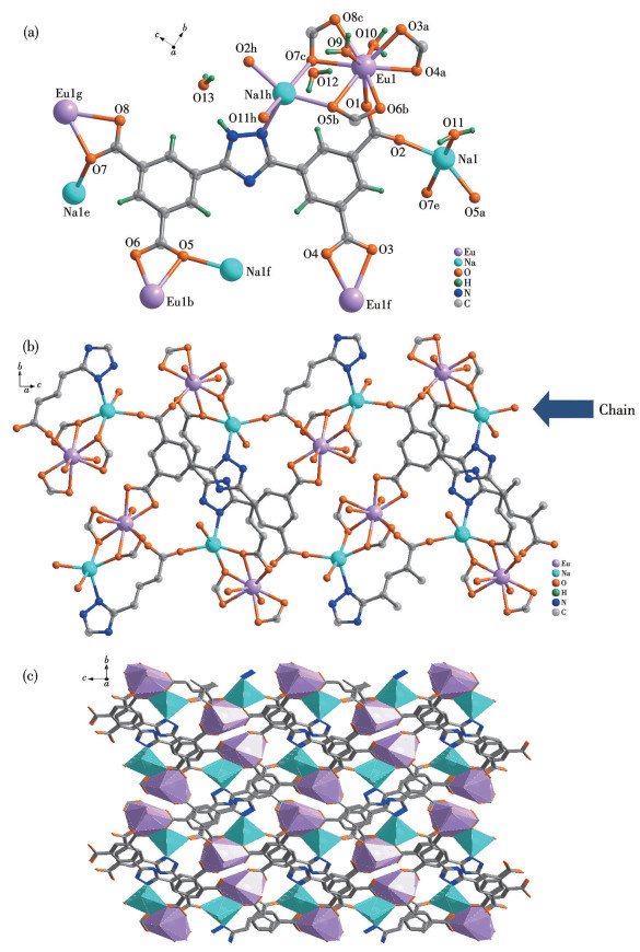
Single-crystal X-ray diffraction analysis reveals that the complex crystallizes in the monoclinic space group P21/n. The asymmetric unit contains one Eu3+ ion, one Na+ ion, one L4- ligand, three coordinated water molecules, and two free water molecules (Fig.1a). Each ligand coordinates to four Eu3+ ions and four Na+ ions.
Figure 1
Figure 1.
Crystal structure of Eu/Na-MOF: (a) basic unit and binary metal unit; (b) 2D chain units; (c) 3D microporous structure
Both Eu3+ and Na+ exhibit only one type of coordination geometry. The Eu3+ ion is nine-coordinated, bonded to seven oxygen atoms from four distinct ligands (O1, O3a, O4a, O5b, O6b, O7c, O8c) and two oxygen atoms from two water molecules (O9, O10). Among these, O3a and O4a, O5b and O6b, O7c and O8c are chelating oxygen atoms, while O1 serves as a monodentate bridging ligand. The Na+ ion is five- coordinated, linked to three oxygen atoms from three distinct ligands (O2, O5a, O7e), one nitrogen atom (N3d), and one oxygen atom from a water molecule (O11). Here, O2, O5a, and O7e act as monodentate bridging donors (Fig.1a)[26].
The Na+ and Eu3+ ions are connected via two bridging oxygen atoms (O5, O7), forming a binuclear metal unit. Notably, both O5 and O7 function as both bridging and chelating oxygen atoms (Fig.1a). The bond length ranges of Eu—O and Na—O are 0.231 3(5)-0.254 9(4) nm and 0.226 4(5)-0.250 5(7) nm, respectively. The bond angles ranges of O—Eu—O and O—Na—O are 51.63(14)°-158.62(18)° and 76.18(16)°-162.4(2)°, respectively. This is consistent with values reported in the literature[27]. Each pair of binuclear units is bridged by two oxygen atoms (O1, O2) from a carboxyl group of the ligand, forming an S-shaped 1D chain (Fig.1b). These chains are further extended by the ligands into a 2D layer (Fig.1b), which is ultimately connected through the ligands to construct a 3D microporous framework (Fig.1c).
2.2
Powder X-ray diffraction, IR, and thermogravimetry analysis
The powder X-ray diffraction (PXRD) pattern (working voltage: 40 kV, current: 50 mA, radiation source: Cu Kα, wavelength: 0.154 06 nm, scanning range: 5°-90°) of Eu/Na-MOF (Fig.S2) showed that the diffraction peaks aligned well with the simulated results, indicating high phase purity of the Eu/Na-MOF crystals. The intensity discrepancies among the peaks may be attributed to preferred crystal orientation.
Fig.S3 shows the infrared spectrum of Eu/Na-MOF powder in a range of 4 000-400 cm-1. The characteristic peak located at 3 575 cm-1 should be the O—H stretching vibration peak of free water, while the peak at 3 357 cm-1 might be the O—H or N—H stretching vibration peak of hydrogen bond association. Peaks at 1 449 and 1 414 cm-1 are attributed to in-plane C—H bending vibrations of the benzene ring, and the peak near 752 cm-1 is the C—H bending vibration peak of the benzene ring. No characteristic absorption peak for —COOH was observed around 1 700 cm-1. Instead, asymmetric and symmetric stretching vibrations of —COO- were present at 1 609, 1 565, and 1 379 cm-1, indicating successful coordination between the metal ions and the oxygen atoms of the carboxyl groups.
The thermogravimetry analysis (TGA) curve of Eu/Na-MOF (Fig.S4) exhibits two distinct weight-loss steps. The first step, occurring below 270 ℃ with a weight loss of 8.49%, corresponds to the removal of coordinated water molecules (Calcd. 8.68%). The second step, between 550 and 650 ℃, involved rapid weight loss due to the gradual collapse of the framework.
2.3
Fluorescence properties of Eu/Na-MOF
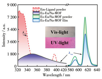
The fluorescence spectra of Eu-MOF powder, Eu/Na-MOF/PMMA film, and ligand powder immersed in aqueous solution (Fig.2) revealed distinct characteristic emission peaks of Eu3+ at 593 and 615 nm (λex=330 nm), attributed to the 5D0→7F1 and 5D0→7F2 transitions of Eu3+, respectively. The strongest emission peak at 615 nm, resulting from an induced electric dipole transition, produced bright red emission under UV light (Fig.2, inset). Notably, Eu/Na-MOF/PMMA retained the luminescent properties of Eu/Na-MOF, indicating that the film-forming technique fully preserves the structure of the complex.
Figure 2
Figure 2.
Fluorescence spectra of the sample
Inset: the photo of Eu/Na-MOF/PMMA film under visible light and ultraviolet light (365nm)
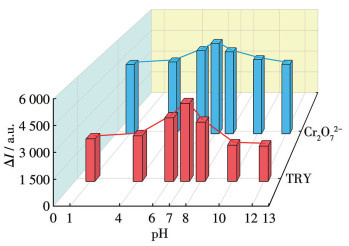
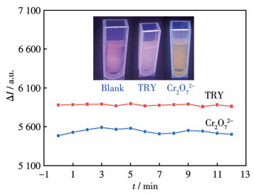
When aqueous solutions of TRY or Cr2O72- were applied to the Eu/Na-MOF/PMMA film, significant fluorescence quenching was observed (Fig.3, inset). To investigate the effect of pH on the sensing recognition of TRY (or Cr2O72-) by Eu/Na-MOF/PMMA, 1 mL of buffer solutions with different pH values (1, 4, 6, 7, 8, 10, 12) was added to 2 mL of the sensing system solutions prepared with HCl and NaOH respectively (Fig.3). The results showed that the variation value (ΔI) of the fluorescence intensity of the system first increased and then decreased with the increase of pH, reaching its maximum near pH=7. Therefore, all subsequent sensing experiments were performed in an aqueous medium (neutral conditions). The ΔI at different response times was also tested (Fig.4), and it was found that ΔI remained relatively stable within 20 min, indicating that sensing time had minimal impact on the detection results.
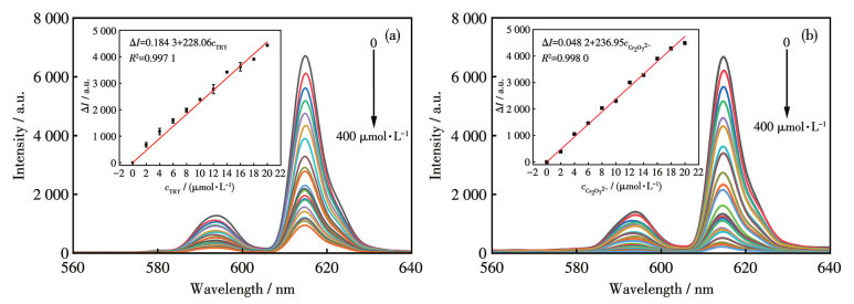
Furthermore, by varying the concentration of TRY (or Cr2O72-) solutions (0-400 μmol·L-1), it was observed that ΔI gradually increased with the concentration of the target analyte (Fig.5). In particular, within the concentration range of 0-20 μmol·L-1, ΔI showed a strong linear relationship with the analyte concentration: ΔI=0.184 31+228.055 97cTRY, R2=0.997 1; ΔI=0.048 15+236.954 2$ {c}_{\text{Cr}_{2}{\text{O}}_{7}^{2-}} $, R2=0.998 0.
Figure 5
Figure 5.
Fluorescence spectra of Eu/Na-MOF/PMMA in TRY (a) or Cr2O72- (b) solutions with different concentrations
Inset: the linear relation between ΔI and the analyte concentration.
The limit of detection (LOD) values were 1.36 μmol·L-1 (TRY) and 1.31 μmol·L-1 (Cr2O72-), respectively (using the LOD=3σ/k method, where σ is the standard deviation of the standard deviation of multiple measurements of the blank signal, and k is the slope). The results demonstrate that this method offers a lower detection limit and a simpler operational procedure.
2.5
Anti-interference performance in sensing TRY and Cr2O72-
The fluorescence intensity of Eu/Na-MOF/PMMA in 1 mL of 0.01 mol·L-1 solutions of different BAs or various potassium salts is shown in Fig.S5. The BAs included β-phenylethylamine (PEA), putrescine (PUT), cadaverine (CAD), tyramine (TA), spermidine (SPD), spermine (SPM), histamine (HA), and TRY. The potassium salts included Cr2O72-, SO32-, IO3-, I-, Cl-, NO3-, Ac-, CO32-, SO42-, SCN-, S2-, Br-, MnO4-. The results indicate that Eu/Na-MOF/PMMA exhibited a specific selective response toward TRY and Cr2O72-. The presence of other BAs does not interfere with TRY sensing (Fig.S5a). Similarly, the presence of other potassium salts does not affect the detection of Cr2O72- (Fig.S5b). These findings demonstrate that Eu/Na-MOF/PMMA is an excellent sensor for the detection of TRY and Cr2O72-[28].
2.6
Reusability of Eu-MOF/PMMA
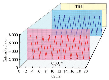
After exposure to TRY (or Cr2O72-), Eu/Na-MOF/PMMA can be easily regenerated by rinsing with distilled water. The fluorescence intensity remained stable even after 10 reuse cycles (Fig.6). Compared to traditional powdered MOF dispersion systems, this reusable fluorescent film significantly reduces consumable consumption and analytical costs. This advantage makes it particularly suitable for high-frequency detection scenarios, such as on-site environmental monitoring and rapid food screening, laying an important foundation for large-scale practical applications.
Figure 6
Figure 6.
Reusability of the Eu-MOF/PMMA sensor towards TRY and Cr2O72-
2.7
Investigation of fluorescence quenching mechanisms
Fluorescence quenching may arise from various mechanisms, including dynamic quenching, static quenching, fluorescence resonance energy transfer (FRET), and the inner filter effect (IFE)[29-33]. As illustrated in Fig.S6, the UV-Vis absorption spectra of TRY and Cr2O72- showed no significant overlap with the excitation or emission spectra of the Eu/Na-MOF/PMMA. This indicates that the quenching mechanism is not attributable to FRET or IFE. Upon the addition of TRY, the quenching constant of Eu/Na-MOF/PMMA increased with rising temperature (Fig.S7a). Moreover, no substantial changes in the position or intensity of the characteristic absorption peaks of Eu/Na-MOF/ PMMA were observed after TRY introduction. Based on these observations, and considering that static quenching involves the formation of a non-fluorescent ground-state complex between the quencher and the fluorophore, it is reasonable to conclude that the fluorescence quenching in this system is primarily governed by a static quenching mechanism. Similarly, when Cr2O72- was introduced, the quenching constant of Eu/Na-MOF/PMMA decreased with increasing temperature (Fig.S7b). Additionally, noticeable changes occurred in the position and intensity of the characteristic absorption peaks of Eu/Na-MOF/PMMA after the addition of Cr2O72-. These spectral responses align with the characteristics of dynamic quenching mechanisms. Therefore, it can be inferred that the fluorescence quenching in this system is mainly caused by dynamic quenching.
3.
Conclusions
In this study, a novel lanthanide-based metal- organic framework (Eu/Na-MOF) was successfully designed and synthesized. Its unique 3D crystal structure, high phase purity, good thermal stability, and remarkable fluorescence emission properties were confirmed by various characterization techniques, including single-crystal X-ray diffraction, PXRD, TGA, and fluorescence spectroscopy.
To address the challenges associated with powdered MOFs in liquid-phase sensing, such as poor solubility and difficult recovery, an innovative polymer embedding strategy was employed. The Eu/Na-MOF powder was uniformly dispersed and immobilized within a PMMA matrix, resulting in a flexible, washable, and easy-to-handle Eu/Na-MOF/PMMA composite fluorescent film. This film not only retained the excellent luminescent properties of the Eu/Na-MOF but also significantly improved the stability and practicality of the material.
Sensing performance tests demonstrated that the prepared composite film exhibits high sensitivity, excellent selectivity, rapid response, good stability, and reversibility toward both Cr2O72- and tryptamine. As a high-performance and practical fluorescent sensing platform, Eu/Na-MOF/PMMA shows great potential for the on-site and rapid detection of environmental pollutants and bioactive molecules, providing valuable insights for the development of novel fluorescent film-based sensors.
Supporting information is available at http://www.wjhxxb.cn
[1]
YANG Y J, LIU D, LI Y H, CUI G H. Two water-stable Zn(Ⅱ)-based MOFs as highly selective luminescent probe for the dual detection of glyoxal and dichromate ions in aqueous solution[J]. J. Solid. State Chem., 2019, 278: 243-256
[2]
CHEN C, WANG X M, ZHANG Y F, LI X Y, GAO H J, WATRRHOUSE G I N, QIAO X G, XU Z X. A molecularly-imprinted SERS sensor based on a TiO2@Ag substrate for the selective capture and sensitive detection of tryptamine in foods[J]. Food Chem., 2022, 394: 133536 doi: 10.1016/j.foodchem.2022.133536
[3]
MARYŠKA M, FOJTÍKOVÁ L, JUROK R, HOLUBOVÁ B, LAPČÍK O, KUCHAŘ M. Use of novel haptens in the production of antibodies for the detection of tryptamines[J]. RSC. Adv., 2018, 8(29): 16243-16250 doi: 10.1039/C8RA02528B
[4]
ŠČAVNIČAR A, ROGELJ I, KOČAR D, KÖSE S, POMPE M. Determination of biogenic amines in cheese by ion chromatography with tandem mass spectrometry detection[J]. J. AOAC Int., 2018, 101(5): 1542-1547 doi: 10.5740/jaoacint.16-0006
[5]
SHI Y, WANG R J, YUAN S, QIANG H S, SHEN M, SHEN B H, DRUMMER O H, YU Z G, ZHAO Y L, XIANG P. UHPLC-MS/MS method for simultaneously detecting 16 tryptamines and their metabolites in human hair and applications to real forensics cases[J]. J. Chromatogr. B, 2020, 1159: 122392
[6]
GIL R L, AMORIM C G, MONTENEGRO M C B S M, ARAÚJO A N. Determination of biogenic amines in tomato by ion-pair chromatography coupled to an amine-selective potentiometric detector[J]. Electrochim. Acta, 2021, 378: 138134 doi: 10.1016/j.electacta.2021.138134
[7]
MUNIR M A, RAHMAWATI F, JAMAL J A, IBRAHIM S, SAID M M, AHAMID M S. Inspecting histamine isolated from fish through a highly selective molecularly imprinted electrochemical sensor approach[J]. ACS Omega, 2023, 8: 13352-13361 doi: 10.1021/acsomega.3c00768
[8]
CHAKRABORTY S, PAUL S, ROY P, RAYALU S. Detection of cyanide ion by chemosensing and fluorosensing technology[J]. Inorg. Chem. Commun., 2021, 128: 108562 doi: 10.1016/j.inoche.2021.108562
[9]
YAN Z, CAI Y, ZHANG J, ZHAO Y. Fluorescent sensor arrays for metal ions detection: A review[J]. Measurement, 2022, 187: 110355 doi: 10.1016/j.measurement.2021.110355
[10]
HE H Y, SUN D W, WU Z H, PU H B, WEI Q Y. On-off-on fluorescent nanosensing: Materials, detection strategies and recent food applications[J]. Trends Food Sci., 2022, 119: 243-256 doi: 10.1016/j.tifs.2021.11.029
[11]
GAN Y L, HUANG K R, LI Y G, QIN D P, ZHANG D M, ZONG Z A, CUI L S. Synthesis, structure and fluorescent sensing for nitrobenzene of a Zn-based MOF[J]. J. Mol. Struct., 2021, 1223: 129217
[12]
KHAILI I E, FONESA J, REITHOFER M R, EDER T, CHIN J M. Tackling orientation of metal-organic frameworks (MOFs): The quest to enhance MOF performance[J]. Coord. Chem. Rev., 2023, 481: 215043 doi: 10.1016/j.ccr.2023.215043
[13]
YAN R K, CHEN X L, REN J, CUI H L, YANG H, WANG J J. Synthesis of highly sensitive and multi-response Eu-MOF, fluorescence sensing properties and anti-counterfeiting applications[J]. Spectroc. Acta Pt. A‒Molec. Biomolec. Spectr., 2024, 322: 124855 doi: 10.1016/j.saa.2024.124855
[14]
SUN T C, WANG P, FAN R Q, CHEN W, HAO S, YANG Y L. Functional microscale single-phase white emission lanthanide MOF for tunable fluorescent sensing and water quality monitoring[J]. J. Mater. Chem., 2019, 7: 3598-3606
[15]
WU Y P, XU G W, DONG W W, ZHAO J, LI D S, ZHANG J, BU X H. Anionic lanthanide MOFs as a platform for iron-selective sensing, systematic color tuning, and efficient nanoparticle catalysis[J]. Inorg. Chem., 2017, 56(3): 1402-1411 doi: 10.1021/acs.inorgchem.6b02476
[16]
LIU S H, HUANG Y F, CUI S C, WANG X X, ZHANG Y F, DENG P Y. Efficient and ultra-stable Zr-MOF membranes for photocatalysis: Synergistic influence of Pt and lattice defects[J]. Int. J. Hydrog. Energy, 2025, 145: 129-138 doi: 10.1016/j.ijhydene.2025.06.076
[17]
LI L L, XIANG Y Y, YANG W F, LIU Z L, CAI M R, MA Z F, WEI Q B, PEI X W, YU B, ZHOU F. Embedded polyzwitterionic brush-modified nanofibrous membrane through subsurface-initiated polymerization for highly efficient and durable oil water separation[J]. J. Colloid Interface Sci., 2020, 575: 388-398 doi: 10.1016/j.jcis.2020.04.117
[18]
ZHANG P F, RAJABZADEH S, VENAULT A, WANG S Y, SHEN Q, JIA Y D, FANG C J, KATO N, CHANG Y, MATSUYAMA H. One-step entrapment of a PS-PEGMA amphiphilic copolymer on the outer surface of a hollow fiber membrane via TIPS process using triple-orifice spinneret[J]. J. Membr. Sci., 2021, 638: 119712 doi: 10.1016/j.memsci.2021.119712
[19]
CĂPRĂRESCU S, TIHAN G T, ZGÂRIAN R G, GRUMEZESCU A M, LAZAU C, BANDAS C, ATANASE L L, NICOLAE C A. Synthesis and characterization of cellulose acetate/polyethylene glycol/poly(styrene)-b-poly(4-vinylpyridine) membrane embedded with hydrothermally activated TiO2 nanoparticles for waste-waters treatment by membrane processes[J]. Polymers, 2025, 17(4): 446 doi: 10.3390/polym17040446
[20]
CHE H C, LI Y, TIAN X K, YANG C, LIU L Q, NIE Y L. A versatile logic detector and fluorescent film based on Eu-based MOF for swift detection of formaldehyde in solutions and gas phase[J]. J. Hazard. Mater., 2020, 410(129): 124624
[21]
XU N, TANG Z H, JIANG Y P, FANG J L, ZHANG L, LAI X F, SUN Q J, FAN J M, TANG X G, LIU Q X, JIAN J K. Highly sensitive ratiometric fluorescent flexible sensor based on the RhB@ZIF-8@PVDF mixed-matrix membrane for broad-spectrum antibiotic detection[J]. ACS Appl. Mater. Interfaces, 2023, 15: 52993-53002
[22]
LI Q Y, LI Y A, GUAN Q, LI W Y, DONG X J, DONG Y B. UiO-68-PT MOF-based sensor and its mixed matrix membrane for detection of HClO in water[J]. Inorg. Chem., 2019, 58: 9890-9896 doi: 10.1021/acs.inorgchem.9b01032
[23]
CHEN W, FAN R Q, FAN J Z, LIU H Y, SUN T C, WANG P, YANG Y L. Lanthanide coordination polymer-based composite films for selective and highly sensitive detection of Cr2O72- in aqueous media[J]. Inorg. Chem., 2019, 58: 15118-15125 doi: 10.1021/acs.inorgchem.9b01841
[24]
CHAI H M, WEI Y Y, SUN X H, BAI W Q, REN Y X, GAO L J. Syntheses, structures of three Ln-MOFs (Ln=La, Ce, Dy), fluorescent sensing Fe3+ and MnO4- of La-MOF film, and magnetic properties of Dy-MOF[J]. J. Mol. Struct., 2023, 1296: 136894
[25]
LI Y, SUN X H, CHAI H M, BAI W Q, REN Y X, GAO L J, ZHANG G Q, ZHANG J. Synthesis, structure of a new bimetallic-organic framework film sensor and fluorescence detection of histamine and metal ions[J]. J. Mol. Struct., 2024, 1321: 139694
[26]
ZHONG W B, LI R X, LV J, HE T, XU M M, WANG B, XIE L H, LI J R. Two isomeric In(Ⅲ)-MOFs: Unexpected stability difference and selective fluorescence detection of fluoroquinolone antibiotics in water[J]. Inorg. Chem. Front., 2020, 7: 1161-1171 doi: 10.1039/C9QI01490J
[27]
GAO L J, JIAO C X, CHAI H M, REN Y X, ZHANG G Q, YU H, TANG L. A highly sensitive multifunctional Eu-MOF sensor with pentacarboxylate for fluorescence detecting acetone, Cu2+ and Cr2O72-, and electrochemical detection of TNP[J]. J. Solid State Chem., 2020, 284: 121199 doi: 10.1016/j.jssc.2020.121199
[28]
WANG B, LV X L, FENG D W, XIE L H, ZHANG J, LI M, XIE Y B, LI J R, ZHOU H C. Highly stable Zr(Ⅳ)-based metal-organic frameworks for the detection and removal of antibiotics and organic explosives in water[J] J. Am. Chem. Soc., 2016, 138: 6204-6216 doi: 10.1021/jacs.6b01663
[29]
YANG Y, CHEN Z H, FU C Y, KUMAR S, SHI W, SUN F Y, YANG X M, REN P. Selective and rapid detection of 4-nitrophenol in river and treated industrial wastewater by a luminescent lanthanide metal-organic framework sensor[J]. Inorg. Chem., 2023, 48: 19565-19572
[30]
SONG X M, HOU X F, ZHAO Q X, MA Z H, REN Y X. Fluorescence-quenching mechanisms of novel isomorphic Zn/Cd coordination polymers for selective nitrobenzene detection[J]. Spectroc. Acta Pt. A‒Molec. Biomolec. Spectr., 2024, 308: 123729 doi: 10.1016/j.saa.2023.123729
[31]
MUBEEN M, KHALID M A, MUKHTAR M, SUMREEN P, TABASSUM M, ASHIQ S, ABBAS S A, AKRAM R, LQBAL A. Elucidating the mechanism of copper-induced photoluminescence quenching in 2-phenylbenzimidazole-5-sulfonic acid[J]. J. Fluoresc., 2025, 35: 2957-2962
[32]
DONG X P, QI H X, ZHAI Z Z, LI W Q, ZHANG P D. Probing the fluorescence quenching mechanism of N-doped carbon quantum dots by inorganic ions[J]. Microchem. J., 2024, 197: 109854 doi: 10.1016/j.microc.2023.109854
[33]
WANG H, LUSTIG W P, LI J. Sensing and capture of toxic and hazardous gases and vapors by metal-organic frameworks[J]. Chem. Soc. Rev., 2018, 47: 4729-4756 doi: 10.1039/C7CS00885F
Figure 1
Crystal structure of Eu/Na-MOF: (a) basic unit and binary metal unit; (b) 2D chain units; (c) 3D microporous structure

 下载:
下载:

 下载:
下载:


