Figure 1.
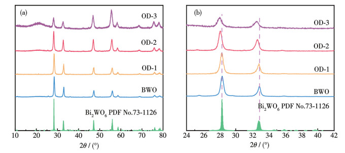
XRD patterns of BWO, OD-1, OD-2, and OD-3
In-situ generated ordered-disordered Bi2WO6 homojunction for enhanced photocatalytic nitrogen fixation performance
Lulu ZHANG , Yahui HOU , Yunfei LU , Rui LI , Jianxin LIU

Since Schrauzer and Guth first reported the light-driven reaction in 1977[1], the technology of photocatalytic nitrogen (N2) fixation has attracted extensive focus from both academic and industrial communities, as it has the potential to replace the industrial process, which operates under harsh conditions (300-550 ℃, 15-25 MPa)[2]. However, the efficiency of this technology is still far from meeting the requirements for practical application. The core bottleneck lies in the difficulty of activating and cleaving the highly stable N≡N bond, which has a bond energy as high as 941 kJ·mol-1 [3-4]. To overcome this limitation, it is necessary to introduce an auxiliary electron-donor center as the initial active site. By capturing molecular N2 and weakening the N≡N bond energy, conditions capable of enabling the photogenerated electrons injection and subsequent reduction reactions are formed. Therefore, the development of a photocatalyst with highly efficient N2 activation active sites has become an urgent need in current research.
According to the differences in crystallinity, solid-state nanomaterials can be classified into three categories: crystals, semi-crystals, and amorphous materials[5]. Different from the long-range ordered atomic arrangements in crystals and semi-crystals, amorphous materials exhibit only short-range ordered but long-range disordered atomic arrangements. Disordered regulation engineering is a novel and effective modification strategy. Relevant research results show that by introducing disordered state defects into the catalyst, an ordered/disordered structure coexistence of disordered semiconductor catalysts can be constructed without introducing external molecules or ions. This enables the regulation of semiconductor band characteristics and photoelectric properties. On one hand, the disordered component can act as an electron acceptor and capture electrons during light excitation, effectively inhibiting the recombination of photogenerated holes and electrons. On the other hand, the disordered structure enhances the visible light response ability of the catalyst, thereby enhancing the photocatalytic activity[6-7]. Due to the unique atomic arrangement with short-range order and long-range disorder and the large number of "dangling bonds" in the amorphous phase[8-9], it usually has more catalytically active sites and shows significant advantages in the catalytic activation of reactant molecules. In fact, research has confirmed that the catalytic performance of amorphous disordered materials is often superior to that of their crystalline counterparts. More importantly, amorphous materials have demonstrated excellent performance in the processes of thermal catalysis and electrocatalytic N2 activation[10-11]. However, there are essential differences between photocatalytic systems and thermal/electrocatalytic systems. Early research suggested that amorphous semiconductors were often regarded as inert or low-activity materials because the lack of long-range atomic order and the presence of many defects were unfavorable for photogenerated electron-hole separation[12]. So, how to improve the electron-hole separation efficiency while retaining the abundant catalytically active centers of amorphous disordered materials has evolved into a key challenge in the studies and development of amorphous disordered photocatalytic materials.
Homojunction refers to an interface junction formed by the same semiconductor material through different construction methods, such as the same semiconductor with different doping types, the same semiconductor with different crystal facets exposed, or the construction of disordered structures by regulating the crystallinity of the material[13]. When the two parts of the semiconductors constituting the junction are in close contact, charge redistribution occurs at the interface due to the difference in carrier concentration, thereby forming a built-in electric field; at the same time, the consistent elemental composition on both sides of the interface can achieve high lattice matching, effectively reducing the resistance of carrier migration. The combined effect of these characteristics can significantly improve the separation efficiency of photogenerated electron-hole pairs. By regulating the carrier behavior near the junction interface, it can provide favorable conditions for chemical reactions on the catalyst surface, ultimately promoting the improvement of catalytic performance.
Bi2WO6 has an appropriate energy band position, a layered structure, excellent optical properties, high thermal excitation conductivity, and outstanding photocatalytic ability[14]. Based on this, we used amorphous Bi2WO6 as a N2 activation center and proposed a strategy for constructing ordered-disordered homojunction Bi2WO6 to achieve synergistic optimization of efficient N2 activation and electron-hole separation. Experimental results showed that compared with the inactive crystalline Bi2WO6, the photocatalytic N2 fixation activity of the ordered-disordered homojunction Bi2WO6 was significantly enhanced. The enhancement mechanism is mainly attributed to: (1) amorphous Bi2WO6 can effectively capture and activate molecular N2; (2) the ordered-disordered homojunction structure significantly improves the carrier separation efficiency.
Sodium tungstate dihydrate (Na2WO4·2H2O) and glyoxal (C2H2O2) were obtained from Shanghai Aladdin Biotechnology Co., Ltd. Bismuth nitrate pentahydrate (Bi(NO3)3·5H2O), ethylene glycol (EG) were obtained from Sinopharm Group Chemical Reagent Co., Ltd. Ethanol (C2H5OH) was bought from Tianjin Guangfu Technology Development Co., Ltd.
The crystalline Bi2WO6 was prepared using a one-step hydrothermal method in a stainless-steel pressure reactor lined with Teflon. The specific steps are as follows: first, 0.97 g of Bi(NO3)3·5H2O was added to 15 mL of deionized water, following which the mixture was sonicated for 10 min to form solution A. Simultaneously, 0.33 g of Na2WO4·2H2O was completely dissolved in 15 mL of deionized water, resulting in the formation of solution B. At room temperature, solution B was gradually added to solution A while continuously stirring for 0.5 h. The mixture was subsequently moved into a 50 mL Teflon-lined stainless steel pressure reactor and heated to 180 ℃ for 12 h. After cooling naturally, the sample was washed multiple times with absolute ethanol and deionized water, and then dried at 60 ℃ for 16 h to obtain Bi2WO6 powder, which was named as BWO.
Ordered-disordered homojunction Bi2WO6 was prepared using EG as the reducing agent in a stainless steel. The specific steps are as follows: different volumes (1.0, 1.5, and 2.0 mL) of EG were added to a mixture of 0.97 g of Bi(NO3)3·5H2O and 15.0 mL of pure water. Ultrasonication was then performed for 10 min to form solution A. The remaining steps were the same as those for the preparation of BWO mentioned above. The resulting photocatalysts were named ordered-disordered Bi2WO6-1 (OD-1), ordered-disordered Bi2WO6-2 (OD-2), and ordered-disordered Bi2WO6-3 (OD-3) when the volumes of EG added were 1, 1.5, and 2.0 mL, respectively.
The crystal structure and composition of the samples were analyzed using X-ray diffraction (XRD) on a Rigaku D/MAX-2500 with Cu Kα radiation (λ=0.154 nm) at 30 mA and 40 kV, and the scanning range of 10° to 80°. The morphological structures and sizes of the catalysts were studied using a scanning electron microscope (SEM, Nanosem 430) and a transmission electron microscope (TEM, JEOL-2011) with an accelerating voltage of 200 kV. The composition and oxidation state of the catalyst elements were analyzed using X-ray photoelectron spectroscopy (XPS, Thermo Fisher Escalab 250Xi, with an Al Kα X-ray source). The catalysts′ microstructure was evaluated using Fourier transform infrared spectroscopy (FTIR, Bruker TENSORII FTIR) and Raman spectroscopy (HORIBA). Photoluminescence (PL) spectra and transient photoluminescence (TRPL) spectra were measured using a British Edinburgh Instruments FLS980 full-function steady-state fluorescence spectrometer. Electrochemical impedance spectroscopy (EIS) was used to characterize the recombination rate of photogenerated electron-hole pairs in the catalyst on the Shanghai Chenhua CHI660E electrochemical workstation. Ultravioletvisible diffuse reflectance spectroscopy (UV-Vis DRS) was conducted on the catalyst using an American Micromeritics ASAP 2020 fully automatic surface area and pore size analyzer for long-term adsorption-desorption tests. N2 temperature-programmed desorption (N2-TPD) test was performed by a automatic chemical adsorption instrument. In-situ diffuse reflectance-Fourier transform infrared technique (DR-FTIR) spectra were recorded by a Bruker Tensor 2 spectrometer.
The photocatalytic N2 fixation to synthesize ammonia (NH4+) performance was tested using a designed closed quartz glass reactor at ambient temperature and pressure. Simulated solar light was provided by an Xe lamp (300 W, PLS-SXE300/300UV, Beijing Perfect Light Technology Co., Ltd.). Typically, 50 mg of the sample and 100 mL of pure water were added to a custom reactor, followed by 10 min of ultrasonic treatment. Then, N2 was introduced into the reactor at a flow rate of 120 mL·min-1, with the aeration maintained for 30 min. Prior to conducting the formal N2 fixation test, a blank experiment was conducted in the dark to eliminate interference. Subsequently, the Xe lamp was illuminated under the full-spectrum condition for 1 h. Finally, 10 mL of the solution in the reactor was filtered through a filter, and the NH4+ concentration was quantified through the combination of Nessler′s reagent and an ultraviolet-visible spectrophotometer (Fig.S1, Supporting information).
The crystallinity and phase composition of the synthesized samples were characterized by XRD. As illustrated in Fig.1a, the diffraction peaks of the BWO, OD-1, OD-2, and OD-3 samples all matched well with the standard card of Bi2WO6 (PDF No.73-1126) and no other impurity diffraction peaks were detected[15], confirming that the pure-phase Bi2WO6 catalyst was successfully synthesized. The enlarged XRD patterns in the 2θ range of 24°-42° (Fig.1b) revealed that, as the amount of EG increased, the diffraction peaks shifted toward lower angles and the full width at half maximum increased slightly. This phenomenon is attributed to the change of crystal compressive stress state and lattice distortion, indicating the presence of the disordered structure[16].
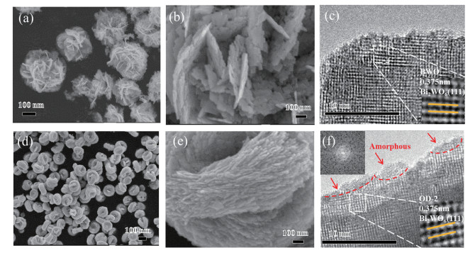
SEM and TEM were employed to observe the morphological features of BWO and OD-2. From Fig. 2a and 2b, BWO was a flower-shaped microsphere with a 2D interlaced nanosheet structure (average particle size of 3-4 μm). OD-2 (Fig. 2d and 2e) had a flat-shaped persimmon-like morphology, and its particle size was significantly smaller compared to BWO, which aligns with the findings from XRD. Further measurements of interplanar spacing of the catalysts revealed that the interplanar spacing of both BWO and OD-2 was 0.375 nm, which is associated with the (111) plane of Bi2WO6, indicating that EG addition only changed the morphology of the catalyst without affecting its crystal planes. Additionally, observations from the TEM and high-resolution TEM (HRTEM) images of BWO revealed that the BWO nanocrystals exhibited a higher degree of crystallinity (Fig. 2c). It is noteworthy that when a certain amount of EG was added during the preparation of its precursor, a small amount of disordered structure appeared on Bi2WO6 crystal surfaces (Fig.2f), with a thickness of approximately 1 nm[17]. This indicates that the addition of EG causes the originally well-crystallized catalyst to gradually undergo a transformation towards disorder.
To verify the structural composition of the amorphous state of OD-2, detection methods sensitive to local (or short-range) structures, such as XPS, FTIR, and Raman, were employed to analyze the microstructures of BWO and OD-2. The XPS survey spectra in Fig.S2 indicated that both BWO and OD-2 were composed of W, Bi, O, and trace C elements (the C element comes from the non-uniform carbon on the sample surface)[18]. Notably, the high-resolution XPS spectra of Bi4f, W4f, and O1s for BWO and OD-2 (Fig.S2b-S2d) showed similar characteristics, further confirming that the composition of the surface disordered structure of OD-2 was Bi2WO6, and indicating the formation of the ordered-disordered Bi2WO6 homojunction. The high-resolution Bi4f XPS spectrum presented in Fig. S2b exhibited characteristic peaks at 159.09 and 158.82 eV (assigned to Bi4f7/2) as well as at 164.41 and 164.14 eV (corresponding to Bi4f5/2)[19]. Such peak distributions indicate the presence of Bi3+ in both samples[20]. As illustrated in Fig. S2c, the high-resolution W4f XPS spectra of BWO and OD-2 exhibited four characteristic peaks: at 37.46 and 37.19 eV (assigned to W4f7/2) and at 35.33 and 35.06 eV (assigned to W4f5/2). Notably, all these peaks are representative of W6+ and W5+ present in the WO6 octahedron[21]. The high-resolution O1s XPS spectrum was deconvoluted into three distinct peaks (Fig. S2d): the characteristic peaks at 529.80 and 529.64 eV, 530.59 and 530.54 eV are attributed to the [Bi2O2]2+ and [WO4]2- lattice oxygen of Bi2WO6, respectively, while those at 532.29 and 531.38 eV belong to the characteristic peaks of adsorbed oxygen, indicating that adsorbed oxygen exists as OH on the catalyst surface[16,22].
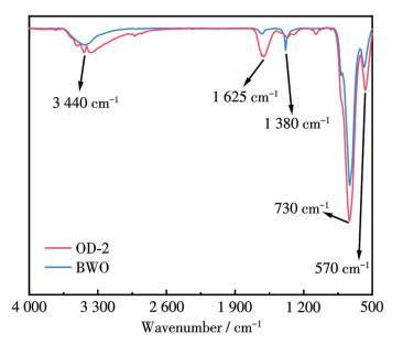
FTIR was also conducted to examine the surface microstructures of BWO and OD-2. As shown in Fig.3, BWO and OD-2 exhibited similar FTIR spectra, which indirectly indicates that the composition of the disordered structure on the OD-2 surface is neither WO3[23] nor Bi2O3[24]. Compared with BWO, OD-2 still retained distinct characteristic peaks of Bi—O stretching vibration and W—O asymmetric stretching vibration[25], corresponding to the sharp peaks around 570 and 730 cm-1, respectively; while the peaks observed at 1 625 and 3 440 cm-1 are associated with the H—OH vibration. Fig. 4 presents the Raman spectra of BWO and OD-2, which also exhibited similarities. Among them, the typical characteristic peaks observed in the 250-350 cm-1 range (Fig. 4a) are assigned to the bending vibration of WO6, as well as to the stretching and bending vibrations of [BiO6] polyhedra[26-27]. The peak near 790 cm-1 in Fig. 4b is associated with the symmetric vibration of the O—W—O, while the peak located near 830 cm-1 is attributed to the asymmetric vibration of the O—W—O. Additionally, the signal at 710 cm-1 can be assigned to the antisymmetric bridging mode of the tungstate chain. In summary, the structural features revealed in XPS, FTIR, and Raman spectra confirm that the amorphous thin layer in the disordered structure on the catalyst surface is an amorphous Bi2WO6 with short-range atomic order and long-range atomic disorder. This result provides clear evidence for the successful fabrication of the ordered-disordered Bi2WO6 homojunction.
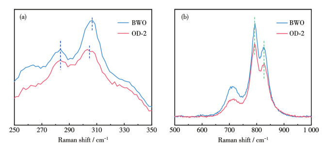
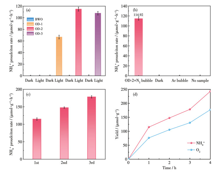
Fig. 5a shows the photocatalytic N2 fixation performance of the prepared samples. No generation of NH4+ was detected without light irradiation for any of the samples, indicating the importance of light in N2 fixation. However, BWO showed no N2 fixation activity even under light irradiation, while all ordered-disordered Bi2WO6 homojunction catalysts (OD-1, OD-2, OD-3) exhibited high-efficiency photocatalytic N2 fixation activity. Among them, OD-2 demonstrated the best N2 fixation performance, reaching a NH4+ production rate of 114.92 μmol·g-1·h-1. A control experiment was conducted to eliminate environmental interference; the results are shown in Fig. 5b. The results indicated that the NH4+ production rate was negligible in the absence of samples, in the dark, or under an Ar atmosphere. Prepared samples were also analyzed via the indophenol blue method correlation. The N2 fixation rates were determined by quantifying the amount of NH4+ generated during the reaction, with the results being 52.37, 101.09, and 88.54 μmol·g-1·h-1 for OD-1, OD-2, and OD-3, respectively (Fig.S3-S4). It is important to note that these values were the average values of three parallel measurements, which ensures the reproducibility and reliability of the activity data. Notably, the N2 fixation activity results obtained in this test showed good consistency with those derived from the Nessler′s reagent method described earlier. The above results suggest that the amorphous layer on the surface of OD-2 is of great significance to the photocatalytic reaction. The continuous NH4+ production test for OD-2 is illustrated in Fig. 5c. O2 can be simultaneously detected during the N2 fixation process, and the amount of produced O2 was close to three-quarters that of NH4+, which agrees with the theoretical value (Fig. 5d). This result further proves that NH3 is generated by coupling the activated N2 with the protons in water. The cycling tests were also used to evaluate the stability of OD-2; it exhibited no obvious activity loss after four cycles, with each run for 1 h (Fig. S5). The results indicate that under full-light irradiation, OD-2 with a disordered surface structure can produce NH4+ stably in an aqueous solution for up to 3 h, reflecting its excellent stability. Fig. S6 shows the crystal structure of OD-2 after the continuous NH4+ production test. There was no significant difference between the crystal structures of the fresh and used samples, which further confirms the stability of OD-2 during the photocatalytic N2 fixation process.
PL, EIS, and TRPL tests were performed to explore the carrier separation and transfer characteristics of the prepared OD-2 and BWO samples. Generally, photocatalysts with high charge separation efficiency exhibited lower PL emission intensity[28]. As shown in Fig. S7a, the PL emission intensity of OD-2 was significantly lower than BWO, confirming a higher efficiency of photogenerated charge separation in OD-2. The EIS Nyquist plots for OD-2 and BWO are displayed in Fig. S7b. A notable feature in the high-frequency range was that OD-2 exhibited a smaller Nyquist arc radius compared to BWO. This observation points to both a faster interface charge transfer rate and more efficient electron-hole pair separation in OD-2[18].
TRPL decay was used to explore the lifetime of the photogenerated carriers in the samples, as shown in Fig.S7c and Table S1. The average decay time of OD-2 was 0.74 ns, lower than BWO (1.00 ns). This shorter lifetime indicates that the order-disorder interface in OD-2 effectively drives the directional transfer of photogenerated electrons: the built-in electric field at the interface promotes electron migration from the ordered region to the disordered region, while defect sites in the disordered region further trap these electrons temporarily——accelerating the separation of photogenerated electron-hole pairs and reducing the probability of radiative/non-radiative recombination[29]. Based on these results, it can be concluded that the surface disordered structures effectively boost carrier separation efficiency. This accelerates the migration of photogenerated carriers from ordered to disordered regions and suppresses carrier recombination, thereby boosting the N2 fixation performance[30].
To further clarify the correlation between the order-disorder interface structure and charge transfer behavior, OD-2 (order-disorder homojunction) exhibited a more optimized interface than BWO (pure ordered structure)——the consistent elemental composition of the order-disorder interface ensures high lattice matching, which reduces the resistance of carrier migration across the interface. Additionally, the built-in electric field formed at the order-disorder interface (driven by carrier concentration differences between ordered and disordered regions) promotes the directional transfer of photogenerated electrons from the ordered region to the disordered region and holes in the opposite direction. This structural advantage directly contributes to the lower PL intensity (weaker recombination) and smaller EIS arc radius (faster transfer) of OD-2 compared to BWO.
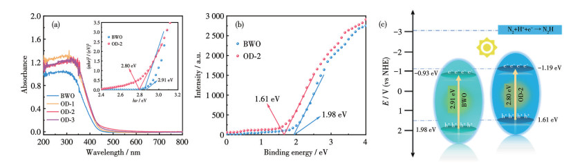
The energy band structure of the synthesized OD-2 and BWO samples was studied by UV-Vis DRS. Fig.6a demonstrates that the absorption edges for OD-2 and BWO were observed at approximately 430 and 438 nm, respectively. The band gap was calculated using the formula αhν=A(hν-Eg)n/2, where A represents a constant, Eg denotes the band gap, α stands for the absorption coefficient, and hν signifies the light frequency. The selection of n value depends on the type of material: n=1 for direct band gap materials, while n=4 for indirect semiconductors. Since Bi2WO6 is a typical indirect band gap semiconductor, n=4 was used here[31]. The band gaps of OD-2 and BWO were calculated as 2.80 and 2.91 eV, respectively, from the tangent line in the inset of Fig.6a. Furthermore, the valence band (VB) energy (EVB) was characterized by the VB-XPS, as shown in Fig. 6b. It showed that the VB of OD-2 and BWO were 1.61 and 1.98 eV, respectively. The conduction band (CB) energies (ECB) of BWO and OD-2 were calculated from the measured band gaps and VB energies using the equation ECB=EVB-Eg, yielding values of -0.93 and -1.19 eV, respectively. Compared with BWO, the CB energy of OD-2 (-1.19 eV) was more negative, indicating its stronger reduction ability; meanwhile, the narrowing of the band gap enhanced its light capture ability, and the introduced surface disorder was found to substantially boost the activity for photocatalytic N2 fixation[17]. Based on the above results, the energy band structure diagram of OD-2 and BWO is plotted in Fig.6c.
Adsorption and activation of N2 are key steps in the photocatalytic N2 fixation process. To elucidate this phenomenon, N2-TPD was employed to examine the adsorption characteristics of N2 on the catalyst surface. As illustrated in Fig.7a, the desorption peak near 90 ℃ corresponds to the physical adsorption of N2, whereas the desorption peaks of BWO and OD-2 within the 250-350 ℃ range are associated with the chemical adsorption of N2[32]. It is worth noting that an obvious desorption peak appeared at 285 ℃ for OD-2, which is attributed to the disordered structure on its surface exposing more active sites, thereby enhancing the chemical adsorption capacity for N2 and facilitating the activation of the adsorbed N2 gas in the photocatalytic reaction.
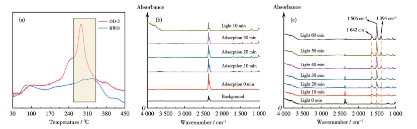
To directly observe the N2 fixation reaction process on the ordered-disordered Bi2WO6 homojunction interface, the in-situ DR-FTIR was employed. As shown in Fig. 7b, 7c, and S8, the infrared spectra of BWO under full light irradiation showed no significant difference compared to those after N2 and H2O adsorption, indicating that the light did not have a remarkable effect on the N2 fixation reaction of BWO. In contrast, after full light irradiation of OD-2 (Fig.7c), the intensities of multiple absorption peaks gradually increased with the light irradiation time (0-60 min). Among them, the band at 1 394 cm-1 is associated with the characteristic absorption of NH4+, the sharp band at 1 506 cm-1 is associated with the bending vibration of N—H, and the band at 1 624 cm-1 is associated with the chemisorbed N2[33]. Notably, N2 is undetectable in conventional FTIR spectroscopy owing to its high N≡N bond symmetry and low dipole moment, whereas the disordered surface structure of OD-2 can perturb the bond symmetry and electronic configuration of the N≡N bond. Consequently, the signal intensity of chemisorbed nitrogen species in the in-situ DR-FTIR spectra was observed to gradually intensify with prolonged light irradiation time. This result strongly proves that the disordered structure on the surface can continuously weaken the N≡N bond until it is finally hydrogenated to form NH4+ [34].
Through a series of N2 fixation activity experiments and characterization analyses, it can be concluded that introducing disordered structures into ordered catalysts can effectively enhance the photocatalytic N2 fixation activity. This is attributed, on the one hand, to the introduction of disordered structures reducing the band gap, and compared with BWO, the CB potential of OD-2 is significantly higher, indicating that the presence of disordered structures improves the catalyst′s reduction ability. Moreover, the electrons in the crystal ordered structure are more likely to transfer to the disordered structure on the surface, which is extremely beneficial for the activation of N2. Furthermore, the formation of special ordered-disordered homojunctions effectively suppresses the recombination of electron-hole pairs. Here, a possible photocatalytic N2 fixation mechanism for the ordered-disordered Bi2WO6 homojunction photocatalyst was proposed: when exposed to simulated sunlight, electrons in the VB of ordered Bi2WO6 are excited by photons of sufficient energy and transition to the CB, then the interface electrons transfer to its surface disordered structure, reacting with the N2 adsorbed on the disordered structure to achieve the reduction of N2 to NH4+. Conversely, the holes remain within the ordered structure of Bi2WO6 and participate in the oxidation of H2O, converting it into O2. In contrast, Bi2WO6, with only a pure ordered structure, lacks the surface disordered structure that can activate the N2 bond, making it difficult to overcome the N≡N bond bottleneck and thus lacking photocatalytic N2 fixation activity.
In summary, an ordered-disordered Bi2WO6 homojunction photocatalyst was fabricated by a one-step hydrothermal method, and its optimal N2 fixation rate could reach 114.92 μmol·g-1·h-1. Based on mechanism studies using HRTEM, FTIR, Raman, and in-situ DR-FTIR, it was clarified that the disordered structure on the surface of OD-2 could promote the adsorption and activation of N2, while the special ordered-disordered homojunction structure was conducive to the transfer of interface electrons to the disordered zone, while holes remained confined in the ordered zone. Such a process markedly suppresses the recombination of photogenerated charge carriers, thus effectively boosting the N2 fixation performance of the Bi2WO6 catalyst. This work not only broadened the ideas for the reasonable construction of efficient catalysts under environmental conditions, but also deepened the understanding of the performance of disordered materials in controlling the performance of photocatalysts.
SCHRAUZER G N, GUTH T D. Photolysis of water and photoreduction of nitrogen on titanium dioxide[J]. J. Am. Chem. Soc., 1977, 9(6): 7189-7193
WEI Y X, JIANG W J, LIU Y, BAI X J, HAO D, NI B J. Recent advances in photocatalytic nitrogen fixation and beyond[J]. Nanoscale, 2022, 14: 2990-2997 doi: 10.1039/D2NR00198E
高美超, 巩云云, 李梓玥, 王百惠, 黄晓清, 于雯娇. 富含光诱导氧空位Bi12O17Br2的制备及其高效光催化固氮性能[J]. 无机化学学报, 2022, 38(3): 542-550GAO M C, GONG Y Y, LI Z Y, WANG B H, HUANG X Q, YU W J. Fabrication of Bi12O17Br2 with efficient photocatalytic N2 fixation boosted by photoinduced oxygen vacancies[J]. Chinese J. Inorg. Chem., 2022, 38(3): 542-550
SONG W, YUE L C, FAN X Y, LUO Y S, YING B W, SUN S J, ZHENG D D, LIU Q, HAMDY M S, SUN X P. Recent progress and strategies on the design of catalysts for electrochemical ammonia synthesis from nitrate reduction[J]. Inorg. Chem. Front., 2023, 10: 3489-3514 doi: 10.1039/D3QI00554B
MANSOORIANFAR M, RAHIGHI R, HOJJATI-NAJAFABADI A, MEI C T, LI D G. Amorphous/crystalline phase control of nanotubular TiO2 membranes via pressure-engineered anodizing[J]. Mater. Des., 2021, 198: 109314 doi: 10.1016/j.matdes.2020.109314
MOU H, WANG J, YU D, ZHANG D L, CHEN W J, WANG Y Q, WANG D B, MU T C. Fabricating amorphous g-C3N4/ZrO2 photocatalysts by one-step pyrolysis for solar-driven ambient ammonia synthesis[J]. ACS Appl. Mater. Interfaces, 2019, 11: 44360-44365 doi: 10.1021/acsami.9b16432
SUN S, SONG P, CUI J, LIANG S H. Amorphous TiO2 nanostructures: Synthesis, fundamental properties and photocatalytic applications[J]. Catal. Sci. Technol., 2019, 9: 4198-4215 doi: 10.1039/C9CY01020C
LV C, YAN C, CHEN G, DING Y, SUN J X, ZHOU Y S, YU G H. An amorphous noble-metal-free electrocatalyst that enables nitrogen fixation under ambient conditions[J]. Angew. Chem. ‒Int. Edit., 2018, 57(21): 6073-6076 doi: 10.1002/anie.201801538
GOLDSMITH B R, PETERS B, JOHNSON J K. Beyond ordered materials: Understanding catalytic sites on amorphous solids[J]. ACS Catal., 2017, 7(11): 7543-7557 doi: 10.1021/acscatal.7b01767
LI S J, BAO D, SHI M M, WULAN B R, YAN J M, JIANG Q. Amorphizing of Au nanoparticles by CeOx-RGO hybrid support towards highly efficient electrocatalyst for N2 reduction under ambient conditions[J]. Adv. Mater., 2017, 29(33): 1700001 doi: 10.1002/adma.201700001
LAN L, LI Y Z, ZENG M, MAO M Y, REN L, YANG Y, LIU H H, YUN L, ZHAO X J. Efficient UV-Vis-infrared light-driven catalytic abatement of benzene on amorphous manganese oxide supported on anatase TiO2 nanosheet with dominant {001} facets promoted by a photothermocatalytic synergetic effect[J]. Appl. Catal. B‒Environ., 2017, 203: 494-504 doi: 10.1016/j.apcatb.2016.10.047
CHEN X B, LIU L, HUANG F Q. Black titanium dioxide (TiO2) nanomaterials[J]. Chem. Soc. Rev., 2015, 44: 1861-1885 doi: 10.1039/C4CS00330F
LIU C, DONG X L, HAO Y C, WANG X Y, MA H C, ZHANG X F. A novel supramolecular preorganization route for improving g-C3N4/g-C3N4 metal-free homojunction photocatalysis[J]. New J. Chem., 2017, 41(20): 11872-118780 doi: 10.1039/C7NJ02639K
ZHANG G X, CHENG D, LI M Y, FENG C Q, WU H M, MEI H. Enhanced the photoelectrochemical performance of Bi2XO6 (X=W, Mo) for detecting hexavalent chromium by modification of CuS[J]. J. Environ. Sci., 2021, 103: 185-195 doi: 10.1016/j.jes.2020.10.019
FEI T, YU L, LIU Z, SONG Y H, XU F, MO Z, LIU C B, DENG J J, JI H Y, CHENG M, LEI Y C, XU H, LI H M. Graphene quantum dots modified flower like Bi2WO6 for enhanced photocatalytic nitrogen fixation[J]. J. Colloid. Interf. Sci., 2019, 557: 498-505 doi: 10.1016/j.jcis.2019.09.011
LIU Y, WEI B, XU L, GAO H, ZHANG M Y. Generation of oxygen vacancy and OH radicals: A comparative study of Bi2WO6 and Bi2WO6-x nanoplates[J]. ChemCatChem, 2015, 7(24): 4076-4084 doi: 10.1002/cctc.201500714
CHEN X, LIU L, YU P Y, MAO S S. Increasing solar absorption for photocatalysis with black hydrogenated titanium dioxide nanocrystals[J]. Science, 2011, 331(6018): 746-750 doi: 10.1126/science.1200448
ZHANG L, YUE X, LIU J, FENG J Q, ZHANG X C, ZHANG C M, LI R, FAN C M. Facile synthesis of Bi5O7Br/BiOBr 2D/3D heterojunction as efficient visible light-driven photocatalyst for pharmaceutical organic degradation[J]. Sep. Purif. Technol., 2020, 231: 115917 doi: 10.1016/j.seppur.2019.115917
HUANG Y, KANG S, YANG Y, QIN H, NI Z, YANG S, LI X. Facile synthesis of Bi/Bi2WO6 nanocomposite with enhanced photocatalytic activity under visible light[J]. Appl. Catal. B‒Environ., 2016, 196: 89-99 doi: 10.1016/j.apcatb.2016.05.022
GNAYEM H, SASSON Y. Nanostructured 3D sunflower-like bismuth doped BiOClxBr1-x solid solutions with enhanced visible light photocatalytic activity as a remarkably efficient technology for water purification[J]. J. Phy. Chem. C, 2015, 119(33): 19201-19209 doi: 10.1021/acs.jpcc.5b05217
YANG J, WANG X H, ZHAO X L, DAI J, MO S R. Synthesis of uniform Bi2WO6-reduced graphene oxide nanocomposites with significantly enhanced photocatalytic reduction activity[J]. J. Phy. Chem. C, 2015, 119(6): 3068-3078 doi: 10.1021/jp510041x
SALARI H. Facile template-free synthesis of 3D flower-like Bi2WO6/MoO3 nanocomposites with ultra-thin sheets and their associated photocatalytic properties under visible light irradiation[J]. J. Photochem. Photobiol. A‒Chem., 2019, 385: 112069 doi: 10.1016/j.jphotochem.2019.112069
LIN F, CHENG J, ENGTRAKUL C, NORDLUND D, MOORE R G, WENG T C, WILLIAMS S K R, RICHARDS R M. In situ crystallization of high performing WO3-based electrochromic materials and the importance for durability and switching kinetics[J]. J. Mater. Chem., 2012, 22(33): 16817-16823 doi: 10.1039/c2jm32742b
WEBER M, RODRIGUEZ R D, ZAHN D R T. γ-Bi2O3-to be or not to be? Comparison of the sillenite gamma Bi2O3 and isomorphous sillenite-type Bi12SiO20[J]. Inorg. Chem., 2018, 57(14): 8540-8549 doi: 10.1021/acs.inorgchem.8b01249
HUANG J, LI X, SU G, GAO R, WANG W, DONG B, CAO L. Construction of layer-by-layer g-C3N4/Ag/Bi2WO6 Z-scheme system with enhanced photocatalytic activity[J]. J. Mater. Sci., 2018, 53(23): 16010-16021
MĄCZYKA M, PTAK M, KĘPIŃSKI L. X-ray, SEM, Raman and IR studies of Bi2W2O9 prepared by Pechini method[J]. Vib. Spectrosc., 2010, 53(2): 199-203 doi: 10.1016/j.vibspec.2010.02.008
ZHOU Y, ANTONOVA E, LIN Y, TOMASZEWSKI P E, HANUZA J. In situ X-ray absorption spectroscopy/energy-dispersive X-ray diffraction studies on the hydrothermal formation of Bi2W1-xMoxO6 nanomaterials[J]. Eur. J. Inorg. Chem., 2012, 2012(5): 783-789 doi: 10.1002/ejic.201101116
LV H, LIU Y M, HU J Y, LI Z J, LU Y. Ionic liquid-assisted hydrothermal synthesis of Bi2WO6-reduced graphene oxide composites with enhanced photocatalytic activity[J]. RSC Adv., 2014, 4(108): 63238-63245 doi: 10.1039/C4RA11276H
MARTHA S, MANSINGH S, PARIDA K M, THIRUMURUGAN A. Exfoliated metal free homojunction photocatalyst prepared by a biomediated route for enhanced hydrogen evolution and rhodamine B degradation[J]. Mater. Chem. Front., 2017, 1: 1641-1653 doi: 10.1039/C7QM00055C
WANG H, SUN X, LI D, ZHANG X D, CHEN S C, SHAO W, TIAN Y P, XIE Y. Boosting hot-electron generation: Exciton dissociation at the order-disorder interfaces in polymeric photocatalysts[J]. J. Am. Chem. Soc., 2017, 139(6): 2468-2473 doi: 10.1021/jacs.6b12878
XIAO X, LIU C, HU R P, ZUO X X, NAN J M, LI L S, WANG L S. Oxygen-rich bismuth oxyhalides: Generalized one-pot synthesis, band structures and visible-light photocatalytic properties[J]. J. Mater. Chem., 2012, 22(43): 22840 doi: 10.1039/c2jm33556e
HOU T T, GUO R H, CHEN L L, XIE Y C, GUO J S, ZHANG W H, ZHENG X S, ZHU W K, TAN X P, WANG L B. Atomic-level insights in tuning defective structures for nitrogen photofixation over amorphous SmOCl nanosheets[J]. Nano Energy, 2019, 65: 104003 doi: 10.1016/j.nanoen.2019.104003
WANG S, HAI X, DING X, CHANG K, XIANG Y G, MENG X G, YANG Z X, CHEN H, YE J H. Light-switchable oxygen vacancies in ultrafine Bi5O7Br nanotubes for boosting solar-driven nitrogen fixation in pure water[J]. Adv. Mater., 2017, 29(31): 1701774 doi: 10.1002/adma.201701774org/guestquery?queryType=xml&restype=unixref&xml=|IEEE/ACM Transactions on Computational Biology and Bioinformatics||20|3|2016|2023|||
DING K, PIERPONT A W, BRENNESSEL W W, LUKAT-RODGERS G, RODGERS K R, CUNDARI T R, BILL E, HOLLAND P L. Cobalt-dinitrogen complexes with weakened N—N bonds[J]. J. Am. Chem. Soc., 2009, 131(27): 9471-9472 doi: 10.1021/ja808783u

Figure 2 SEM images of (a, b) BWO and (d, e) OD-2; TEM and HRTEM images of (c) BWO and (f) OD-2

Figure 4 Raman spectra of as-synthesized BWO and OD-2 in the range of (a) 250-350 cm-1 and (b) 500-1 000 cm-1

Figure 5 (a) Photocatalytic NH4+ production activities of BWO, OD-1, OD-2, and OD-3; (b) NH4+ production rates of OD-2 in different contrast conditions; (c) Continuous NH4+ production test diagram of OD-2; (d) Photocatalytic yield of NH4+ and O2 during the N2 fixation over OD-2

Figure 6 (a) UV-Vis DRS of as-synthesized samples; (b) VB-XPS spectra of BWO and OD-2; (c) Schematic diagram of the energy band structure of BWO and OD-2

 扫一扫看文章

扫一扫关注我们
 下载:
下载:

 下载:
下载:

