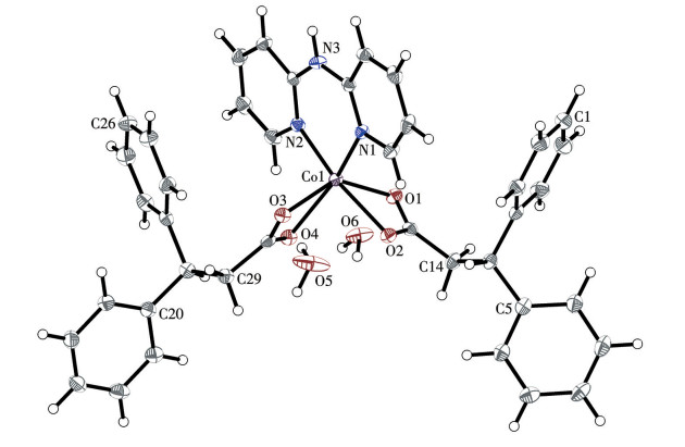
Figure 1.
ORTEP view of complex 1 with 50% probability ellipsoids

Short-chain phenylcarboxylic acid ligand complexes, characterized by their high structural flexibility and extensive structural derivatization, consistently hold a significant position within the family of functional complexes, such as magnetic materials[1-2], fluorescent molecules[3], catalysts[4-5], molecular switches[6], and chiral compounds[7]. The synthesis and characterization of such complexes are both common and significant in the field of coordination chemistry. In bioinorganic chemistry, short-chain phenylcarboxylic acid ligand complexes also hold significant importance[8-9].
Short-chain phenylcarboxylic acids and their derivative building blocks have long been favored by researchers in molecular biology and medicinal chemistry due to their structurally simple yet highly efficient physiologically active properties[10-11]. The core structure of short-chain phenylcarboxylic acids, specifically the short-chain carboxylic acid moiety, mainly derives from microbial metabolic and fermentation processes. It is ubiquitously present in nature and plays a significant role in modulating biological immunity, energy metabolism, carbon cycling, and various other physiological processes[12]. Using propionic acid as a representative C3 backbone molecule, this short-chain fatty acid is abundantly produced in fermented dairy products and mammalian digestive systems through gut microbiota metabolism[13]. As a key metabolic intermediate, it undergoes conversion to propionyl-CoA, transforms into succinyl-CoA, and finally enters the tricarboxylic acid (TCA) cycle. Propionate metabolism serves as a primary energy source for ruminants and contributes to cellular ATP production[14-15]. Recent studies have increasingly highlighted its regulatory roles in diverse physiological processes, including glucose homeostasis, cholesterol metabolism, inflammatory responses, obesity pathogenesis, and neurodevelopment[16-18]. Structurally analogous to propionic acid, phenylpropanoic acid derivatives exhibit multifaceted bioactivities[19-21]. Beyond their well-established therapeutic efficacy in anti-inflammatory applications, exemplified by nonsteroidal anti-inflammatory drugs (NSAIDs) such as ibuprofen and ketoprofen[22], these compounds demonstrate significant potential as eco-friendly antifungal and antibacterial agents in crops[23]. Furthermore, diphenyl-substituted derivatives show promise as highly efficient antioxidants with potent bioactivity and low toxicity[24].
Cobalt, an essential trace element for sustaining life processes, exhibits versatile redox activity due to its accessible oxidation states[25]. The 3d electrons of cobalt ions can engage in synergistic interactions with accessible σ-/π-orbitals, generating electronic effects analogous to σ-radicals[26]. This unique characteristic underpins cobalt′s critical role in mediating biological free radical reactions[27-28]. Vitamin B12, an essential agent in the prevention of pernicious anemia, is a cobalt-containing coordination complex. The central cobalt ion is coordinated to a corrin ring, a cyanide group, and a 5, 6-dimethylbenzimidazole nucleotide (Dmb), which is covalently linked to the nucleotide loop[29]. In certain B12-dependent enzymes, a histidine residue displaces Dmb coordination to configure the active site, as seen in the corrinoid iron-sulfur protein (CFeSP) of CO dehydrogenase/acetyl-CoA synthase systems[30, 31]. Furthermore, cobalt plays a vital role in the metabolism of fatty acids and amino acids and has a profound impact on neurotransmitter processes, cell division, and cell generation[32].
Nickel is ubiquitously distributed in the biosphere and commonly localizes in microbial enzymes such as urease, [NiFe]-hydrogenase, and acetyl-CoA synthase[33]. These enzymatic systems drive critical steps in global biogeochemical cycling of carbon and nitrogen[34]. The physiological role and underlying mechanisms of nickel in the human body remain partially controversial and not yet fully elucidated[35]. Excessive exposure to nickel may interfere with essential physiological processes, including the regulation of key elements such as calcium and the integrity of DNA repair systems[36]. Although the precise mechanisms are still under investigation, some studies indicate that trace amounts of nickel might contribute to the maintenance of normal physiological functions[37]. Nevertheless, excessive intake can lead to adverse effects, such as alterations in the composition of the gut microbiota and a reduction in populations of beneficial probiotic bacteria[38]. It is unequivocally established that trace-level nickel residues persist in major human organs due to the ubiquitous environmental dispersion and industrial utilization of nickel compounds[39].
Co2+ and Ni2+ ions can be readily chelated by various 2,2′-bipyridine ligands to form stable coordination complexes[40]. More importantly, with the assistance of such ligands, the electron density and lipophilicity of the resulting complexes are enhanced, primarily due to increased π-interactions with biomolecules induced by the 2,2′-bipyridine framework. Particularly in cases where 2,2′-bipyridine ligands coordinate synergistically with carboxylate groups, the resulting mixed-ligand complexes often exhibit enhanced biological activities compared to the original bioactive small-molecule building blocks or single-ligand complexes[41-42]. These enhancements include improved DNA-binding affinity[43], antimicrobial properties[44], anti-inflammatory effects[45], anticancer activity[46], and enzymatic catalytic performance[47].
In summary, we designed and synthesized mixed-ligand complexes of the biocompatible transition metal ions Co2+ and Ni2+, using 3,3-diphenylpropionic acid (HDPA) as the primary ligand and 2,2′-dipyridylamine (PAm) as a functional co-ligand, namely [Co(DPA)2(PAm)]·2H2O (1) and [Ni(DPA)2(PAm)]·2H2O (2). The structures and biological activities of the two resulting complexes were investigated in detail, with preliminary discussions regarding potential structure-activity relationships and underlying biochemical mechanisms.
The reagents and solvents were purchased from commercial suppliers and utilized without any additional treatment. The structures of the complexes were investigated via single-crystal X-ray diffraction in a low-temperature environment of 100 K. A Bruker SMART APEX2 CCD diffractometer, which was equipped with a Mo Kα irradiation source (λ=0.071 073 nm), was employed for this analysis. The Shimadzu 6100 diffractometer was used to carry out the powder X-ray diffraction (PXRD) experiment. The elemental composition (including C, H, and N) of the complexes was analyzed using the model Elementar Vario Macro cube. The FTIR spectra were obtained by means of a Bruker Equinox 55 FT-IR spectrophotometer. Moreover, an Agilent Cary Eclipse FLSP920 fluorescence spectrofluorometer was utilized to acquire the emission spectra.
The synthesis of complex 1 was carried out as follows: Co(CH3COO)2·4H2O (1 mmol) was used as the metal source and dissolved together with an equimolar amount of HDPA in a mixed ethanol/water solvent system (7∶3, V/V). After thorough stirring, an equimolar amount of 2, 6-lutidine was added to deprotonate the HDPA ligand. The mixture was then refluxed at 80 ℃ for 2 h. Subsequently, 1 mmol of PAm was added to the reaction mixture, and refluxing was continued for an additional hour under the same conditions. Upon completion, the heating was stopped, and the reaction mixture was allowed to cool to room temperature naturally. The solution was then filtered, and the filtrate was left undisturbed under dark and stable conditions for crystallization. By the following day, abundant purplish-red needle crystals were obtained. The complex exhibited good water solubility. Elemental analysis Calcd. for C40H39CoN3O6(%): C 67.03; H 5.48; N 5.86; Found(%): C 66.85; H 5.70; N 6.04. Selected FTIR peaks (KBr, cm-1): ν(—OH) 3 442(br), ν(O—C—O)asym 1 637(m), ν(O—C—O)sym 1 482(m) (Δν=νasym-νsym=155 < 200), ν(C=N) 1 550(m), ν(C—H) 1 425(m), 1 109(s) ν(C—O), ν(Co—O) 619(s) (Fig.S1a, Supporting information).
The synthesis of complex 2 followed the same procedure as that of complex 1, with the only difference being the substitution of the cobalt salt with Ni(CH3COO)2·4H2O. However, the crystallization process for complex 2 required several months, which was significantly longer than that observed for complex 1. Elemental analysis Calcd. for C40H39NiN3O6(%): C 67.06; H 5.49; N 5.86; Found(%): C 66.93; H 5.58; N 5.73. Selected FTIR peaks (KBr, cm-1): ν(—OH) 3 440(br), ν(O—C—O)asym 1 634(m), ν(O—C—O)sym 1 481(m) (Δν=153 < 200), ν(C—H) 1 421(m), 1 107(s) ν(C—O), ν(Ni—O) 619(s) (Fig.S1b).
Single-crystal X-ray diffraction data were collected on a Bruker SMART APEX2 CCD diffractometer with Mo Kα radiation (λ=0.071 073 nm) at 100 K. For complex 1, a crystal with dimensions 0.30 nm×0.25 nm×0.10 mm was selected, while a crystal measuring 0.16 nm×0.13 nm×0.12 mm was chosen for complex 2. The diffraction data were integrated and reduced using the SAINT program, and absorption corrections were applied with SADABS. The structure solution was carried out by direct methods, followed by full-matrix least-squares refinement on F 2, using SHELXL-2014 implemented in the OLEX2 1.3 software suite. All non-hydrogen atoms were refined anisotropically, and hydrogen atoms were introduced in calculated positions and refined with a riding model. Single-crystal XRD confirmed that both complexes crystallize in the triclinic space group P1. Single-crystal XRD analysis gave 21 448 reflections (θ=1.876°-29.190°) for complex 1 and 38 941 reflections (θ=1.868°-29.336°) for complex 2. The crystal phase composition of the samples was analyzed using PXRD (Cu Kα radiation, λ=0.154 056 nm) under operating conditions of 40 kV and 30 mA, with data collected over a 2θ range of 5°-75°. A comparison of the simulated and experimental PXRD patterns is shown in Fig. S2, confirming good agreement between the peak positions. Crystallographic refinement data are summarized in Table S1.
Hirshfeld surface analysis is an effective approach for identifying and predicting intermolecular interaction patterns, thereby providing insights into structure-activity relationships. The CIF files of the complexes were processed using CrystalExplorer 21.5, which enabled the generation of high-resolution Hirshfeld surfaces for further analysis. The 2D fingerprint plots derived from Hirshfeld surface analysis allow for the quantitative evaluation of intermolecular contacts and the contribution of specific atomic interactions, offering supportive data for predicting molecular interaction behavior. For both Co(Ⅱ) and Ni(Ⅱ) complexes, the 2D fingerprint plots were generated using the expanded mode, with the distance range set between 0.06 and 0.28 nm.
The crystal structure of jack bean urease (PDB entry 3LA4) was acquired from the Protein Data Bank. Using PyMOL, co-crystallized ligands and water molecules were removed to prepare the protein. Molecular docking simulations between the complexes and the target protein were executed in AutoDock Vina. A cubic docking space measuring 2.5 nm3 was positioned at the active pocket coordinates (-38.2, -45.2, 75.2)[48]. AutoDock Vina generated docking poses ranked by binding energy, with the optimal conformation selected based not only on minimal energy values but also on favorable orientation within the catalytic pocket and the presence of key interactions. Discovery Studio was employed to visualize hydrogen bonding and hydrophobic contacts in 2D, while PyMOL rendered the principal 3D docking pose for manuscript figures.
Fluorescence quenching resulting from the competitive interaction between fluorescent dyes and DNA-binding agents is a widely used strategy to evaluate the binding affinity of active compounds to DNA. In this assay, calf thymus DNA (ct-DNA) was pre-stained with ethidium bromide (EB) to enable fluorescence-based detection. A series of eight complex-containing solutions in DMSO was prepared with concentrations ranging from 0.1 to 0.7 μmol·L-1, increasing in 0.1 μmol·L-1 increments. These solutions were gradually introduced into the EB-DNA system in Tris buffer under light-protected conditions and incubated for 2 h. Fluorescence measurements were then conducted using a 350 nm excitation wavelength to monitor the degree of quenching for further analysis.
All materials were sterilized before experimentation. Staphylococcus aureus (Gram-positive) and Escherichia coli (Gram-negative) were cultivated in 3 mL LB broth within 12 mL tubes. Single colonies were inoculated and cultured at 37 ℃, 200 r·min-1 for 15 h, then diluted to 106 CFU·mL-1 with LB medium. The tested complexes were dissolved in 5 mL LB medium to yield 2 mg·mL-1 solutions. For each test, 0.5 mL of the diluted bacterial suspension was added. A positive control using penicillin sodium (1 mg·mL-1 in 5 mL LB) and a blank control without compounds were also included. All groups were incubated statically at 37 ℃ for 24 h. Afterward, 200 μL of each culture was transferred to a 96-well microplate, and OD600 was measured using a microplate reader. Each sample was tested in triplicate, with three parallel wells per replicate.
The tested complexes were initially dissolved in DMSO to prepare a stock solution of 25 mg·mL-1. For the reference drugs, cisplatin and carboplatin, stock solutions were prepared by dissolving them in 0.9% sodium chloride injection at 2 mg·mL-1. These were then diluted with complete culture medium to obtain working solutions with final mass concentrations of 0.625, 1.25, 2.5, 5, and 10 μg·mL-1. Similarly, the complex stock solutions were diluted in complete medium to generate a series of test mass concentrations: 0.016, 0.8, 4, 20, and 100 μg·mL-1. HepG2 cells were treated with both the complex and reference drug solutions for 72 h. Cell viability was then measured using the MTT assay, and the inhibitory effects of the different mass concentrations were analyzed to calculate the corresponding IC50 values.
The tested complexes were prepared as H2O/DMSO (1∶1, V/V) solutions at concentrations of 40, 20, 10, 5, 2.5, and 1.25 μmol·L-1 using a two-fold serial dilution method. Urease solution (15 kU·L-1, pre-frozen) and phosphate-buffered saline (PBS, pH=6.8) were incubated in a 37 ℃ water bath for 30 min before use. Subsequently, 25 μL of urease solution was added to each well of a 96-well microplate. Following this, 25 μL of the test solutions were added sequentially to the wells in descending order of concentration. A blank control group was included, in which 25 μL of DMSO was added instead of the test compound. The plate was sealed with a membrane and incubated at 37 ℃ for 1 h. After incubation, PBS was added to each well, and the microplate was placed in a plate reader. Phenol red was used as a pH indicator to monitor the reaction endpoint. Absorbance at 570 nm was recorded once the pH of the PBS rose from 6.8 to 7.7, indicating the formation of ammonium carbonate in the reaction system. The urease inhibition rate (r) was calculated using the following formula: r=(tS-t0)/t0, where tS is the time required for the test sample to reach the endpoint, and t0 is the time for the blank control group.
It is not coincidental that the Co(Ⅱ) and Ni(Ⅱ) complexes exhibit similar coordination patterns due to their identical valence states and comparable ionic radii. Both complexes adopt a hexacoordinate geometry, in which the central metal ion is coordinated by the carboxylate oxygen atoms from two distinct DPA- ligands and the nitrogen atoms of PAm. The coordination bond parameters of the complexes in this series are all within a reasonable range, as detailed in Table S2. The fundamental structure of the complexes is depicted in Fig. 1. Due to their structural isomorphism, only complex 1 is presented and discussed in detail. Additionally, in the crystal structures of both complexes, each molecular unit incorporates two lattice water molecules introduced from the crystallization environment via hydrogen bonding interactions.
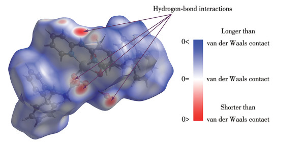
Hirshfeld surface analysis revealed common intermolecular interaction patterns shared by both complexes. The dnorm surface mapping of complex 1 clearly displayed distinct red spots near the carboxylate oxygen atoms of the DPA- ligands and the N—H groups of the PAm moieties[49]. These red regions suggest the potential formation of intermolecular hydrogen bonds, with the corresponding O or H atoms likely serving as interaction nodes (Fig. 2). A similar dnorm distribution was observed in the Hirshfeld surface of complex 2, indicating comparable hydrogen-bonding capabilities and involving the same types of functional groups as in complex 1. The detailed dnorm surface features of complex 2 are presented in Fig.S3.
It is widely recognized that hydrogen bonds exhibit directionality and saturation, which guide the arrangement of molecules or ions in a specific manner during crystal formation and help maintain defined crystal structures. Consequently, these interactions influence the physical and chemical properties of the crystal, including mechanical strength[50], solubility[51], optical behavior[52], chemical reactivity[53], and proton conductivity[54]. In many molecular crystals, hydrogen bonds form distinct structural motifs, such as chains, layers, or 3D networks, which can even impact the space group and unit cell parameters of the crystal lattice[55]. The two complexes display these characteristic structural features in their crystalline forms.
Taking complex 2 as an example, crystal growth along the a-axis is initially driven by hydrogen bonding interactions between the carboxylate oxygen atoms (O3, O4#1, and O1#3; Symmetry codes: #1: 1-x, 1-y, 1-z; #3: -x, 1-y, 1-z) from three different complex molecules and two lattice water molecules. Subsequently, one of the lattice water molecules (O6) extends along the b-axis and forms a hydrogen bond with the amine hydrogen atom (H2#2; Symmetry code: #2: x, 1-y, z) of the PAm ligand in a fourth complex molecule. In addition, the two lattice water molecules are interconnected through hydrogen bonding. These interactions collectively facilitate the formation of a 2D supramolecular layer parallel to the ab plane through an orderly hydrogen-bonding network. This hydrogen-bonded layered assembly is illustrated in Fig. 3, which also details the specific hydrogen-bonding interactions involved. A similar 2D layered structure arising from cooperative hydrogen bonding is also observed in complex 1, with structural details provided in Fig.S4.
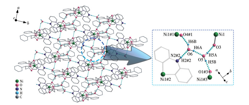
Symmetry codes: #1: 1-x, 1-y, 1-z; #2: x, 1-y, z; #3:-x, 1-y, 1-z.
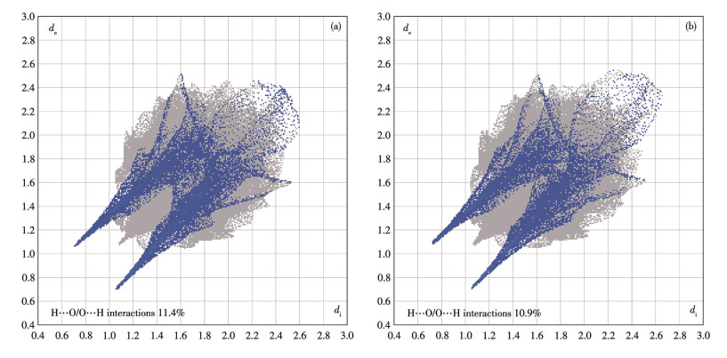
Furthermore, quantitative 2D fingerprint plots analysis revealed that O/H interactions contribute 11.4% in complex 1 and 10.9% in complex 2. In both plots, the O⋯H interaction regions appear sharp, bright, and symmetrical (Fig.4). The relatively high proportion of these contacts, together with their distinctive shape, confirms the strong hydrogen-bonding capacity of both complexes[56]. These findings also highlight the involvement of key hydrogen-bonding donor and acceptor sites, such as the carboxylate oxygen atoms of the DPA- ligands and the amino hydrogen atoms of the PAm moieties. Detailed hydrogen bonding parameters and the contributions of all intermolecular contacts are provided in Table S3-S5.
Unit: nm.
In addition, π⋯π interactions between the complex molecules were identified through auxiliary calculations using the PLATON software. Taking complex 1 as an example, two PAm ligands from different molecules exhibit π⋯π stacking interactions between their opposing pyridyl rings [Cg(C31—C35, N1) to Cg#1(C31—C35, N1), Symmetry code: #1: 1-x, 2-y, 1-z] along the a- and c-axis, with a centroid-to-centroid distance of 0.350 3 nm and an interplanar dihedral angle of 0.02°. The centroid coordinates of the pyridyl ring Cg(C31—C35, N1) are x=0.622 81(6), y=0.982 35(6), and z=0.587 48(4). However, this π⋯π interaction appears localized and does not extend continuously or propagate throughout the structure, existing only between two isolated complex molecules. This conclusion is further supported by the Hirshfeld surface analysis. The shape index mapping reveals pronounced red concave patches at the aromatic rings of the PAm ligands, which clearly indicate π⋯π interactions (Fig.5). Moreover, the curvedness mapping displays broad, flat surface regions in the same areas, tightly aligned with corresponding surfaces of neighboring molecules[57]. This morphological match suggests significant π⋯π interaction strength between complex molecules (Fig.S5).
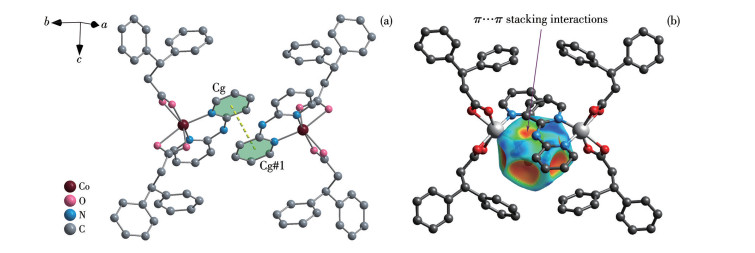
Symmetry code: #1: 1-x, 2-y, 1-z.
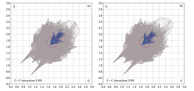
Finally, 2D fingerprint plots reveal that C⋯C contacts account for 3.0% in both the Co(Ⅱ) and Ni(Ⅱ) complexes. The corresponding regions in the plots are closed, symmetric, and brightly centered, which are characteristic features of π⋯π stacking (Fig.6). These observations collectively imply the pyridyl groups in both complexes exhibit a considerable propensity for engaging in π⋯π interactions[58]. The intermolecular π interactions in complex 2 are closely similar to those in complex 1, with specific representations and parameters provided in Fig.S6.
Unit: nm.
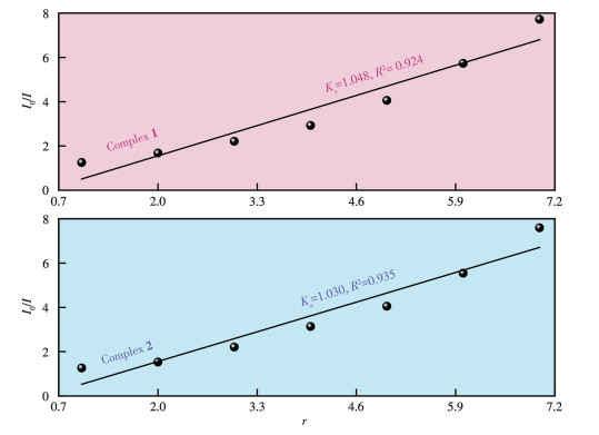
Fluorescence spectral analysis revealed that the EB/DNA complex exhibited a maximum emission wavelength at 630 nm. A significant quenching of fluorescence intensity was observed upon the introduction of metal complexes. Compared to the emission intensity of the blank control group, the addition of the complexes to the EB/DNA system resulted in gradual fluorescence quenching, which became more pronounced with increasing concentrations of the complexes. This initial observation suggests that both complexes possess notable binding affinity toward ct-DNA. Based on the Stern-Volmer equation, the quenching behavior of the Co(Ⅱ) and Ni(Ⅱ) complexes yielded linear fitting plots with calculated slopes of 1.048 and 1.030, respectively. The relatively large slope values imply that the complexes are capable of effectively displacing EB from its binding site on DNA to a significant extent, thereby demonstrating a certain level of interaction with DNA molecules[59]. Detailed fluorescence quenching profiles and corresponding linear fitting plots are provided in Fig.7 and 8.
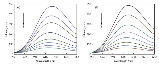
1: cDNA=5.0 μmol·L-1, cEB=1.0 μmol·L-1; 2-8: complex+EB-DNA, ccomplex=1.0, 2.0, 3.0, 4.0, 5.0, 6.0, 7.0 μmol·L-1, respectively.
Urease is a nickel-dependent enzyme widely found in bacteria, fungi, plants, and certain invertebrates. It plays a crucial role in the nitrogen cycle. Many pathogenic microorganisms, such as Helicobacter pylori and Proteus mirabilis, enhance their survival within host organisms by producing urease. Urease inhibitors have shown considerable potential in a range of applications, including improving the efficiency of nitrogen fertilizer utilization in agricultural soils, regulating nutrient absorption in ruminants, and serving as promising alternatives to conventional antibiotics.
In terms of enzyme inhibition, both complexes exhibited enhanced urease inhibitory activity compared to the free ligand, attributed to the synergistic coordination between the metal ions and the ligands. In the assays, complex 1 demonstrated superior inhibitory performance relative to complex 2, with IC50 values of (20.85±0.81) μmol·L-1 and (28.37±2.14) μmol·L-1, respectively (Table 1). Furthermore, complex 1 showed stronger inhibition than the positive control, acetohydroxamic acid, whereas the activity of complex 2 was comparable to that of the control. The similar coordination geometries and molecular conformations of the two complexes likely contribute to their urease inhibitory properties. In addition, the better water solubility of complex 1 may enhance its hydrophilicity and biological affinity, which could explain its stronger enzyme inhibition activity compared to complex 2.
 下载:
导出CSV
下载:
导出CSV
| Compound | IC50 / (μmol·L-1) |
| HDPA | > 100 |
| PAm | > 100 |
| 1 | 20.85±0.81 |
| 2 | 28.37±2.14 |
| Acetohydroxamic acid | 27.73±2.93 |
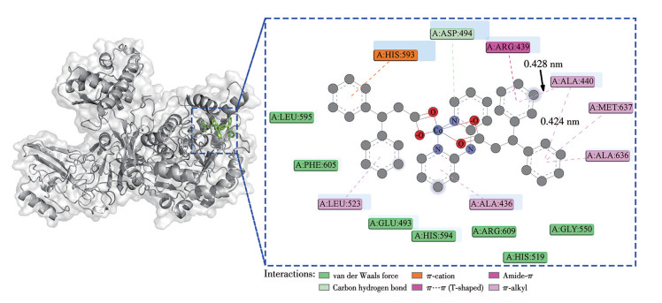
Through examining the possible and reasonable binding configurations of the series of complexes with jack bean urease, a deeper insight can be gained into the active effects that their molecular structures may produce. Based on docking simulations, both complexes successfully enter the active site and form stable interactions with the kinase, exhibiting similar binding orientations and affinities due to their structural similarity.
The binding affinities of the Co(Ⅱ) and Ni(Ⅱ) complexes with urease were calculated to be -173.3 and -171.5 kJ·mol-1, respectively. Among the various interaction modes observed, π interactions involving the aromatic rings of the DPA- ligands were particularly significant. For complex 1, the two aromatic rings of the DPA- ligand simultaneously form strong π-alkyl interactions with the ALA: 440 residue (Fig. 9). Similarly, complex 2 exhibits the same type of π interactions with the same residue through identical aromatic moieties. In both cases, the π interaction distances are less than 0.43 nm (Fig. S7). These docking results should be interpreted as indicative of relative binding trends rather than absolute affinities. The consistency observed between the Co(Ⅱ) and Ni(Ⅱ) complexes supports the reliability of the simulations, while experimental binding data in future studies would provide valuable complementary validation.
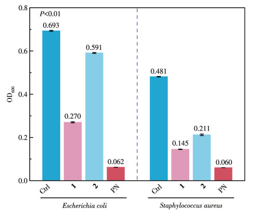
In the subsequent antibacterial activity validation experiments, Fig.10 illustrates the differential inhibitory effects exhibited by the two complexes. Compared with the positive control, sodium penicillin, complex 1 demonstrated moderate broad-spectrum antibacterial activity, effectively inhibiting both Gram-negative Escherichia coli and Gram-positive Staphylococcus aureus to a certain extent. In contrast, complex 2 showed only weak antibacterial activity, which was limited to Staphylococcus aureus, and its efficacy was notably lower than that of complex 1. These findings are consistent with the results of urease inhibition assays and binding affinity calculations, where complex 2 also displayed lower activity than complex 1. This discrepancy may still be attributed to differences in water solubility, as lower solubility could restrict the bioavailability and thereby the biological efficacy of complex 2. The biological activities observed for the Co(Ⅱ) and Ni(Ⅱ) complexes can be tentatively attributed to their coordination environments and ligand properties. The dipyridylamine ligand provides a bidentate N, N′-chelating framework that stabilizes the octahedral geometry of the complexes and creates additional hydrogen-bonding opportunities with biological targets. Meanwhile, the aromatic diphenylpropionate anion contributes extended π-conjugation, which is favorable for π⋯π stacking interactions with nucleic acid bases or aromatic amino acid residues in proteins. These features are consistent with the Hirshfeld surface and molecular docking analyses, where hydrogen bonding and π⋯π contacts were found to dominate. Such interactions may underlie the ability of the complexes to bind DNA, inhibit urease activity, and interfere with bacterial growth. Furthermore, the slightly different electronic configurations and ionic radii of Co(Ⅱ) and Ni(Ⅱ) may influence solubility and bioavailability, providing a plausible explanation for the consistently higher activities of Co(Ⅱ) complex compared to its Ni(Ⅱ) analogue.
The cytotoxic potential of the complexes was assessed in vitro using the MTT assay. The results demonstrated that the complexes displayed significant inhibitory activity against HepG2 cells, with a clear increase in effectiveness as their concentration was raised. According to Table 2, both complexes exhibited cytotoxic effects against HepG2 cells that were comparable to the positive control, carboplatin, and more potent than those of cisplatin. It is noteworthy that despite the structural similarity between the two complexes, complex 2 consistently showed lower biological activity than complex 1 in all biological assays and simulations. This observation suggests that the ligand system adopted in these complexes, which comprises an oxygen-containing carboxylate and a bipyridyl-type ligand, represents a classical coordination model commonly associated with significant biological activity. When the central metal ion is cobalt, a metal that generally possesses higher bioavailability and stronger biological affinity, the resulting complex tends to demonstrate improved bioactivity and broader applicability. In contrast, the cytotoxicity data indicate that for metal centers such as Ni(Ⅱ), which often exhibit higher biological toxicity and lower biocompatibility, the use of suitable ligands enabling saturated coordination environments may enhance overall biological performance, particularly in cytotoxic effects. These mechanistic differences may be attributed to the distinct coordination preferences and electronic properties of Co(Ⅱ) and Ni(Ⅱ). Co(Ⅱ), with its higher bioavailability and stronger biological affinity, affords complexes with superior solubility, stability, and biomolecular interactions, thereby exhibiting consistently stronger activity. In contrast, the lower biocompatibility of Ni(Ⅱ) may limit its performance, although appropriate ligand environments can partially compensate by enhancing coordination saturation and biological response.
下载:
导出CSV
| Compound | IC50 / (μmol·L-1) |
| HDPA | > 100 |
| PAm | > 100 |
| 1 | 1.453±0.624 |
| 2 | 1.547±0.139 |
| Cisplatin | 3.158±0.266 |
| Carboplatin | 1.486±0.196 |
Two complexes, [Co(DPA)2(PAm)]·2H2O (1) and [Ni(DPA)2(PAm)]·2H2O (2), were synthesized and structurally characterized by single-crystal X-ray diffraction. Hirshfeld surface analysis and molecular docking revealed key intermolecular interactions, including hydrogen bonding and π interactions from the carboxylate ligands, which may be associated with their enzymatic inhibition properties. Both complexes exhibited measurable urease inhibitory activity and notable DNA-binding affinity as evidenced by fluorescence spectroscopy. In antibacterial assays, complex 1 displayed moderate broad-spectrum activity against Gram-negative Escherichia coli and Gram-positive Staphylococcus aureus, whereas complex 2 showed only weak inhibition against Staphylococcus aureus. Cytotoxicity evaluation indicated that both complexes outperformed cisplatin and carboplatin against HepG2 cells. These findings on mixed-ligand Co(Ⅱ) and Ni(Ⅱ) complexes with N, N′-chelating agents and propionic acid derivatives contribute a representative structural framework and new bioactivity data that compare favorably with recently reported transition-metal carboxylate complexes with similar ligands[60-62]. Given that related systems are rarely documented, our results help fill a gap in structure-activity relationships, and they support further development of metal-based therapeutics with broader application potential.
PAKULA R J, BERRY J F. Cobalt complexes of the chelating dicarboxylate ligand "esp": A paddlewheel-type dimer and a heptanuclear coordination cluster[J]. Dalton Trans., 2018, 47(39): 13887-13893 doi: 10.1039/C8DT03030H
BIÇER F A, ARICI M, YEŞILEL O Z. Syntheses, characterization of three new cobalt(Ⅱ) complexes with 2-phenylsuccinic acid and flexible bis(imidazole) linkers[J]. J. Mol. Struct., 2023, 1284: 135444 doi: 10.1016/j.molstruc.2023.135444
YEŞILEL O Z, GÜLER U, ÇIFTÇI E, ARICI M. A series of coordination polymers constructed by 2-phenylsuccinic acid and flexible bis(imidazole) ligands: Syntheses, structures, and photoluminescent properties[J]. J. Mol. Struct., 2022, 1262: 132991 doi: 10.1016/j.molstruc.2022.132991
HO P H, HUNG C C, WANG Y H, CHUANG G J. Intermolecular nitrene insertion by bimetallic catalysts[J]. Asian J. Org. Chem., 2019, 8(2): 275-278 doi: 10.1002/ajoc.201800641
PAKULA R J, MARTINEZ A M, NOTEN E A, HARRIS C F, BERRY J F. New chromium, molybdenum, and cobalt complexes of the chelating esp ligand[J]. Polyhedron, 2019, 161: 93-103 doi: 10.1016/j.poly.2018.12.045
SKIPPER H E, MAY C V, RHEINGOLD A L, DOERRER L H, KAMENETSKA M. Hard-soft chemistry design principles for predictive assembly of single molecule-metal junctions[J]. J. Am. Chem. Soc., 2021, 143(40): 16439-16447 doi: 10.1021/jacs.1c05142
ZAVAKHINA M S, SAMSONENKO D G, DYBTSEV D N, FEDIN V P. Rigid 1D coordination polymers with tunable metal cation and chiral pendant moieties[J]. Z. Anorg. Allg. Chem., 2015, 641(3/4): 590-595
FERNÁNDEZ C Y, ALVAREZ N, ROCHA A, MENDES L F S, COSTA-FILHO A J, ELLENA J, BATISTA A A, FACCHIN G. Phenanthroline and phenyl carboxylate mixed ligand copper complexes in developing drugs to treat cancer[J]. J. Inorg. Biochem., 2024, 260: 112700 doi: 10.1016/j.jinorgbio.2024.112700
ARUN P P, PATEL R R, SINGH S K, PARMAR K, SINGH M. Exploring metal complexes for cancer treatment: Mechanistic insights and therapeutic potential[J]. J. Organomet. Chem., 2025, 1035: 123682 doi: 10.1016/j.jorganchem.2025.123682
HAMER H M, JONKERS D, VENEMA K, VANHOUTVIN S, TROOST F J, BRUMMER R J. Review article: The role of butyrate on colonic function[J]. Aliment. Pharmacol. Ther., 2008, 27(2): 104-119 doi: 10.1111/j.1365-2036.2007.03562.x
PAUTOVA A K, BURNAKOVA N A, BELOBORODOVA N V, REVELSKY A I. Simultaneous determination of aromatic, short-chain fatty and dicarboxylic acids in blood serum and cerebrospinal fluid by gas chromatography-mass spectrometry[J]. J. Anal. Chem., 2023, 78(14): 1942-1954 doi: 10.1134/S1061934823140058
HAYS K E, PFAFFINGER J M, RYZNAR R. The interplay between gut microbiota, short-chain fatty acids, and implications for host health and disease[J]. Gut Microbes, 2024, 16(1): 2393270 doi: 10.1080/19490976.2024.2393270
AL-LAHHAM S H, PEPPELENBOSCH M P, ROELOFSEN H, VONK R J, VENEMA K. Biological effects of propionic acid in humans; metabolism, potential applications and underlying mechanisms[J]. Biochim. Biophys. Acta Mol. Cell Biol. Lipids, 2010, 1801(11): 1175-1183
COLLADO M S, ARMSTRONG A J, OLSON M, HOANG S A, DAY N, SUMMAR M, CHAPMAN K A, REARDON J, FIGLER R A, WAMHOFF B R. Biochemical and anaplerotic applications of in vitro models of propionic acidemia and methylmalonic acidemia using patient-derived primary hepatocytes[J]. Mol. Genet. Metab., 2020, 130(3): 183-196 doi: 10.1016/j.ymgme.2020.05.003
YANG Y L, LIU C M, ZHAO W Y, MAZARJI M, REN L H, LIU C, PAN J T, YAN B H. Anaerobic propionic acid production via succinate pathway at extremely low pH[J]. Chem. Eng. J., 2024, 486: 150190 doi: 10.1016/j.cej.2024.150190
LOREFICE L, ZOLEDZIEWSKA M. Propionic acid impact on multiple sclerosis: Evidence and challenges[J]. Nutrients, 2024, 16(22): 3887 doi: 10.3390/nu16223887
WANG Y N, LI M J, GUO J Y, KANG S G, HUANG K L, TONG T. Olfr78, a novel short-chain fatty acid receptor, regulates glucose homeostasis and gut GLP-1 secretion in mice[J]. Food Frontiers, 2023, 4(4): 1893-1912 doi: 10.1002/fft2.301
FOLEY K A, MACFABE D F, VAZ A, OSSENKOPP K P, KAVALIERS M. Sexually dimorphic effects of prenatal exposure to propionic acid and lipopolysaccharide on social behavior in neonatal, adolescent, and adult rats: Implications for autism spectrum disorders[J]. Int. J. Dev. Neurosci., 2014, 39(1): 68-78 doi: 10.1016/j.ijdevneu.2014.04.001
RETA G F, TONN C E, RÍOS-LUCI C, LEÓN L G, PÉREZ-ROTH E, PADRÓN J M, DONADEL O J. Cytotoxic bioactivity of some phenylpropanoic acid derivatives[J]. Nat. Prod. Commun., 2012, 7(10): 1341-1346
KINRA M, JOSEPH A, NAMPOOTHIRI M, ARORA D, MUDGAL J. Inhibition of NLRP3-inflammasome mediated IL-1β release by phenylpropanoic acid derivatives: In-silico and in-vitro approach[J]. Eur. J. Pharm. Sci., 2021, 157: 105637 doi: 10.1016/j.ejps.2020.105637
AYYADURAI G K, JAYAPRAKASH R, SHAJAHAN A, RATHIKA S. Studies on 2-((2, 4-dihydroxybenzylidene) amino)-3-phenylpropanoic acid include antimicrobial, antidiabetic, antioxidant, anticancer, hemolysis, and theoretical QSAR[J]. J. Biomol. Struct. Dyn., 2025, 43(6): 2864-2876 doi: 10.1080/07391102.2023.2294383
GENÇER H K, ÇEVIK U A, KAYA ÇAVUŞOĞLU B K, SAĞLIK B N, LEVENT S, ATLI Ö, ILGIN S, ÖZKAY Y, KAPLANCIKLI Z A. Design, synthesis, and evaluation of novel 2-phenylpropionic acid derivatives as dual COX inhibitory-antibacterial agents[J]. J. Enzym. Inhib. Med. Chem., 2017, 32(1): 732-745 doi: 10.1080/14756366.2017.1310726
YEHIA R S, OSMAN G H, ASSAGGAF H, SALEM R, MOHAMED M S M. Isolation of potential antimicrobial metabolites from endophytic fungus Cladosporium cladosporioides from endemic plant Zygophyllum mandavillei[J]. S. Afr. J. Bot., 2020, 134: 296-302 doi: 10.1016/j.sajb.2020.02.033
URBANI P, RAMUNNO A, FILOSA R, PINTO A, POPOLO A, BIANCHINO E, PIOTTO S, SATURNINO C, DE PRISCO R, NICOLAUS B, TOMMONARO G. Antioxidant activity of diphenylpropionamide derivatives: Synthesis, biological evaluation and computational analysis[J]. Molecules, 2008, 13(4): 749-761 doi: 10.3390/molecules13040749
SWANNER E D, PLANAVSKY N J, LALONDE S V, ROBBINS L J, BEKKER A, ROUXEL O J, SAITO M A, KAPPLER A, MOJZSIS S J, KONHAUSER K O. Cobalt and marine redox evolution[J]. Earth Planet. Sci. Lett., 2014, 390: 253-263 doi: 10.1016/j.epsl.2014.01.001
DING J, WEI Z M, LI F H, ZHANG J C, ZHANG Q, ZHOU J, WANG W J, LIU Y H, ZHANG Z, SU X Z, YANG R Z, LIU W, SU C L, YANG H B, HUANG Y Q, ZHAI Y M, LIU B. Atomic high-spin cobalt(Ⅱ) center for highly selective electrochemical CO reduction to CH3OH[J]. Nat. Commun., 2023, 14(1): 6550 doi: 10.1038/s41467-023-42307-1
LEONARD S, M. GANNETT P, ROJANASAKUL Y, SCHWEGLER-BERRY D, CASTRANOVA V, VALLYATHAN V, SHI X L. Cobalt-mediated generation of reactive oxygen species and its possible mechanism[J]. J. Inorg. Biochem., 1998, 70(3/4): 239-244
MANIKANDAN R, VISWANATHAMURTHI P, VELMURUGAN K, NANDHAKUMAR R, HASHIMOTO T, ENDO A. Synthesis, characterization and crystal structure of cobalt complexes containing 2-acetylpyridine thiosemicarbazones: DNA/protein interaction, radical scavenging and cytotoxic activities[J]. J. Photochem. Photobiol. B‒Biol., 2014, 130: 205-216 doi: 10.1016/j.jphotobiol.2013.11.008
BANERJEE R, GHERASIM C, PADOVANI D. The tinker, tailor, soldier in intracellular B12 trafficking[J]. Curr. Opin. Chem. Biol., 2009, 13(4): 484-491 doi: 10.1016/j.cbpa.2009.07.007
GIEDYK M, GOLISZEWSKA K, GRYKO D. Vitamin B12 catalysed reactions[J]. Chem. Soc. Rev., 2015, 44(11): 3391-3404 doi: 10.1039/C5CS00165J
GRUBER K, PUFFER B, KRÄUTLER B. Vitamin B12-derivatives—Enzyme cofactors and ligands of proteins and nucleic acids[J]. Chem. Soc. Rev., 2011, 40(8): 4346-4363 doi: 10.1039/c1cs15118e
GREGOROWICZ W, PAJCHEL L. The role of cobalt ions in angiogenesis—A review[J]. Int. J. Mol. Sci., 2025, 26(15): 7236 doi: 10.3390/ijms26157236
BOER J L, MULROONEY S B, HAUSINGER R P. Nickel-dependent metalloenzymes[J]. Arch. Biochem. Biophys., 2014, 544: 142-152 doi: 10.1016/j.abb.2013.09.002
RAGSDALE S W. Nickel-based enzyme systems[J]. J. Biol. Chem., 2009, 284(28): 18571-18575 doi: 10.1074/jbc.R900020200
LIU Y C, LUO X M, PENG Y D, CAI L. Cardio-metabolic effects of nickel: A narrative review[J]. Cardiovasc. Toxicol., 2025, 25(7): 944-954 doi: 10.1007/s12012-025-10014-6
ZAMBELLI B, UVERSKY V N, CIURLI S. Nickel impact on human health: An intrinsic disorder perspective[J]. BBA‒Proteins Proteomics, 2016, 1864(12): 1714-1731 doi: 10.1016/j.bbapap.2016.09.008
SRIVASTAVA A K, SNAPPER D M, ZHENG J W, YILDRIM B S, SRIVASTAVA S, WOOD S C. Examining the role of nickel and NiTi nanoparticles promoting inflammation and angiogenesis[J]. J. Immunotoxicol., 2022, 19(1): 61-73 doi: 10.1080/1547691X.2022.2080307
YANG J F, FENG P Y, LING Z M, KHAN A, WANG X, CHEN Y L, ALI G, FANG Y T, SALAMA E S, WANG X M, LIU P, LI X K. Nickel exposure induces gut microbiome disorder and serum uric acid elevation[J]. Environ. Pollut., 2023, 324: 121349 doi: 10.1016/j.envpol.2023.121349
MARET W. Metalloproteomics, metalloproteomes, and the annotation of metalloproteins[J]. Metallomics, 2010, 2(2): 117-125 doi: 10.1039/B915804A
张莉, 蒋畅, 郭林峰, 张小玲, 林雄强, 康杰, 孙伟明. 含吡啶-2, 6-二羧酸的钴(Ⅱ)配合物的合成、表征、抗肿瘤活性和理论计算[J]. 无机化学学报, 2021, 37(2): 368-374ZHANG L, JIANG C, GUO L F, ZHANG X L, LIN X Q, KANG J, SUN W M. Synthesis, characterization, antitumor activity, and theoretical calculations of Co(Ⅱ) complex based on pyridine-2, 6-dicarboxylic acid[J]. Chinese J. Inorg. Chem., 2021, 37(2): 368-374
LOUBALOVÁ I, KOPEL P. Coordination compounds of Cu, Zn, and Ni with dicarboxylic acids and N donor ligands, and their biological activity: A review[J]. Molecules, 2023, 28(3): 1445 doi: 10.3390/molecules28031445
RUTA L L, FARCASANU I C, BACALUM M, RĂILEANU M, ROSTAS A M, DANILIUC C, CHIFIRIUC M C, MĂRUȚESCU L, POPA M, BADEA M, IORGULESCU E E, OLAR R. Biological activity of triazolopyrimidine copper(Ⅱ) complexes modulated by an auxiliary N-N-chelating heterocycle ligands[J]. Molecules, 2021, 26(22): 6772 doi: 10.3390/molecules26226772
陶钦, 吴健, 葛超, 王萌萌, 吕梦迪, 薛旭玲, 刘红科. 反式阿魏酸钌配合物的合成、表征、荧光光谱及其与DNA/蛋白作用[J]. 无机化学学报, 2020, 36(10): 1853-1864TAO Q, WU J, GE C, WANG M M, LÜ M D, XUE X L, LIU H K. Synthesis, characterization, fluorescence and interactions with DNA/BSA properties of ruthenium ferulate complexes[J]. Chinese J. Inorg. Chem., 2020, 36(10): 1853-1864
JAHANI S, KHORASANI-MOTLAGH M, NOROOZIFAR M. DNA interaction of europium complex containing 2,2′-bipyridine and its antimicrobial activity[J]. J. Biomol. Struct. Dyn., 2016, 34(3): 612-624 doi: 10.1080/07391102.2015.1048481
PERONTSIS S, DIMITRIOU A, FOTIADOU P, HATZIDIMITRIOU A G, PAPADOPOULOS A N, PSOMAS G. Cobalt(Ⅱ) complexes with the non-steroidal anti-inflammatory drug diclofenac and nitrogen-donor ligands[J]. J. Inorg. Biochem., 2019, 196: 110688 doi: 10.1016/j.jinorgbio.2019.04.002
DU L Q, CHEN Z X, WEI Q C, CHEN Z L, YANG Y. Chlorquinaldol-zinc(Ⅱ)-bipyridine complexes: Design, synthesis, structure and anticancer evaluation[J]. Inorg. Chem. Commun., 2023, 156: 111238 doi: 10.1016/j.inoche.2023.111238
MESSINA M A, MAUGERI L, FORTE G, RUGGIERI M, PETRALIA S. A highly sensitive colorimetric approach based on tris (bipyridine) ruthenium (Ⅱ/Ⅲ) mediator for the enzymatic detection of phenylalanine[J]. Front. Chem., 2023, 11: 1164014 doi: 10.3389/fchem.2023.1164014
CHEN Z Q, ZHANG X B, PENG C, WANG J A, XU Z J, CHEN K X, SHI J Y, ZHU W L. D3Pockets: A method and web server for systematic analysis of protein pocket dynamics[J]. J. Chem. Inf. Model., 2019, 59(8): 3353-3358 doi: 10.1021/acs.jcim.9b00332
CHEN T, LIAO X, GAO L E. Hydrogen bonding and Hirshfeld surface analysis of gallic acid and its monohydrate based on terahertz spectroscopy[J]. Chem. Phys., 2024, 578: 112159 doi: 10.1016/j.chemphys.2023.112159
YUAN H, XUE B, YANG D Y, RENCUS-LAZAR S, CAO Y, GAZIT E, TAN D, YANG R S. Rational design of biological crystals with enhanced physical properties by hydrogen bonding interactions[J]. Research, 2023, 6: 0046 doi: 10.34133/research.0046
PHAN C Y, SHEN J, YU K X, LIU J Y, TANG G P. Hydrogen bonds, topologies, energy frameworks and solubilities of five sorafenib salts[J]. Int. J. Mol. Sci., 2021, 22(13): 6682 doi: 10.3390/ijms22136682
SARANYA K, MUHILDHARANI E, RAJA C R. Growth, structural confirmation and the effect of hydrogen bond on the third-order nonlinear optical properties of diisopropylammonium 4-aminobenzenesulfonate crystal[J]. J. Mater. Sci. ‒Mater. Electron., 2023, 34(7): 661 doi: 10.1007/s10854-023-10088-4
PRAJAPATI P, PANDEY J, TANDON P, SINHA K, SHIMPI M R. Molecular structural, hydrogen bonding interactions, and chemical reactivity studies of ezetimibe-L-proline cocrystal using spectroscopic and quantum chemical approach[J]. Front. Chem., 2022, 10: 848014 doi: 10.3389/fchem.2022.848014
李成娟, 李荣云, 初芷同, 刘厚亭, 卢静, 王素娜, 李允伍. 一个含有连续氢键的晶态Ni(Ⅱ)-MOF的合成、结构及质子传导性能[J]. 无机化学学报, 2021, 37(4): 645-652LI C J, LI R Y, CHU Z T, LIU H T, LU J, WANG S N, LI Y W. Synthesis, structure and proton conduction of a crystalline Ni(Ⅱ)-MOF with continuous hydrogen bonds[J]. Chinese J. Inorg. Chem., 2021, 37(4): 645-652
PUNIYA R R, TAKHAR P, CHHAPOLIYA M, DEKA R, KALITA D J, SINGH D. Development of photoluminescent hydrogen-bonded frameworks based on pyromellitic diimide-tethered carboxylic acid hosts and multi-bonding solvent guests[J]. Mater. Adv., 2024, 5(19): 7817-7829 doi: 10.1039/D4MA00634H
MAHAPATRA A D, SHAIK A, THIRUVENKATAM V, DATTA B. Supramolecular architecture in sulfonylurea, sulfonyldiurea and sulfonyltriurea drugs: Synthesis, X-ray structure and Hirshfeld surface analysis[J]. J. Mol. Struct., 2021, 1233: 130158 doi: 10.1016/j.molstruc.2021.130158
LEÓN A C D, ECHEVERRÍA G A, PIRO O E, ULIC S E, JIOS J L, TAPIA L C A, GUZMÁN M M F. New thiourea and urea derivatives containing trifluoromethyl- and bis-triflouromethyl-4H-chromen-3-yl substituents[J]. Mol. Phys., 2019, 117(3): 368-381 doi: 10.1080/00268976.2018.1514132
FEIJOO M G, FERNÁNDEZ-LIENCRES M P, GIL D M, GÓMEZ M I, ALTABEF A B, NAVARRO A, TUTTOLOMONDO M E. A detailed study of intermolecular interactions, electronic and vibrational properties of the metal complex bis(uracilato)diammine copper(Ⅱ) dihydrate[J]. J. Mol. Struct., 2018, 1155: 424-433 doi: 10.1016/j.molstruc.2017.11.030
魏强, 董建方, 李文彬, 赵培然, 丁菲菲, 李连之. 两个组氨酸希夫碱镍(Ⅱ)配合物的合成、晶体结构和DNA相互作用及SOD活性[J]. 无机化学学报, 2016, 32(05): 789-798WEI Q, DONG J F, LI W B, ZHAO P R, DING F F, LI L Z. Syntheses, crystal structures, DNA interactions and SOD activities of two nickel(Ⅱ) complexes with L-histidine Schiff base[J]. Chinese J. Inorg. Chem., 2016, 32(05): 789-798
FERNÁNDEZ C Y, ROCHA A, AZAM M, ALVAREZ N, MIN K, BATISTA A A, COSTA-FILHO A J, ELLENA J, FACCHIN G. Synthesis, characterization, DNA binding and cytotoxicity of copper(Ⅱ) phenylcarboxylate complexes[J]. Inorganics, 2023, 11(10): 398 doi: 10.3390/inorganics11100398
ALEM M B, DAMENA T, DESALEGN T, KOOBOTSE M, ESWARAMOORTHY R, NGWIRA K J, OMBITO J O, ZACHARIAH M, DEMISSIE T B. Cytotoxic mixed-ligand complexes of Cu(Ⅱ): A combined experimental and computational study[J]. Front. Chem., 2022, 10: 1028957 doi: 10.3389/fchem.2022.1028957
PEÑA Q, SCIORTINO G, MARÉCHAL J D, BERTAINA S, SIMAAN A J, LORENZO J, CAPDEVILA M, BAYÓN P, IRANZO O, PALACIOS Ò. Copper(Ⅱ) N, N, O-chelating complexes as potential anticancer agents[J]. Inorg. Chem., 2021, 60(5): 2939-2952 doi: 10.1021/acs.inorgchem.0c02932

Figure 3 Layered hydrogen bond network and details of complex 2
Symmetry codes: #1: 1-x, 1-y, 1-z; #2: x, 1-y, z; #3:-x, 1-y, 1-z.

Figure 4 Two-dimensional fingerprint plots of complexes 1 (a) and 2 (b) showing H⋯O/O⋯H interactions
Unit: nm.

Figure 5 Intermolecular π interactions in complex 1: (a) details of the π⋯π interactions; (b) Hirshfeld surface shape index of the π⋯π interactions
Symmetry code: #1: 1-x, 2-y, 1-z.

Figure 6 Two-dimensional fingerprint plots of complexes 1 (a) and 2 (b) showing C⋯C interactions
Unit: nm.

Figure 7 Fluorescence spectra for complexes 1 (a) and 2 (b) binding to EB-DNA
1: cDNA=5.0 μmol·L-1, cEB=1.0 μmol·L-1; 2-8: complex+EB-DNA, ccomplex=1.0, 2.0, 3.0, 4.0, 5.0, 6.0, 7.0 μmol·L-1, respectively.
Table 1. Jack bean urease inhibitory activity of the compounds
| Compound | IC50 / (μmol·L-1) |
| HDPA | > 100 |
| PAm | > 100 |
| 1 | 20.85±0.81 |
| 2 | 28.37±2.14 |
| Acetohydroxamic acid | 27.73±2.93 |
 下载: 导出CSV
Table 2. Cytotoxicity of tested compounds against tumor cells after 72 h incubation
| Compound | IC50 / (μmol·L-1) |
| HDPA | > 100 |
| PAm | > 100 |
| 1 | 1.453±0.624 |
| 2 | 1.547±0.139 |
| Cisplatin | 3.158±0.266 |
| Carboplatin | 1.486±0.196 |
下载: 导出CSV

 扫一扫看文章

扫一扫关注我们