Shaanxi Key Laboratory of Chemical Reaction Engineering, College of Chemistry and Chemical Engineering, Yan′an University, Yan′an, Shaanxi 716000, China
Received Date:
01 July 2025 Revised Date:
24 September 2025 Available Online:
10 February 2026
Abstract:
In this study, using 3, 5-di(3′, 5′-dicarboxylphenyl)-1H-1, 2, 4-triazole (H4L) as ligands, a gadolinia-based organic framework complex {[GdNa(L)(H2O)3]·2H2O}n (Gd-Na-MOF) was successfully designed and synthesized by hydrothermal method. The structure and properties were systematically characterized and tested by techniques such as single-crystal X-ray diffraction, powder X-ray diffraction, thermogravimetric analysis, infrared spectroscopy, and fluorescence spectroscopy. The results indicate that this complex has a unique 3D structure, excellent thermal stability, and outstanding luminescent performance. Based on its luminescent properties, a polymer-embedding method was employed to fabricate the Gd-Na-MOF into a flexible, washable composite fluorescent film, Gd-Na-MOF@PMMA/BMA (PMMA=polymethyl methacrylate, BMA=butyl methacrylate). This fluorescent film exhibited highly sensitive recognition capability for tyramine, with a low detection limit of 1.66 μmol·L-1. It was used for the detection of tyramine in bananas, with a recovery rate of 96.92%-100.26%.
Tyramine (TA) is a common biogenic amine that is widely present in fermented foods, dairy products, and some fruits (such as bananas)[1]. As a core metabolite of catecholamine neurotransmitters, TA participates in physiological activities like mood regulation and cardiovascular function[2]. However, excessive intake of TA may trigger adverse reactions such as headaches and hypertension, and even lead to intoxication[3-4]. Therefore, developing efficient and sensitive methods for TA detection is crucial for food safety monitoring. Currently, TA detection primarily relies on traditional techniques such as high-performance liquid chromatography (HPLC) and mass spectrometry. However, these methods usually require complex sample pretreatment, expensive instruments, and professional operation, which limits their application in rapid detection[5-7]. Fluorescence sensing technology, with its advantages of simple operation, rapid response, and high sensitivity, has emerged as a powerful tool for detecting hazardous substances in food[8-9].
Lanthanide metal-organic frameworks (Ln-MOFs) are crystalline porous materials constructed from organic ligands coordinated with lanthanide metal ions/ clusters. Due to their tunable pore structures, high specific surface areas, and excellent luminescent properties, Ln-MOFs show significant potential in the field of fluorescence sensing[10-11]. However, powdered MOFs face limitations in practical liquid-phase applications due to difficulties in dissolution, poor dispersion, and challenges in recovery[12-13]. To address these issues, researchers have recently explored compounding MOFs with polymer materials to fabricate thin-film sensors, enhancing their stability and practicality[14].
Current strategies for preparing MOF-based fluorescent sensing films mainly include sol-gel embedding, Langmuir-Blodgett (LB) film technique, layer-by-layer assembly, and polymer embedding[15-16]. The sol-gel method is simple but involves long preparation times, and the resulting films are prone to shrinkage, cracking, and poor stability[17]. While the LB technique allows precise control over film structure and is suitable for biosensing, its stringent experimental conditions and low efficiency limit its application[18]. The layer-by-layer assembly method can achieve precise regulation of the film composition and multi-component detection, but it is time-consuming and highly sensitive to the solution environment[19]. In contrast, polymer embedding is highly favored due to its low cost, simple operation, and good stability[20]. Among polymers, polyvinylidene fluoride (PVDF), polymethyl methacrylate (PMMA), and polyvinyl alcohol (PVA) are ideal polymeric matrices for constructing MOF-supporting frameworks, owing to their excellent film-forming ability and chemical stability[21-22]. For instance, Ali et al. fabricated a Cu-MOF film using PVA for H2S gas detection[23]. Kazemi et al. prepared a Cu-Zn-MOF film with PVA for ethylene adsorption to extend the shelf life of fresh produce[24]. Fang et al. utilized PMMA to create a UiO-66 film for the selective separation of the dye rhodamine B[25]. Nevertheless, challenges such as achieving uniform dispersion of MOF crystals within the film, controlling film thickness, and overcoming the inherent mechanical brittleness of the films remain obstacles to their practical application.
In this study, a 3D porous gadolinium-sodium metal-organic framework {[GdNa(L)(H2O)3]·2H2O}n (Gd-Na-MOF) was successfully synthesized by the hydrothermal method using 3, 5-bis(3′, 5′-dicarboxyphenyl)-1H-1, 2, 4-triazole (H4L) as the ligand. Its structure, thermal stability, and luminescent properties were systematically studied. Based on its luminescent properties, a flexible Gd-Na-MOF@PMMA/BMA (PMMA=polymethyl methacrylate, BMA=butyl methacrylate) composite film was successfully fabricated by the polymer embedding method. This composite film ingeniously combines the highly efficient luminescent properties of Gd-Na-MOF with the excellent flexibility and processability of the polymeric material (PMMA-BMA). Experiments show that this flexible sensing film can achieve efficient and sensitive fluorescence detection of TA in banana samples, providing a highly promising new sensing platform for rapid monitoring of food safety.
1.
Experimental
The details of reagents and methods can be found in the Supporting information.
1.1
Preparation of Gd-Na-MOF
First, GdCl3·6H2O (0.05 mmol), H4L (0.05 mmol), and 4, 4′-bipyridine (0.05 mmol) were added successively into a glass bottle. Subsequently, HAc-NaAc buffer solution (pH=6, 5 mL) and isopropanol (1 mL) were added. The mixture was stirred to make it evenly dispersed. After continuous stirring for 15 min, the glass bottle was directly placed into a 25 mL stainless steel autoclave with a polytetrafluoroethylene liner. The autoclave was sealed, and then the temperature was increased to 160 ℃ in a programmed manner, and a constant temperature reaction was maintained at this temperature for 72 h. After the reaction was completed, the autoclave was cooled down to 30 ℃ at a rate of 5 ℃·h-1. Finally, the reaction product was filtered and separated, the solid was washed with deionized water and ethanol in sequence, and colorless rod-shaped crystals were obtained after drying.
1.2
Single-crystal X-ray analysis
The Gd-Na-MOF crystals with good crystallization and appropriate size were selected, and placed on a single-crystal diffractometer for single-crystal testing. Single-crystal data were collected at room temperature. We performed diffraction operations using Mo Kα radiation (λ=0.071 073 nm). All data were restored using the Saint program and subjected to multi-scan absorption correction by SADABS[26]. The structure was solved by the partial structure expansion method of SHELXS and refined by the full matrix least squares method[27]. All non-hydrogen atoms were refined using anisotropic temperature factors, hydrogen atoms were determined by theoretical hydrogenation, and disordered solvent molecules in the structure were removed by the PLATON/SQUEEZE program. The crystallographic data of Gd-Na-MOF are listed in Table S1, and the main bond lengths and bond angles are listed in Table S2.
1.3
Preparation of Gd-Na-MOF@PMMA/BMA
PMMA (0.8 g) was added to 3 mL of tetrahydrofuran to form a saturated solution. Then, 0.07 g of colorless rod-shaped crystal Gd-Na-MOF was ground into powder and added to the above saturated solution. Under the stirring of a magnetic stirrer, 3 mL of BMA was added to enhance the flexibility of the solution. When it became viscous, it was evenly smeared onto a clean glass slide (24.5 mm×76.2 mm) and allowed air dry naturally at room temperature to obtain a composite fluorescent film (Gd-Na-MOF@PMMA/BMA).
1.4
Fluorescence characteristics and sensing experiments of Gd-Na-MOF
At room temperature, we used a fluorescence spectrophotometer (voltage: 700 V, slit width: 5 nm) to measure the fluorescence spectra of Gd-Na-MOF powder, Gd-Na-MOF@PMMA/BMA immersed in an aqueous solution, and the ligand H4L, respectively.
In a 1 cm quartz cuvette with four-sided light transmission, we added 1 mL of deionized water and 1 mL of tyramine solution (or the sample to be tested) with different concentrations (0, 2, 4, 6, 8, 10, 12, 14, 16, 18, 20 μmol·L-1), respectively. The solution was shaken well, then Gd-Na-MOF@PMMA/BMA was immersed in it. the fluorescence intensity F was recorded at the optimal emission wavelength (λem=351 nm) under the optimal excitation wavelength (λex=300 nm), and the value of ΔF=F0-F (F0 is the fluorescence value when the concentration of the tyramine solution is zero) was calculated.
1.5
Preparation of banana samples
The banana middle section samples (30 g, purchased from China Resources Vanguard Supermarket in Baota District, Yan′an City, Shaanxi Province) were cut into pieces and frozen. The samples were ground into a homogenate in an ice bath. The banana homogenate (5 g) was put into a 50 mL centrifuge tube. 20 mL HCl (0.01 μmol·L-1)was added to inhibit the inactivation of tyrosine decarboxylase, thus preventing its catalytic degradation of tyramine[28]. After ultrasonic treatment in ice water for 10 min, the mixture was centrifuged at 5 000 r·min-1 for 20 min at 4 ℃. The supernatant was filtered through a 0.22 μm microporous filter membrane to obtain a clear banana extract, which was ready for use.
2.
Results and discussion
2.1
Crystal structure
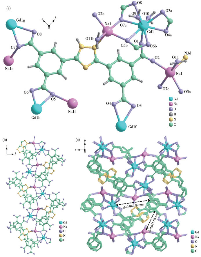
Single crystal X-ray studies indicate that the Gd-Na-MOF crystallizes in the P21/n space group of the monoclinic system, presenting a 3D framework structure. The asymmetric unit of the complex contains one Gd3+ ion, one Na+ ion, one L4- ligand, three coordinated water molecules, and two free water molecules (Fig.1a). Gd3+ coordinates with seven oxygen atoms (O1, O3a, O4a, O6b, O5b, O7c, O8c) from four L4- ligands and two oxygen atoms (O9, O10) from two water molecules, forming a nine-coordination mode; Na+ coordinates with three oxygen atoms (O2, O7e, O5b) from three L4- ligands, one oxygen atom (O11) from one water molecule, and a nitrogen atom (N3d) from one L4- ligand, forming a five-coordination mode. The bond lengths of Gd—O range from 0.234 9(4) to 0.254 6(4) nm, and the bond lengths of Na—O range from 0.224 1(4) to 0.249 3(4) nm, which are consistent with the literature reports[29-30] (Fig.1a). Moreover, Gd3+ and Na+ are connected through bridging oxygen atoms (such as O5b, O7c) to form a binary metal unit (Fig.1a). The O1 and O2 of the L4- ligand, as bridging carboxyl groups, connect the heteronuclear dimetal units into an S-shaped 1D chain (Fig.1b). Then it is extended into a 3D microporous structure through the L4- ligand (Fig.1c).
Figure 1
Figure 1.
Crystal structure of the complex: (a) coordination environment; (b) 1D chain of dual-core units; (c) 3D microporous structure
2.2
Phase purity, infrared spectra, and thermal stability
The powder X-ray diffraction (PXRD) pattern of Gd-Na-MOF is shown in Fig.S1. By comparison, it can be seen that the diffraction peaks of Gd-Na-MOF measured in the experiment had a high degree of agreement with the simulated diffraction peaks, indicating that the prepared Gd-Na-MOF crystal has good phase purity.
The IR spectrum of Gd-Na-MOF powder in a range of 4 000-400 cm-1 is presented in Fig.S2. The peak at 3 567 cm-1 likely corresponds to the O—H stretching vibration of crystalline water. The band at 3 381 cm-1 is attributed to the N—H vibration of the triazole ring, confirming the presence of nitrogen-containing functional groups. The peak at 1 627 cm-1 arises from the asymmetric stretching vibration of carboxylate groups (—COO-), which indicates that the carboxyl group undergoes coordination during the formation of MOF. The peak at 1 540 cm-1 may be the stretching vibration of the aromatic ring (C=C) of the ligand or the N—H bending vibration peak. The peaks at 1 482 and 1 462 cm-1 are the in-plane bending vibration peaks of the aromatic ring (benzene ring) C—H, indicating that the molecule contains a benzene ring. The peak at 1 314 cm-1 is the symmetric stretching vibration of the carboxylate group (—COO-), further confirming its participation in the coordination to form MOF. The peak near 769 cm-1 is due to C—H bending vibrations of aromatic rings (benzene rings), consistent with the ligand composition.
The TGA curve of Gd-Na-MOF (Fig.S3) exhibited a two-stage weight loss: the first stage was a gradual weight loss of 14.44% occurring below 550 ℃, which is attributed to the removal of solvent molecules and coordinated water (Calcd. 13.57 %). The second stage involved rapid weight loss between 550 and 650 ℃, which is attributed to the gradual collapse of the framework.
2.3
Luminescent properties of Gd-Na-MOF
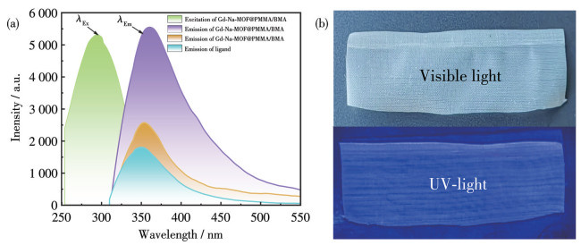
Fluorescence spectra of H4L and Gd-Na-MOF solid powder and Gd-Na-MOF@PMMA/BMA immersed in aqueous solution (Fig.2a) showed distinct characteristic emission at 351 nm (λex=300 nm), indicating that the emission of the complex originated from the ligand. Moreover, the emission intensity of Gd-Na-MOF@ PMMA/BMA was more pronounced, so this peak was used as the detection peak for fluorescence sensing of the composite film. And the physical images of Gd-Na-MOF@PMMA/BMA film under visible and UV-light radiation are shown in Fig.2b.
Figure 2
Figure 2.
(a) Fluorescence spectra of H4L, Gd-Na-MOF solid powder, and Gd-Na-MOF@PMMA/BMA; (b) Physical images of Gd-Na-MOF@PMMA/BMA film under visible- and UV-light radiation
2.4
Fluorescence sensing of TA by Gd-Na-MOF@PMMA/BMA
Following the sensing experimental protocol, 1 mL of deionized water was replaced with buffer solutions of varying pH values (2, 4, 6, 7, 8, 10, 12). Acetate buffers (HAc-NaAc) were used for pH 2-6, deionized water for pH 7, and ammonia buffers (NH3·H2O-NH4Cl) for pH 8-12. The results revealed that the fluorescence intensity variation (ΔF) initially increased and then decreased with rising pH, reaching its maximum near 7 (Fig.S4). Consequently, no buffer medium was added in subsequent sensing experiments. Further experiments tested the fluorescence intensity at different response times (Fig.S5). The ΔF remained essentially stable within 20 min, indicating that the sensing time has minimal impact on the detection results.
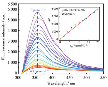
When the concentration of TA (0-400 μmol·L-1) was changed, it was found that ΔF gradually increased with the increase of cTA (Fig.3). Notably, within the cTA range of 0-20 μmol·L-1, ΔF exhibited a strong linear correlation with cTA: ΔF=16.1887+197.90cTA, R2= 0.994 3. The limit of detection (LOD) was 1.66 μmol·L-1 (LOD=3σ/k, where σ is the standard deviation of the blank signal and k=197.90 L·μmol-1). The performance of fluorescence detection for TA with the method was compared with the existing literature (Table 1). The results demonstrate that this method offers a lower LOD and a more straightforward operational procedure.
Figure 3
Figure 3.
Fluorescence spectra of Gd-Na-MOF@PMMA/BMA in TA solutions with different concentrations
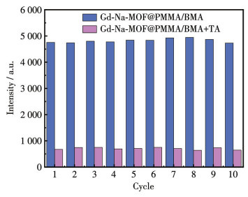
The reusability of fluorescent film sensors is crucial for reducing costs in practical applications. After detection of TA by Gd-Na-MOF@PMMA/BMA, the TA was easily eluted with water (Fig.4), and after repeated use for ten times, the fluorescence response performance remained stable, and the fluorescence intensity recovery rate remained above 90% after each cycle. This characteristic demonstrates the reusability of Gd-Na-MOF@PMMA/BMA for TA sensing detection. Compared with traditional disposable sensors or powder MOF dispersion systems, the reusability of this fluorescent film significantly reduces consumable consumption and analytical costs. This type of sensor has significant advantages in scenarios that require high-frequency detection, such as on-site environmental monitoring and rapid food screening, and also lays an important foundation for large-scale practical applications.
Figure 4
Figure 4.
Reusability of the Gd-Na-MOF@PMMA/BMA sensor
The fluorescence quenching mechanisms of MOFs can be primarily attributed to the following four aspects.
(1) Structural destruction: quenchers induce the collapse or dissociation of the MOF framework, leading to the destruction of its inherent luminescent centers.
(2) Quencher-ligand interaction: quenchers directly interact with the luminescent ligands within the MOF. This interaction alters or perturbs the electronic states or energy level structure of the ligands, thereby suppressing their luminescence process.
(3) Energy transfer: collisions occur between the fluorescent species in the singlet excited state and quencher molecules. This facilitates the transfer of excitation energy from the donor to the acceptor, preventing the donor from undergoing radiative de-excitation and resulting in fluorescence quenching.
(4) Inner filter effect (IFE): quenchers absorb the excitation light and/or the emission light of the system. This absorption reduces the detectable fluorescence intensity, manifesting as fluorescence attenuation or even complete quenching[36].
From the UV absorption spectrum of TA (Fig.S6) and the fluorescence spectra (excitation and emission spectra) of the complex, it can be seen that the absorption spectrum of TA significantly overlapped with the excitation spectrum of Gd-Na-MOF@PMMA/BMA around 310 nm, suggesting that the quenching mechanism may be caused by IFE.
2.7
Analysis of actual samples
2.7.1
Influence of coexisting ions
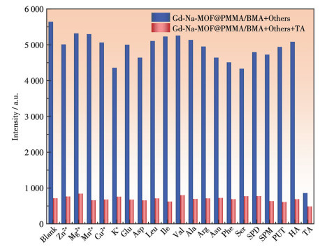
Bananas usually contain various chemical substances such as Zn2+, Mg2+, Mn2+, Cu2+, K+, glutamic acid (Glu), aspartic acid (Asp), leucine (Leu), isoleucine (Ile), valine (Val), alanine (Ala), arginine (Arg), asparagine (Asn), phenylalanine (Phe), serine (Ser), histamine (HA), TA, as well as spermidine (SPD), spermine (SPM), putrescine (PUT), etc[37-39]. To evaluate selectivity, Gd-Na-MOF@PMMA/BMA was respectively immersed in 1 mL of a 0.01 mol·L-1 solution containing the above species. Only TA induced a significant and specific fluorescence quenching effect on the film, achieving a quenching efficiency of up to 85% (Fig.5).
Figure 5
Figure 5.
Selective recognition performance of Gd-Na-MOF@PMMA/BMA for common substances in bananas
Furthermore, when 0.3 mL of a 1.0 mmol·L-1 TA solution was added to a system containing the above coexisting substances, the degree of fluorescence quenching was the same as when TA was present alone (Fig.5). This result confirms that the presence of other coexisting substances does not interfere with the fluorescence detection of TA by Gd-Na-MOF@PMMA/BMA, demonstrating its excellent anti-interference capability and selective recognition for the target analyte.
2.7.2
Detection of TA in banana samples
The banana stock solution (1 mL) was taken, and six parallel determinations (n=6) of TA in bananas were conducted according to the sensing experiment. Meanwhile, TA solutions of different concentrations (5, 10, and 15 μmol·L-1) were added for the spiked recovery experiments. The average recovery rate was 96.92%-100.26%, and the relative standard deviation (RSD) was 0.98%-2.61%. The results are listed in Table 2.
Table 2
Table 2.
Determination of TA in actual samples (n=6)
In this study, a lanthanide metal-organic framework complex (Gd-Na-MOF) was successfully designed and synthesized. Through systematic characterizations such as single-crystal X-ray diffraction, PXRD, TGA, IR, and fluorescence spectroscopy, it was confirmed that this material has a unique 3D crystal structure, excellent phase purity, good thermal stability, and significant fluorescence emission properties. To address the problems of poor solubility and difficult recovery of powdered MOFs in liquid-phase sensing, this study innovatively adopted a polymer embedding strategy. The Gd-MOF powder was compounded with a polymer material (PMMA-BMA) to prepare a flexible, washable, and easy-to-operate Gd-Na-MOF@PMMA/BMA composite film. This film was used for the sensing and detection of TA in bananas. The results show that the composite film exhibits advantages such as high sensitivity, excellent selectivity, rapid response, strong stability, good reusability, and reliable practical application ability for TA in banana samples, demonstrating broad practical application prospects in the field of fluorescence sensing. This research strategy provides new ideas for the application of metal-organic framework materials in food detection.
Supporting information is available at http://www.wjhxxb.cn
[1]
SHENG W, SUN C C, FANG G Z, WU X N, HU G S, ZHANG Y, WANG S. Development of an enzyme-linked immunosorbent assay for the detection of tyramine as an index of freshness in meat and seafood[J]. J. Agric. Food Chem., 2016, 64(46): 8944-8949 doi: 10.1021/acs.jafc.6b04422
[2]
MA L, CHEN Z C, LI X, LIU W W, YU Z C, LI C G, YU G, XU Q Y. De novo synthesis of tyramine in engineered Escherichia coli using two-stage dissolved oxygen-controlled fermentation[J]. J. Agric. Food Chem., 2025, 73(7): 4174-4184 doi: 10.1021/acs.jafc.4c11385
[3]
KAPOOR A, RAJPUT J K. Electroactive core-shell chitosan-coated lanthanum iron oxide as a food freshness level indicator for tyramine content determination[J]. ACS Sustainable Chem. Eng., 2022, 10(35): 11666-11679 doi: 10.1021/acssuschemeng.2c03747
[4]
WANG T T, LIU F N, CHEN C X, LU Y Z. Fluorometric "AND" logic gate for detection of tyramine and tyrosinase based on in-situ formation of silicon-containing nanoparticles[J]. Anal. Chim. Acta, 2024, 1298: 342415 doi: 10.1016/j.aca.2024.342415
[5]
FAN L H, ZHANG J Y, ZHAO Y, SUN C Y, LI W J, CHANG Z D. A robust Eu-MOF as a multi-functional fluorescence sensor for detection of benzaldehyde, Hg2+, and Cr2O72-/CrO42-[J]. Microchem. J., 2024, 196: 109712 doi: 10.1016/j.microc.2023.109712
[6]
QI D Y, SI X, GUO L L, YAN Z P, SHAO C Y, YANG L R. Two novel and high-efficiency optical chemosensors of detecting Fe3+ and CrO42- based on metal-organic frameworks of Cd(Ⅱ)[J]. J. Solid State Chem., 2022, 314: 123400 doi: 10.1016/j.jssc.2022.123400
[7]
YANG C L, ZHANG H W, HOU C H, SUN F F, XU G J. Self-assembly fluorescent copper nanoclusters in alginate-based hydrogel sensor for histamine detection and visual monitoring of food spoilage[J]. Talanta, 2025, 294: 128292 doi: 10.1016/j.talanta.2025.128292
[8]
YAN Y, GUO H, YU Z, YANG Z Y, ZHUANG D K, MA Y Y, WANG M Y, YANG W. A rapid on-site fluorescence sensing platform for malachite green in an aqueous phase based on lanthanide-functionalized MOF[J]. Microchim. Acta, 2025, 192: 253 doi: 10.1007/s00604-025-07113-0
[9]
PAPAGEORGIOU M, LAMBROULOU D, MORRISON C, KŁODZIŃSKA E, NAMIEŚNIK J, PŁOTKA-WASYLKA J. Literature update of analytical methods for biogenic amines determination in food and beverages[J]. Trac‒Trends Anal. Chem., 2018, 98: 128-142 doi: 10.1016/j.trac.2017.11.001
[10]
GU M Y, SUN M R, LU Y, ZENG D C, ZHAO J H, CHANG Z H, XIN D X, ZHOU X Y, HU Y Y, DU E D, ZHANG Y Q, PENG M G. A facile fluorescence Zr-MOF probe for selective sensing tetracycline in water[J]. Inorg. Chem. Commun., 2025, 178: 114548 doi: 10.1016/j.inoche.2025.114548
[11]
LU Y Y, ZHOU H J, YANG H, ZHOU Z, JIANG Z C, PANG H. Anisotropy of metal-organic framework and their composites: Properties, synthesis, and applications[J]. J. Mater. Chem. A, 2024, 12(15): 6243-6260
[12]
GAO Y X, ZHU Y Y, WANG Y P, BI J H. Water-stable Ln-MOF as a multi-emitting luminescent sensor for the detection of metal ions and pharmaceuticals[J]. Spectroc. Acta Pt. A‒Molec. Biomolec. Spectr., 2024, 323: 124915 doi: 10.1016/j.saa.2024.124915
[13]
YAN R K, CHEN X L, REN J, CUI H L, YANG H, WANG J J. Design and synthesis of a new highly efficient adjustable Ln-MOF for fluorescence sensing and information encryption[J]. Spectroc. Acta Pt. A‒Molec. Biomolec. Spectr., 2025, 330: 125669 doi: 10.1016/j.saa.2024.125669
[14]
中国科学院宁波材料技术与工程研究所. 提高MOFs在聚合物溶液中分散性的方法以及MOFs/聚合物复合膜的制备方法: CN201810294450.4[P]. 2021-03-26.NINGBO INSTITUTE OF MATERIALS TECHNOLOGY AND ENGINEERING, CHINESE ACADEMY OF SCIENCES. Method for improving dispersibility of MOFs in polymer solution and preparation method of MOFs/polymer composite membrane: CN201810294450.4[P]. 2021-03-26.
[15]
APPELHAN N L, HUGHES L, MCKENZIE B, RODRIGUEZ M, GRIEGO J, BRISCOE J, MOORMAN M, FREDERICK E, WRIGHT B J. Facile microwave synthesis of zirconium metal-organic framework thin films on gold and silicon and application to sensor functionalization[J]. Microporous Mesoporous Mat., 2021, 323: 111133 doi: 10.1016/j.micromeso.2021.111133
[16]
SABZEHEMEIDANI M M, GAFARI S, JAMALI S, KAZEMZAD M. Concepts, fabrication and applications of MOF thin films in optoelectronics: A review[J]. Appl. Mater. Today, 2024, 38: 102153 doi: 10.1016/j.apmt.2024.102153
[17]
WANG X, WANG Y, CHEN L, XIE X F, SUN J. Gel-state MOFs for environmental decontamination: Synthesis, application and optimization[J]. Chem. Eng. J., 2024, 499: 156241 doi: 10.1016/j.cej.2024.156241
[18]
MAKIURA R. Creation of metal-organic framework nanosheets by the Langmuir-Blodgett technique[J]. Coord. Chem. Rev., 2022, 469: 214650 doi: 10.1016/j.ccr.2022.214650
[19]
SUN Z H, ZHAO G K, TANG G Q, ZHAO Z H, LI P. Preparation of high-performance pervaporation membranes for ethanol dehydration using a layer-by-layer self-assembly method[J]. Adv. Membr., 2025, 5: 100132 doi: 10.1016/j.advmem.2025.100132
[20]
CEVIK E, IQBAL A, MUSTAFA A, QAHAN F T, ZEEZHAN M, ISIK O. Metal organic frameworks embedded polymeric membranes: A comprehensive review on application in water purification and seawater desalination[J]. J. Environ. Chem. Eng., 2025, 13(3): 116215 doi: 10.1016/j.jece.2025.116215
[21]
ZHANG Z H, MA W P, YAN B. Multi-step tandem functionalization assembly of MOFs-based hybrid polymeric films for color tuning luminescence and responsive sensing on organic vapors[J]. Colloid Surf. A‒Physicochem. Eng. Asp., 2022, 648: 129416 doi: 10.1016/j.colsurfa.2022.129416
[22]
MUTMAINNA I, TAHIR D, GARESO L P, SURYANI S. Development of PVA-chitosan based smart packaging with the addition of red cabbage (Brassica Oleracea Var. capitata F. rubra) anthocyanin extract and copper-based metal-organic material (Cu-MOF)[J]. Int. J. Biol. Macromol., 2025, 313: 14420
[23]
ALI A, ALTAKROORI H H D, GREISH Y E, ALZAMLY A, SIDDIG L A, QAMHIEH N, MAHMOUD S T. Flexible Cu3(HHTP)2 MOF membranes for gas sensing application at room temperature[J]. J. Nanomater., 2022, 12(6): 913 doi: 10.3390/nano12060913
[24]
KAZEMI A, MOHAMMADI M, PORDSARI M A, TAMTAJI M, BAESMAT H, KESHAVARZ S, ZEINALI F, MANTEGHI F, FASIHI M, GHAEMI A, ROHANI S, GODDARD A W. Eco-friendly mixed-metal MOF embedded in PVA-based packaging films for ethylene adsorption and enhancing fresh produce shelf-life[J]. Food Packaging Shelf Life, 2025, 49: 101517 doi: 10.1016/j.fpsl.2025.101517
[25]
FANG M, DROBEK M, COT D, MONTORO C, SEMSARILAR M. A straightforward method to prepare MOF-based membranes via direct seeding of MOF-polymer hybrid nanoparticles[J]. J. Membr. Sci., 2023, 13(1): 65
[26]
KRAUSE L, HERBST-IRMER R, SHELDRICK G M, STALKE D. Comparison of silver and molybdenum microfocus X-ray sources for single-crystal structure determination[J]. J. Appl. Crystallogr., 2015, 48(1): 3-10 doi: 10.1107/S1600576714022985
[27]
SHELDRICK G M. Crystal structure refinement with SHELXL[J]. Acta Crystallogr. Sect. A, 2015, A71(1): 3-8
[28]
KIRKAN B. Enzyme inhibition: Mechanisms, types, and applications in drug development[J]. J. Cell. Mol. Pharmacol., 2024, 8(6): 249
[29]
ALLWARDT J R, STEBBINS J F, SCHMIDT B C, FROST D J, WITHERS A C, HIRSCHMANN M M. Aluminum coordination and the densification of high-pressure aluminosilicate glasses[J]. Am. Miner., 2005, 90(7): 1218-1222 doi: 10.2138/am.2005.1836
[30]
GAGNÉ O C, HAWTHORNE F C. Bond-length distributions for ions bonded to oxygen: Results for the transition metals and quantification of the factors underlying bond-length variation in inorganic solids[J]. IUCrJ, 2020, 7(4): 581-629 doi: 10.1107/S2052252520005928
[31]
ESTEBAN T G, SÁNCHEZ L B, EXPÓSITO L, RODRÍGUEZ-SAN-MIGUEL D, ZAMORA F, PARIENTE F, GUTIÉRREZ-SÁNCHEZ C, LORENZO E. Synergistic enhancement of electrochemiluminescence through hybridization of α-Ge nanolayers and gold nanoparticles for highly sensitive detection of tyramine[J]. Sens. Actuator B‒Chem., 2023, 396: 134649 doi: 10.1016/j.snb.2023.134649
[32]
SINGH R, ZHANG W, LIU X C, ZHANG B Y, KUMAR S. WaveFlex biosensor: MXene-immobilized W-shaped fiber-based LSPR sensor for highly selective tyramine detection[J]. Opt. Laser Technol., 2024, 171: 110357 doi: 10.1016/j.optlastec.2023.110357
[33]
AHMAD M W, DEY B, KIM B H, SARKHEL G, YANG D J, HOSSAIN S S, KAMAL T, CHOUDHURY A. Bimetallic copper-cobalt MOFs anchored carbon nanofibers hybrid mat based electrode for simultaneous determination of dopamine and tyramine[J]. Microchem. J., 2023, 193: 109074 doi: 10.1016/j.microc.2023.109074
[34]
TORRE R, CERRATO-ALVAREZ M, NOUWS P A H, DELERUE-MATOS C M, FERNÁNDEZ-ABEDUL T, COSTA-RAMA E. A hand-drawn graphite electrode for affordable analysis: Application to the enzymatic determination of tyramine in fish[J]. Sens. Actuator B‒Chem., 2025, 423: 136705 doi: 10.1016/j.snb.2024.136705
[35]
ZHANG D W, ZHANG Y H, LI K X, WANG S N, MA Y C, LIAO Y H, WANG F H, LIU H. A smartphone-combined ratiometric fluorescence molecularly imprinted probe based on biomass-derived carbon dots for determination of tyramine in fermented meat products[J]. Food Chem., 2024, 454: 139759 doi: 10.1016/j.foodchem.2024.139759
[36]
SU Y, YU J H, LI Y B, PHUA S F Z, LIU G F, LIM W Q, YANG X Z, GANGULY R. Versatile bimetallic lanthanide metal-organic frameworks for tunable emission and efficient fluorescence sensing[J]. Commun. Chem., 2018, 1: 12 doi: 10.1038/s42004-018-0016-0
[37]
傅院霞, 王莉, 徐丽, 周彧, 宫昊. 水果中微量元素的LIBS检测[J]. 蚌埠学院学报, 2020, 9(5): 71-75FU Y X, WANG L, XU L, ZHOU Y, GONG H. LIBS detection of trace elements in fruits[J]. Journal of Bengbu University, 2020, 9(5): 71-75
[38]
GAO H J, LIU P, HE W D, BI F C, HU C H, DENG G M, DOU T X, YANG Q S, LI C Y, YI G J, SHENG O, DONG T. Ripening-stage variations in small metabolites across six banana cultivars: A metabolomic perspective[J]. Food Chem., 2025, 478: 143658 doi: 10.1016/j.foodchem.2025.143658
[39]
BORGES C V, BELIN M A F, AMORIM E P, MINATEL I O, MONTEIRO G C, GOMEZ H A G, MONAR G R S, LIMA G P P. Bioactive amines changes during the ripening and thermal processes of bananas and plantains[J]. Food Chem., 2019, 298: 125020 doi: 10.1016/j.foodchem.2019.125020
Figure 1
Crystal structure of the complex: (a) coordination environment; (b) 1D chain of dual-core units; (c) 3D microporous structure
Figure 2
(a) Fluorescence spectra of H4L, Gd-Na-MOF solid powder, and Gd-Na-MOF@PMMA/BMA; (b) Physical images of Gd-Na-MOF@PMMA/BMA film under visible- and UV-light radiation

 下载:
下载:

 下载:
下载:


