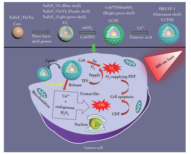
Scheme 1.
Schematic fabrication process of the UCTSH and their NIR-triggered PDT with self-supplying O2 and pH-responsive CDT for synergistic therapy

Cancer treatment strategy based on the special properties of reactive oxygen species (ROS) is a new science rising gradually in recent years[1]. ROS with strong oxidation activity mainly include the superoxide anion (·O2-), the hydroxyl radical (·OH), and singlet oxygen (¹O2). And, the continuously elevated ROS level can lead to organelle damage and stress-induced apoptosis of cancer cells, thereby inhibiting cancer progression. On this basis, a variety of ROS-mediated nanotherapeutic strategies have been gradually developed. Among the various cancer therapy methods, chemodynamic therapy (CDT), photodynamic therapy (PDT), and some combined treatments have garnered significant attention[2-4]. CDT is a cancer treatment strategy that utilizes Fenton or Fenton-like catalytic reactions to kill tumors or cancer cells by converting hydrogen peroxide (H2O2) into highly cytotoxic ·OH. PDT is a highly selective and noninvasive treatment that combines three elements: light source, photosensitizers, and oxygen (O2). Its therapeutic principle is that the photosensitizer absorbs the energy of an external light source and converts O2 to highly toxic 1O2 to the targeted tissues for tumor ablation[5]. In order to obtain a high level of ROS, designing nanotherapeutic systems based on the two methods has great significance for improving the efficiency of cancer treatment.
At present, there are mainly three nanotherapeutic systems based on CDT and PDT: (1) the nanostructures that can catalyze metallic materials to generate ·OH. Many such nanoparticles have been extensively developed, i.e., iron nano-metallic glasses[6-7], FePt[8-9], and Fe-ZIF-8[10]. However, using this kind of material alone cannot supply adequate ·OH because of the insufficient H2O2 content (100 μmol·L-1-1 mmol·L-1) in the tumor cells[11-14]. To solve this problem, some materials have been used to self-supply H2O2 in an acidic environment[15-16]. However, the therapeutic effect of these materials alone is still not good. (2) The nanostructures that can convert O2 to highly toxic 1O2. Due to the limitation of the oxygen content of anaerobic tumors, the therapeutic effect of these single PDT platforms is not ideal for the treatment of cancer[17-19]. (3) The nanotherapeutic systems with synergistic CDT and PDT functions. Compared with the first two nanostructures, this therapeutic strategy has a good comprehensive treatment effect on cancer, and, thus, attracts much attention in the construction of nanotherapeutic systems. At present, the nanoplatforms that integrate these two methods include metal-organic frameworks (MOFs)[20], nanodots[21], nanogels[22], upconversion nanoparticles[23], and so on. Among them, upconversion nanoparticles have received lots of attention since they possess the outstanding advantages: a narrow emission peak, large Stokes shift, low toxicity, deep tissue penetration, and good photostability[24-25].
Considering the therapeutic effect and the advantages of upconversion nanomaterials, we designed a multifunctional nanosystem: a Cu-based MOF and a mesoporous silica (mSiO2) coated upconversion nanoplatform with self-supplying O2, which can improve the level of ROS in the tumor microenvironment, thereby enhancing the combination therapy of CDT and PDT. As shown in Scheme 1, a Nd3+-sensitized core-shell structured upconversion nanoparticles (NaErF4: Yb/Tm@ NaLuF4: Yb@NaLuF4: Nd/Yb@NaLuF4, noted as UC) was coated with a mSiO2 shell, which had chlorin e6 (Ce6) and triethoxy(1H, 1H, 2H, 2H-nonafluorohexyl) silane (TFS) covalently loaded. Thereafter, HKUST-1, a kind of Cu-based MOF shell, was grown on mSiO2 using a facile way at room temperature to obtain UC@Ce6/TFS@mSiO2@HKUST-1 (UCTSH). In this nanosystem, the core-shell structure allows UC nanoparticles to harvest 808 nm photons and achieve red emission through a multiphoton process, which indicates that the photosensitizer can be activated in the therapeutic window. At the same time, TFS has the function of carrying oxygen, which can increase the content of O2 and promote the production of more 1O2. Furthermore, the MOF shell of UCTSH would be quickly degraded in the acidic tumor microenvironment to realize the responsive release of Cu2+. The released Cu2+ participates in a Fenton-like reaction to generate ·OH to kill cancer cells. The ROS generators achieved by Cu2+ and Ce6 were used for metal ion-triggered ·OH production and NIR-induced 1O2 production. Noticeably, the strategy upregulated the intracellular ROS level. Compared with CDT or PDT alone, the rapid increase of the ROS concentration in combined CDT/PDT with self-supplying O2 can lead to cancer cell apoptosis.
All the chemicals were used without further purification. Rare-earth acetates [thulium(Ⅲ)acetate hydrate, erbium(Ⅲ)acetate tetrahydrate, neodymium(Ⅲ)acetate hydrate, and lutetium(Ⅲ)acetate hydrate], oleic acid (OA), 1-octadecene (ODE), sodium oleate (NaOA), and 1,3-diphenylisobenzofuran (DPBF) were purchased from Sigma-Aldrich. TFS was purchased from TCI. Ce6 was purchased from Frontier Scientific. Cetyltrimethylammonium bromide (CTAB) was purchased from Adamas. 1-Ethyl-3-(3-dimethylaminopropyl)-carbodiimide, N-hydroxysuccinimide sodium, tetraethoxysilane (TEOS), and (3-aminopropyl)triethoxysilane (APTES) were purchased from Aladdin Company. Copper acetate was purchased from Tianjin Fuchen Chemical Reagent Company. Trimesic acid was purchased from Shanghai Chemical Corporation. 2′,7′-Dichlorodihydrofluorescein diacetate (DCFH-DA) was obtained from Macklin Biochemical Company. Calcein-AM/propidium iodide (PI) cell viability/cytotoxicity assay kit and 3-(4,5-dimethylthiazol-2-yl)-2,5-diphenyltetrazolium bromide (MTT) were purchased from Beyotime Biotechnology.
The absorption spectra were recorded with an ultraviolet-visible (UV-Vis)-2450 spectrometer (Shimadzu, Japan). The upconversion emission spectra were obtained on a Raman Fluorescence (RF)-5301 spectrofluorometer (Shimadzu, Japan) equipped with an external 808 nm laser excitation source. X-ray photoelectron spectroscopy (XPS) measurements were carried out on an ULVAC-PHI 5000 Versa Probe Ⅲ instrument. Transmission electron microscopy (TEM) and elemental mapping analysis were performed on a Thermo Scientific Talos F200X microscope (200 kV). Powder X-ray diffractometer (PXRD, BRUKER D8 Advance, Germany, Cu Kα irradiation, λ=0.154 06 nm, U=60 kV, I=80 mA, 2θ=5°-140°) was used for the phase composition analysis of the sample. Fourier-transform infrared (FTIR) spectra were obtained with a Tensor27 FTIR infrared spectrometer (BRUKER, Germany). The specific surface area porosity analyzer (TriStar Ⅱ 3020, USA) was used to measure the specific surface area of the sample. An INESA JPSJ-605F dissolved O2 analyzer was used to determine the O2-carrying capacity of UCTSH. The confocal laser scanning microscope (CLSM) images were collected on a Nikon A1 equipment microscope (Japan).
NaErF4: Yb/Tm was prepared by using a robust binary sodium strategy[26]. 0.6 mmol of the rare-earth acetates (molar fractions of Er, Yb, and Tm were 30%, 69%, and 1%, respectively) were added to a 50 mL flask containing 3.6 mL of OA and 9 mL of ODE. The mixture was heated at 160 ℃ for 30 min under an argon environment and then cooled down to 35 ℃. Subsequently, NaOA (395.8 mg) was added, followed by stirring for 10 min. NH4F (88.8 mg) and NaOH (8.0 mg) were dissolved in methanol (6 mL) and added dropwise. The solution was stirred for 30 min, and then heated to 100 ℃ to remove methanol. Finally, the reaction solution was heated at 300 ℃ for 1 h and cooled to room temperature. NaErF4: Yb/Tm core was collected by centrifugation using anhydrous ethanol, washed with cyclohexane and anhydrous ethanol, and then dispersed in cyclohexane.
Coating the NaLuF4: Yb shell onto NaErF4: Yb/Tm core was realized by the seed-mediated epitaxial growth strategy[27]. 0.3 mmol of the corresponding rare-earth acetates (molar fractions of Lu and Yb were 85% and 15%) were added to a 50 mL flask containing 3.0 mL of OA and 7.0 mL of ODE. The mixture was heated at 150 ℃ for 60 min under an argon environment and then cooled down to 50 ℃. Corresponding NaErF4: Yb/Tm core (0.3 mmol) was added to the reaction solution. Subsequently, NH4F (44.4 mg) and NaOH (30.0 mg) were dissolved in methanol (5 mL) and added dropwise. The solution was stirred for 30 min, and then heated to 100 ℃ to remove methanol. Finally, the reaction solution was heated at 300 ℃ for 1 h and cooled to room temperature. The NaErF4: Yb/Tm@NaLuF4: Yb were purified and dispersed in cyclohexane.
A sensitized shell NaLuF4: Nd/Yb was grown on the surface of NaErF4: Yb/Tm@NaLuF4: Yb. 0.3 mmol of the corresponding rare-earth acetates (molar fractions of Lu, Nd, and Yb were 60%, 30%, and 10%, respectively) was added to a 50 mL flask containing 3.0 mL of OA and 7.0 mL of ODE. The mixture was heated at 150 ℃ for 60 min under an argon environment and then cooled down to 50 ℃. NaErF4: Yb/Tm@NaLuF4: Yb (0.3 mmol), methanol (5 mL) containing NaOA (0.375 mmol), NH4F (1.2 mmol), and NaOH (0.375 mmol) were injected into the flask and maintained for 30 min, and then heated to 100 ℃ to remove methanol. Finally, the reaction solution was heated at 300 ℃ for 1 h and cooled to room temperature. The product NaErF4: Yb/Tm@NaLuF4: Yb@NaLuF4: Nd/Yb was purified and dispersed in cyclohexane.
The growth method of the outermost inert shell was consistent with the method for preparing NaLuF4: Yb. 0.3 mmol of lutetium(Ⅲ)acetate hydrate was added to a 50 mL flask containing 3.0 mL of OA and 7.0 mL of ODE. The mixture was heated at 150 ℃ for 60 min under an argon environment and then cooled down to 50 ℃. The product obtained in the previous step (NaErF4: Yb/Tm@NaLuF4: Yb@NaLuF4: Nd/Yb) was added to the reaction solution. Subsequently, NH4F (44.4 mg) and NaOH (30.0 mg) were dissolved in methanol (5 mL) and added dropwise. The solution was stirred for 30 min, and then heated to 100 ℃ to remove methanol. Finally, the reaction solution was heated at 300 ℃ for 1 h and cooled to room temperature. The final upconversion nanomaterials NaErF4: Yb/Tm@NaLuF4: Yb@NaLuF4: Nd/Yb@NaLuF4 were labeled UC.
UCTS was synthesized by the sol-gel method using CTAB as a template. First, Ce6 was modified with APTES by mixing 250 μL of dimethyl sulfoxide with 3.0 mg of 1-ethyl-3-(3-dimethylaminoprop-yl)-carbodiimide, 2.0 mg of N-hydroxysuccinimide sodium, 1.0 mg of Ce6, and 6 μL of APTES. The mixture was stirred at room temperature for 2 h and stored at 4 ℃ until further use. Thereafter, UC in chloroform (150 μL, 0.1 mol·L-1) was added dropwise to 2.5 mL CTAB aqueous solution (15 mg·mL-1) to form a milky oil-in-water microemulsion. The resulting solution was sonicated for 40 min, and then the chloroform solvent was evaporated in a 60 ℃ water bath. Then the solution was added to 22.5 mL of water and heated to 70 ℃. After 10 min of stirring, 150 μL of NaOH (2 mol·L-1), 70 μL of TEOS, 45 μL of TFS in 200 μL of acetonitrile, and 1.25 mg of APTES-premodified Ce6 were slowly added; then, 0.5 mL of ethyl acetate was injected quickly. The mSiO2 co-doped with Ce6/TFC was grown at 70 ℃ for 3 h. Next, the solution was centrifuged and washed sequentially with water and ethanol for one cycle. UCTS was obtained by removing CTAB from the mSiO2 layer with an ethanol solution of NH4NO3 (0.075 mol·L-1, 60 ℃, 2 h), and washing sequentially with deionized water and ethanol for two cycles, dispersing in pure water, and storing at 4 ℃ until further use.
UC@mSiO2 (US) and UC@Ce6@mSiO2 (UCS) were synthesized and denoted similarly to UCTS, except that the corresponding precursors were used.
For the HKUST-1 coating, copper acetate (10 mmol, 2 mL), UCTS (4 mg), and 3 mL ethanol were added into a 25 mL flask and stirred for 30 min. Then, the product was centrifuged and washed twice with ethanol to remove the excess Cu2+, and following, dispersed in 2 mL of ethanol. Trimesic acid (10 mmol) in 2 mL of ethanol was poured into the above solution. After reaction for 30 min, nanoparticles were centrifuged and washed with ethanol and water, and finally dispersed in 2 mL water for further use.
The UCTSH (4 mg·mL-1) dispersed in water (2.0 mL) was first saturated with O2 and then injected into of deoxygenated water (10.0 mL), which was prepared in a sealed dissolved O2 analyzer equipped with a glass bottle by bubbling N2. The concentration of dissolved O2 was monitored under gentle magnetic stirring. The oxygen-carrying detection method of UCS and UCTSH was the same as the above method.
1O2 generation was measured by using a probe of DPBF. UCTSH dispersion (400.0 μL, 100.0 μg·mL-1) was mixed with a dimethyl sulfoxide solution of DPBF (6.0 μL, 1 mg·mL-1). The UV spectra were recorded at a predetermined point in time during the 808 nm laser irradiation (2.0 W·cm-2).
Measurements were carried out in 1 mL of pH 5.5 acetate buffer or pH 7.4 phosphate buffer (PBS) containing UCTSH (100.0 μg·mL-1), H2O2 (5 mmol·L-1), and 3,3′,5,5′-tetramethylbenzidine (TMB, 1.0 mmol·L-1) at room temperature for 1 h. The absorbance spectra were observed using a UV-Vis spectrometer.
ROS content was measured by using a probe of DPBF and DCFH-DA. The UCTSH dispersion (400.0 μL, 100.0 μg·mL-1) was mixed with a dimethyl sulfoxide solution of DPBF (6.0 μL, 1 mg·mL-1) or a DCFH-DA (10 μmol·L-1, 20 μL) solution containing or without a certain amount of H2O2 at a pH 5.5 acetate buffer. The UV spectra (417 nm) or fluorescence spectra (λEx=435 nm, λEm=520 nm) were recorded during the 808 nm laser irradiation (2.0 W·cm-2).
The uptake of UCTSH by cancer cells was evaluated. 5×10³ of MCF-7 cells were first seeded in 20 mm glass-bottom cell culture dishes. After incubation at 37 ℃ for 24 h, the culture medium was then replaced with 125.0 μg·mL-1 of UCTSH suspended in fresh medium and continued to culture for different times. After different times of incubation, cell nuclei were stained with DAPI. Finally, MCF-7 cells were observed by CLSM.
To evaluate the cytotoxic effect of different nanomaterials, the MTT assays were conducted. MCF-7 cells (5×10³ cells per well) were seeded in 96-well plates and cultured for 24 h at a volume fraction of 5% CO2 and 37 ℃. After removal of the original culture media, the cells were treated with UCS, UCTS, or UCTSH at various concentrations. The cells were divided into two groups. For one group without NIR laser irradiation treatment, the cells were incubated for 24 h before typical MTT assays. For another group, the cells were incubated for 12 h and then were exposed to NIR laser irradiation (2.0 W·cm-2) for 5 or 10 min, followed by a further 12 h incubation before MTT assays.
The ROS inside MCF-7 cells was measured with DCFH-DA. To experiment, MCF-7 cells (5×10³ cells per dish) were seeded in 20 mm glass-bottom cell culture dishes and incubated for 24 h. Then, the culture medium was replaced by fresh medium containing PBS, UCTS (125.0 μg·mL-1), or UCTSH (125.0 μg·mL-1). After 12 h of incubation, the cells were washed with PBS three times. Then, DCFH-DA solution (20.0 μmol·L-1) was added to the cells for another 15 min at 37 ℃. Finally, the cells were washed with PBS several times and exposed to 808 nm laser irradiation (2.0 W·cm-2) for 5 min before the 2′,7′-dichlorofluorescein (DCF) fluorescence imaging observations by CLSM.
To show the in vitro synergistic therapy efficiency of UCTSH, the killing effect of UCTSH towards cancer cells was visualized by a live/dead cell double staining kit and CLSM observation. The cell culture procedures were similar to those in the above cellular uptake studies. Then, the original culture media were replaced with fresh media containing PBS or the as-designed nanoconstructs (UCS, UCTS, or UCTSH, 250.0 μg·mL-1 in PBS). After 12 h, the cells were exposed to NIR laser irradiation (2.0 W·cm-2) for 10 min and further incubated for 12 h. Then, the cells were incubated with Calcein-AM/PI cell viability/cytotoxicity assay kit for 30 min at 37 ℃. The cells were washed with PBS for fluorescence imaging via a CLSM. The groups without NIR laser irradiation were used as controls.
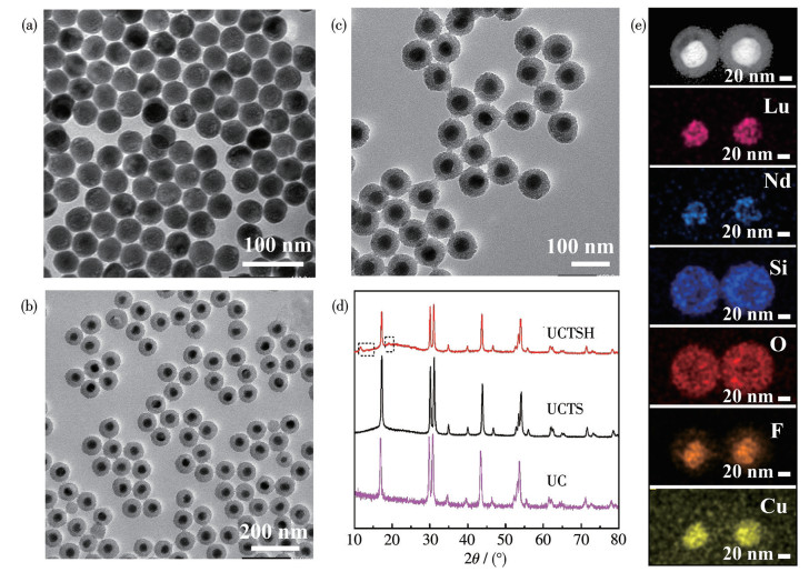
The synthetic procedure of UCTSH is presented in Scheme 1. First, UC was fabricated via a robust binary sodium strategy[26] and an epitaxial growth way[27] for the first time. The core-shell structure UC can be excited by 808 nm light to effectively avoid the thermal effect caused by excitation because of the minimum absorption of biological tissue at this NIR wavelength. When excited by an 808 nm laser, UC performed an efficient upconversion, which is very beneficial for the subsequent application of resonance energy transfer. The UC nanoparticles were monodispersed and uniform with an average diameter of 41.2 nm (Fig. 1a). The premodified Ce6 and TFS were covalently conjugated on the mSiO2 layer along with coating the mSiO2 shell on UC, and after that, a Cu-based MOF shell (HKUST-1) was directly grown on the mSiO2 layer through a facile room temperature synthesis. Fig. 1b showed that the particle size increased to around 80.0 nm after coating with mSiO2. However, the particle size did not increase significantly after coating the MOF′s shell (Fig. 1c). Besides, the XRD patterns of the as-prepared core-shell UC, UCTS, and UCTSH are displayed in Fig. 1d. The diffraction peaks of UC were kept well in all the samples. In the XRD pattern of UCTS, there was a broad diffraction peak at 2θ=22° besides the characteristic sharp peaks of UC, implying the successful mSiO2 coating[28]. In comparison with UCTS, the XRD peaks of UCTSH at 11.6°, 13.4°, and 19.2° can be indexed to the (222), (422), and (731) planes of Cu-MOFs, respectively[29-30]. This proves the successful preparation of Cu-MOFs. The even distribution of Lu, Nd, Si, O, F, and Cu element signals further suggests the successful fabrication of mSiO2 and Cu-MOFs coated upconversion nanomaterials in Fig. 1e.
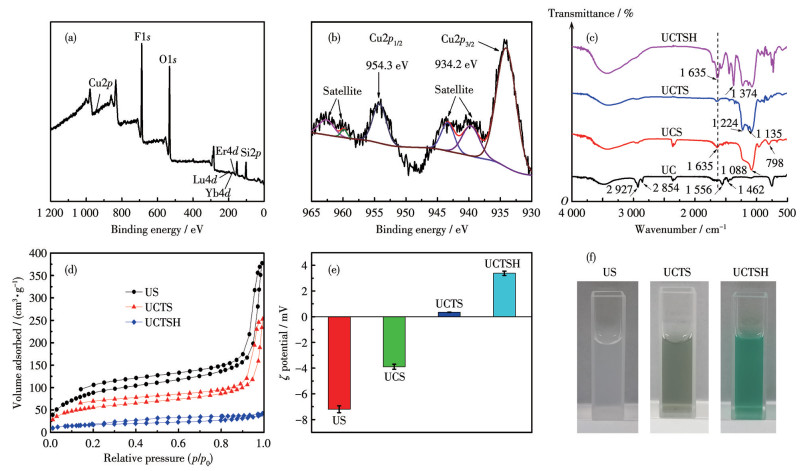
As shown in Fig. 2a and 2b, XPS uncovered the Cu signal originating from coated Cu-MOFs. Specifically, the high-resolution XPS spectrum of Cu2p indicates that Cu existed mainly in the form of Cu2+ (Cu2p3/2: 934.2 eV, Cu2p1/2: 954.3 eV) with shakeup satellite peaks (Fig. 2b). FTIR spectra of the samples in every step were tested to verify the functional groups on the sample surface. As depicted in Fig. 2c, the OA-capped UC exhibited a broad band at around 3 450 cm-1 associated with the stretching vibration of O—H. The strong transmission bands at 2 927 and 2 854 cm-1 are respectively caused by the symmetric and asymmetric stretching vibrations of CH2, and the bands at 1 556 and 1 462 cm-1 derive from the vibrations of the carboxylic groups. After coating the mSiO2 shell, the FTIR spectrum of UCS poses typical Si—O—Si deformation (798 cm-1) vibrations and asymmetric stretching (1 088 cm-1). Besides, the C=O bond peak at 1 635 cm-1 confirms that Ce6 was modified on the nanoparticles successfully. Compared with UCS, the FTIR spectrum of UCTS at 1 224 and 1 135 cm-1 showed the stretching vibration peaks of C—F, which also proved that TFS was successfully doped in mSiO2. In addition, on the FTIR spectrum of UCTSH, it is observed that the enhanced C=O bond peak at 1 635 cm-1 and the band of the aromatic ring skeleton stretching vibration at 1 374 cm-1, which indicate the existence of trimesic acid[31-32]. As can be seen in Fig. 2d, UCTSH showed a relative decrease in surface area (14 m2·g-1) and pore size (3.28 nm), while the US and UCTS had a higher surface area (312 and 190 m2·g-1, respectively). This may be because the synthesized MOFs efficiently fill the pores of the mSiO2 shell. Specifically, when a single Ce6 was combined with the empty mSiO2, the ζ potential increased from -7.20 mV of US to -3.89 mV of UCS; and when Ce6 and TFS were combined, the ζ potential increased to 0.35 mV of UCTS. After Cu-MOFs were wrapped, the ζ potential of UCTSH increased to 3.38 mV (Fig. 2e). In addition, the color of the dispersion changed from milk white of US to blue of UCTSH due to the decoration of HKUST-1 (Fig. 2f). All the above results support the successful preparation of UCTSH.
Based on the previous research[33], the Nd3+/Yb3+/Tm3+ series sensitized strong red emission upconversion nanomaterials were successfully prepared under the irradiation of an 808 nm laser. The upconversion luminescence (UCL) spectra of UC were analyzed (Fig. 3a). The peaks were mainly situated at 654 nm, which are attributed to electronic transitions from the Nd3+/Yb3+/Tm3+ series sensitization process. Fig. 3b presents the energy-transfer mechanism in UC. Specifically, as a sensitizer, Nd3+ ions absorb the energy of 808 nm excitation light through the 4F5/2 (Nd3+) energy level, transfer the energy to bridge ions Yb3+ (2F5/2), and then transfer the energy to luminous crystal nucleus NaErF4: Yb/Tm through bridge ions Yb3+ (2F5/2). Yb3+ ions in the luminous crystal nucleus first absorb the energy of Yb3+ (2F5/2) of the transition shell and transfer it to the bridge ion Tm3+, and realize strong red light emission under 808 nm excitation through an efficient Yb3+→Tm3+→Er3+ sensitization process. In the constructed multi-shell structure of upconversion nanomaterials, the strong red-light emission under 808 nm excitation is realized through an efficient series sensitization process of Nd3+→Yb3+→Tm3+→Er3+.

To implement upconversion guided PDT, wavelengths matching between upconversion luminescence and photosensitizer absorption play an important role. As shown in Fig. 3c, the absorption peak of photosensitizer Ce6 remained basically consistent with the UC emission, which reveals that the Förster resonance energy transfer (FRET) from UC to Ce6 was viable. Moreover, the emission peaks of UC between 600 and 700 nm decreased sharply after Ce6 conjugation. This can be ascribed to the FRET process between UC and Ce6 (Fig. 3d). Next, we utilized DPBF as a probe to detect the generation of ROS caused by the nanomaterial exposed to an 808 nm laser. 1O2 can react with DPBF instantly and then induce obvious DPBF fading. In order to produce more 1O2, thereby enhancing PDT, an oxygen carrier (TFS) was introduced into the UCTS, and the amount of Ce6 was also optimized. As can be seen from Fig.S1-S6 (Supporting information), the dosage of 1.25 mg Ce6 made the PDT effect the best by the UCTS exposed to an 808 nm laser. Therefore, 1.25 mg Ce6 was fed to obtain efficient PDT. Then, we discussed the effect of adding TFS or not on the efficiency of PDT. It can be seen from Fig.S4 that the UCS with TFS decreased the absorbance of DPBF by 50.4% within 10 min, whereas the UCS only decreased the absorbance of DPBF by 27.3% (Fig.S7). This proves that the addition of TFS plays a great role in enhancing the efficiency of PDT. In addition, it is reported in the literature that the addition of TFS can stabilize the fluorescence intensity of upconversion nanoparticles in water[34]. Therefore, when UCTS in water was placed for three months, the material could still reduce the absorbance of DPBF by 49.8% (Fig.S8). This fact showed that the fluorescence of UCTS in water was hardly quenched, which is conducive to stable and efficient PDT. At the same time, we also examined the PDT effect of UCTSH, reducing the absorbance of DPBF by 46.6%, which was lower than the value of UCTS due to the decline of the luminous intensity of UCTSH than UCTS (Fig. 3d and 3e). Yet, without UCTSH by 808 nm NIR laser irradiation (Fig.S9), the absorbance of DPBF showed a slight decrease by 9.2%. Besides, the oxygen-carrying capacity of UCTSH was lower than the value of UCTS due to the coating of HKUST-1 (Fig.S10). This may also be the reason for the slightly lower PDT efficiency of UCTSH. However, the oxygen storage capacity of UCTSH was still higher than that of UCS. These indicate that the UCTSH can produce 1O2 efficiently to enhance the PDT effect under 808 nm NIR laser irradiation.
To demonstrate that UCTSH can produce ·OH in the presence of H2O2, TMB was employed as a probe to monitor ·OH generation. We expect the Cu2+ released from UCTSH to react with H2O2 to form ·OH[16], and the colorless TMB can be oxidized by ·OH to chromogenic TMB cation-free radicals with the characteristic absorbance at 654 nm as shown in Fig. 4a[31]. Next, nanoparticles were dipped in PBS at pH=7.4 and acetate buffer at 5.5, respectively, to evaluate the ability to produce ·OH. As expected, the absorbance at pH 5.5 was much higher than that at pH 7.4 under the same mass concentration (100.0 μg·mL-1) of UCTSH in 1 h, which indicated that the system produced more ·OH at pH 5.5, favoring the CDT of cancer in the tumor microenvironment (Fig. 4b). This also reflects that Cu-MOFs in UCTSH was easier to release Cu2+ in acidic solution.

We further investigated the ability of UCTSH to generate extracellular ROS using by DPBF probe. When UCTSH (100.0 μg·mL-1) acted with DPBF and H2O2 (2 mmol·L-1 at pH 5.5), the absorbance of DPBF decreased by 44.7% without light, indicating that a large amount of ·OH was generated. Then, under the condition of light, compared with the control experiments, the produced ·OH and 1O2 completely degraded DPBF within 10 min, suggesting that the material is an excellent nanoagent for producing ROS (Fig. 4c and S11-S14). The phenomenon was also confirmed by using a non-fluorescent probe DCFH-DA, which can be oxidized by ROS to form fluorescent DCF (Fig. 4d). In a word, UCTSH can enhance the ROS level via a mild acid tumor microenvironment.
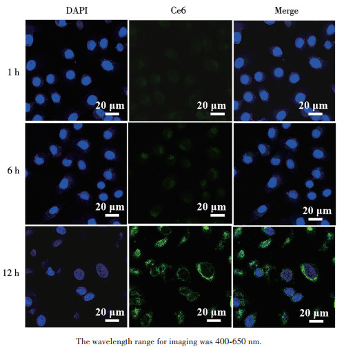
Before the in vitro anticancer assay, the cell uptake behavior of the prepared UCTSH was studied. The MCF-7 cells were incubated with UCTSH for 1, 6, and 12 h, respectively, and the related CLSM images are shown in Fig. 5. The cell nuclei were labeled with 4′,6-diamidino-2-phenylindole with blue fluorescence; the green fluorescence was derived from the loaded Ce6 molecules, and the merged channel was also provided. After UCTSH (125.0 μg·mL-1) interacted with the cells for 1 h, green fluorescence of Ce6 was not seen around the nucleus. With the increase in time, the apparent green fluorescence of Ce6 was observed around the nucleus after incubation of MCF-7 cells with UCTSH for 12 h, showing the efficient uptake of UCTSH nanocomposites by cells.
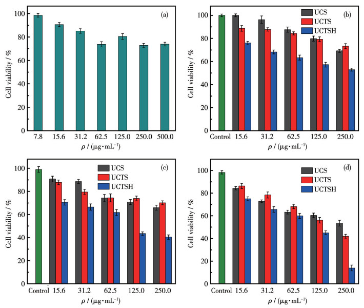
Next, the typical MTT assay was executed on mouse fibroblasts (L929) to assess the biocompatibility of UCTSH with normal cells. As shown in Fig. 6a, the L929 cell viability was not influenced even for mass concentrations up to 500.0 μg·mL-1, indicating that UCTSH damage to healthy tissues is negligible due to the lower amount of H2O2 in healthy cells. Subsequently, the antitumor capability of UCTSH with CDT and PDT was evaluated. Fig. 6b shows the cell viability of three groups of MCF-7 cells treated with different nanomaterials for 24 h without light. With the increase in concentration, the cell viability of the group treated with UCTS was slightly higher than that of UCS. This may be because the incorporation of TFS reduces the toxicity of UCTS. However, the cell viability of the group treated with UCTSH was much lower than that of UCTS and UCS, which proves that UCTSH has excellent ·OH-mediated CDT effect. Then, the synergistic therapeutic effects of PDT and CDT under light were investigated. In NIR groups, the viability of cancer cells treated with 808 nm light alone, indicating that the NIR light is not harmful to the cells. As shown in Fig. 6c, compared with UCS, the viability of cancer cells treated with UCTS was higher and did not show the advantage of oxygen carrying under 808 nm laser irradiation for 5 min. However, Fig. 6d showed the viability of cancer cells treated with UCTS was lower than that of UCS under the irradiation condition of 10 min, which suggests UCTS had excellent enhanced 1O2-mediated PDT performance. More importantly, the cell viability of cancer cells treated with UCTSH (250.0 μg·mL-1) was 14.1%, lower than 40.1% after 5 min irradiation (Fig. 6c) for synergistic PDT and CDT and 52.8% for single CDT (Fig. 6b). This reveals that the longer irradiation time under the same conditions, the better the PDT effect of UCTSH, and the bimodal combined treatment of PDT and CDT was better than that of single CDT on killing cancer cells. Therefore, the as-proposed UCTSH showed low cytotoxicity for the normal cells; however, good synergistic enhanced PDT/CDT capability and tumor microenvironment-responsive therapeutic characteristics.
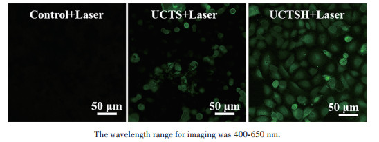
Finally, the contribution of synergistic PDT/CDT to the high antitumor efficiency of UCTSH was further explored by monitoring the ROS generation in MCF-7 cells. The level of ROS in MCF-7 cells was evaluated by using the non-fluorescent probe DCFH-DA. Compared with the cells without treatment by UCTS and UCTSH, both UCTS and UCTSH-treated cells showed bright DCF fluorescence under an 808 nm laser irradiation for 5 min (Fig. 7). More importantly, as the cells were treated with UCTSH, DCF fluorescence became even stronger compared with the cells treated with UCTS. The ROS evolutions in MCF-7 cells confirm the contribution of the synergistic PDT/CDT to the high antitumor efficiency of UCTSH.
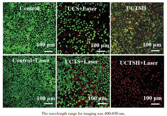
Furthermore, the killing efficiency of UCTSH to MCF-7 cells was assessed by Calcein-AM/PI cell viability/cytotoxicity assay kit to distinguish live/dead cells after treatment. The green fluorescent spots in Fig. 8 represent living cells, and the red fluorescent spots are dead cells. Obviously, most of the MCF-7 cells were still living after being treated by UCS or UCTS, whereas a large number of cells were dead upon being treated by UCTSH under 808 nm NIR laser irradiation, indicating the stronger killing efficiency of UCTSH. The results further validate the synergistic effects of enhanced PDT/CDT in achieving high antitumor efficiency of UCTSH.
In terms of material preparation, so far, there have been many PDT-based nano therapeutic platforms developed by relying on the unique advantages of upconversion nanomaterials. Among them, the photosensitizers in most therapeutic agents were physically adsorbed[23-24, 35]. The nanostructure we designed was covalently connected photosensitizers Ce6 to prevent the leakage of photosensitizers from harming normal tissues. In addition to joining Ce6, TFS was added. The TFS can not only improve the oxygen content, but also stabilize the light of upconversion nanomaterials and enhance the effect of PDT. These advantages are not available in other oxygen storage agents, such as manganese dioxide[28] and hemoglobin[36]. Secondly, the outermost layer of our nanostructure was added with Cu-MOFs (HKUST-1) for CDT. As far as we know, this is the first time to use this material has been used for upconversion binding photodynamic nanoplatform. HKUST-1 coated on mSiO2 can reduce the degradation of Ce6 by visible light and maintain a stable PDT effect. Moreover, the Fenton-like reaction rate of copper ions was higher than that of ferrous ions[37-38]. Therefore, this nanoconstruction was theoretically an excellent nanoplatform for collaborative PDT and CDT. On evaluating the effect of killing cancer cells by MTT experiments, two excellent treatment methods of PDT and CDT were perfectly combined. Compared with a single CDT[31] or PDT[39] nanoagent, the dosage of this bifunctional nanomaterial was smaller, and the death rate of cells was higher. It can achieve almost the same therapeutic effect as the three treatment modes. For instance, under NIR radiation, the survival rate of cancer cells treated with UMNOCC-PEG (250 μg·mL-1) was ca. 13.0% for PDT/CDT/gas therapy[24]; the cell viability of cancer cells cultured with PEG/Y-UCSZ & DOX (250 μg·mL-1)[25] and UCSM-PEG-DOX (250 μg·mL-1)[28] were ca. 15.0% and ca. 10.0% respectively with PDT/CDT/chemotherapy; furthermore, the survival rate of cancer cells cultured with UCSP-LDH (250 μg·mL-1) was ca. 30.0% for PDT/CDT/photothermal therapy[40]. Whereas this work shows that the cell viability of cancer cells treated by UCTSH was only 14.1% with additional light. In conclusion, this UCTSH is an excellent nanotherapeutic agent with PDT combined with CDT for killing cancer cells.
In summary, we report functionalized upconversion nanoparticles (UCTSH) as a rising ROS level nanoreactor for synergistically enhanced PDT and CDT against cancer. On the one hand, the nanoconstruction enters cancer cells through efficient endocytosis and provides a stable and efficient PDT effect with supplied O2 due to the amount of 1O2 by Ce6 under 808 nm laser irradiation. On the other hand, Fenton-like reaction is triggered to produce a large number of highly toxic ·OH in the tumor microenvironment, realizing efficient CDT. Therefore, the nanocomposite can rapidly and efficiently promote the ROS level in cancer cells and accelerate the apoptosis of cancer cells. Meanwhile, the UCTSH demonstrated low toxicity to normal cells. Finally, the dual-mode nanoplatform for collaborative PDT/CDT puts forward a feasible strategy to achieve a facile tumor nanotherapeutic system with enhanced bio-safety and simultaneous high therapeutic efficiency compared with single PDT or CDT and other multifunctional combination therapies.
YANG B, CHEN Y, SHI J L. Reactive oxygen species (ROS)-based nanomedicine[J]. Chem. Rev., 2019, 119(8): 4881-4985 doi: 10.1021/acs.chemrev.8b00626
TANG Z, LIU Y, HE M Y, BU W B. Chemodynamic therapy: Tumour microenvironment-mediated Fenton and Fenton-like reactions[J]. Angew. Chem.‒Int. Edit., 2019, 58(4): 946-956 doi: 10.1002/anie.201805664
WANG K N, LIU L Y, MAO D, HOU M X, TAN C P, MAO Z W, LIU B. A nuclear-targeted AIE photosensitizer for enzyme inhibition and photosensitization in cancer cell ablation[J]. Angew. Chem.‒Int. Edit., 2022, 61(15): e202114600 doi: 10.1002/anie.202114600
WANG Z J, YU L, WANG Y H, WANG C L, MU Q C, LIU X J, YU M, WANG K N, YAO G Y, YU Z Q. Dynamic adjust of non-radiative and radiative attenuation of AIE molecules reinforces NIR-Ⅱ imaging mediated photothermal therapy and immunotherapy[J]. Adv. Sci., 2022, 9(8): 2104793 doi: 10.1002/advs.202104793
XIN H T, LIN Q W, SUN S M, WANG Y Y, LIU B, WANG W J, MAO Z W, WANG K N. Gram-negative bacteria targeting AIE photosensitizer for selective photodynamic killing of vibrio vulnificus[J]. Aggregate, 2025, 6(3): e709 doi: 10.1002/agt2.709
ZHANG C, BU W B, NI D L, ZHANG S J, LI Q, YAO Z W, ZHANG J W, YAO H L, WANG Z, SHI J L. Synthesis of iron nanometallic glasses and their application in cancer therapy by a localized Fenton reaction[J]. Angew. Chem.‒Int. Edit., 2016, 55(6): 2101-2106 doi: 10.1002/anie.201510031
CHEN X Y, ZHANG H L, ZHANG M, ZHAO P R, SONG R X, GONG T, LIU Y Y, HE X H, ZHAO K L, BU W B. Amorphous Fe-based nanoagents for self-enhanced chemodynamic therapy by re-establishing tumor acidosis[J]. Adv. Funct. Mater., 2019, 30(6): 1908365
XU C J, YUAN Z L, KOHLER N, KIM J M, CHUNG M A, SUN S H. FePt nanoparticles as an Fe reservoir for controlled Fe release and tumor inhibition[J]. J. Am. Chem. Soc., 2009, 131(42): 15346-15351 doi: 10.1021/ja905938a
YANG B C, LIU Q Y, YAO X X, ZHANG D S, DAI Z C, CUI P, ZHANG G R, ZHENG X W, YU D X. FePt@MnO-based nanotheranostic platform with acidity-triggered dual-ions release for enhanced MR imaging-guided ferroptosis chemodynamic therapy[J]. ACS Appl. Mater. Interfaces, 2019, 11(42): 38395-38404 doi: 10.1021/acsami.9b11353
DU T Y, ZHAO C Q, REHMAN F U, LAI L M, LI X Q, SUN Y, LUO S H, JIANG H, GU N, SELKE M, WANG X M. In situ multimodality imaging of cancerous cells based on a selective performance of Fe2+-adsorbed zeolitic imidazolate framework-8[J]. Adv. Funct. Mater., 2017, 27(5): 1603926 doi: 10.1002/adfm.201603926
HALLIWELL B, CLEMENT M V, LONG L H. Hydrogen peroxide in the human body[J]. FEBS Lett., 2000, 486(1): 10-13 doi: 10.1016/S0014-5793(00)02197-9
KIM J, CHO H R, JEON H, KIM D, SONG C, LEE N, CHOI S H, HYEON T. Continuous O2-evolving MnFe2O4 nanoparticle-anchored mesoporous silica nanoparticles for efficient photodynamic therapy in hypoxic cancer[J]. J. Am. Chem. Soc., 2017, 139(32): 10992-10995 doi: 10.1021/jacs.7b05559
LÓPEZ-LÁZARO M. Dual role of hydrogen peroxide in cancer: Possible relevance to cancer chemoprevention and therapy[J]. Cancer Lett., 2007, 252(1): 1-8
SZATROWSKI T P, NATHAN C F. Production of large amounts of hydrogen peroxide by human tumor cells[J]. Cancer Res., 1991, 51(3): 794-798
LIN L S, WANG J F, SONG J B, LIU Y J, ZHU G Z, DAI Y L, SHEN Z Y, TIAN R, SONG J, WANG Z T, TANG W, YU G C, ZHOU Z J, YANG Z, HUANG T, NIU G, YANG H H, CHEN Z Y, CHEN X Y. Cooperation of endogenous and exogenous reactive oxygen species induced by zinc peroxide nanoparticles to enhance oxidative stress-based cancer therapy[J]. Theranostics, 2019, 9(24): 7200-7209 doi: 10.7150/thno.39831
LIN L S, HUANG T, SONG J B, OU X Y, WANG Z T, DENG H Z, TIAN R, LIU Y J, WANG J F, LIU Y, YU G C, ZHOU Z J, WANG S, NIU G, YANG H H, CHEN X Y. Synthesis of copper peroxide nanodots for H2O2 self-supplying chemodynamic therapy[J]. J. Am. Chem. Soc., 2019, 141(25): 9937-9945 doi: 10.1021/jacs.9b03457
YUE J, LI L, JIANG CY, MEI Q, DONG W F, YAN R H. Riboflavin-based carbon dots with high singlet oxygen generation for photodynamic therapy[J]. J. Mater. Chem. B, 2021, 9(38): 7972-7978 doi: 10.1039/D1TB01291F
FENG L L, HE F, DAI Y L, GAI S L, ZHONG C N, LI C X, YANG P P. Multifunctional UCNPs@MnSiO3@g-C3N4 nanoplatform: Improved ROS generation and reduced glutathione levels for highly efficient photodynamic therapy[J]. Biomater. Sci., 2017, 5(12): 2456-2467 doi: 10.1039/C7BM00798A
LIU C, LIU B, ZHAO J, DI Z H, CHEN D Q, GU Z J, LI L L, ZHAO Y L. Nd3+-sensitized upconversion metal-organic frameworks for mitochondria-targeted amplified photodynamic therapy[J]. Angew. Chem.‒Int. Edit., 2020, 59(7): 2634-2638 doi: 10.1002/anie.201911508
XIE Z X, LIANG S, CAI X C, DING B B, HUANG S S, HOU Z Y, MA P A, CHENG Z Y, LIN J. O2-Cu/ZIF-8@Ce6/ZIF-8@F127 composite as a tumor microenvironment-responsive nanoplatform with enhanced photo-/chemodynamic antitumor efficacy[J]. ACS Appl. Mater. Interfaces, 2019, 11(35): 31671-31680 doi: 10.1021/acsami.9b10685
CUI Y Y, CHEN X, CHENG Y, LU X Y, MENG J J, CHEN Z W, LI M K, LIN C C, WANG Y L, YANG J. CuWO4 nanodots for NIR-induced photodynamic and chemodynamic synergistic therapy[J]. ACS Appl. Mater. Interfaces, 2021, 13(19): 22150-22158 doi: 10.1021/acsami.1c00970
QIN X, WU C, NIU D C, QIN L M, WANG X, WANG Q G, LI Y S. Peroxisome inspired hybrid enzyme nanogels for chemodynamic and photodynamic therapy[J]. Nat. Commun., 2021, 12: 5243 doi: 10.1038/s41467-021-25561-z
ZOU M, ZHAO Y J, DING B B, JIANG F, CHEN Y Q, MA P A, LIN J. NIR-triggered biodegradable MOF-coated upconversion nanoparticles for synergetic chemodynamic/photodynamic therapy with enhanced efficacy[J]. Inorg. Chem. Front., 2021, 8(10): 2624-2633 doi: 10.1039/D1QI00252J
LIU S K, LI W T, DONG S M, ZHANG F M, DONG Y S, TIAN B S, HE F, GAI S L, YANG P P. An all-in-one theranostic nanoplatform based on upconversion dendritic mesoporous silica nanocomposites for synergistic chemodynamic/photodynamic/gas therapy[J]. Nanoscale, 2020, 12(47): 24146-24161 doi: 10.1039/D0NR06790C
DONG S M, XU J T, JIA T, XU M S, ZHONG C N, YANG G X, LI J R, YANG D, HE F, GAI S L, YANG P P, LIN J. Upconversion-mediated ZnFe2O4 nanoplatform for NIR-enhanced chemodynamic and photodynamic therapy[J]. Chem. Sci., 2019, 10(15): 4259-4271 doi: 10.1039/C9SC00387H
SHEN J W, WANG Z Q, LIU J W, LI H. Nano-sized NaF inspired intrinsic solvothermal growth mechanism of rare-earth nanocrystals for facile control synthesis of high-quality and small-sized hexagonal NaYbF4: Er[J]. J. Mater. Chem. C, 2017, 5(37): 9579-9587 doi: 10.1039/C7TC02573D
ABEL K A, BOYER J C, VAN VEGGEL F C J M. Hard proof of the NaYF4/NaGdF4 nanocrystal core/shell structure[J]. J. Am. Chem. Soc., 2009, 131(41): 14644-14645 doi: 10.1021/ja906971y
XU J T, HAN W, YANG P P, JIA T, DONG S M, BI H T, GULZAR A, YANG D, GAI S, HE F, LIN J, LI C X. Tumor microenvironment-responsive mesoporous MnO2-coated upconversion nanoplatform for self-enhanced tumor theranostics[J]. Adv. Funct. Mater., 2018, 28(36): 1803804 doi: 10.1002/adfm.201803804
WU H S, CHEN F H, GU D H, YOU C Q, SUN B W. A pH-activated autocatalytic nanoreactor for self-boosting Fenton-like chemodynamic therapy[J]. Nanoscale, 2020, 12(33): 17319-17331 doi: 10.1039/D0NR03135F
ZHAO M, ZHANG X M, DENG C H. Rational synthesis of novel recyclable Fe3O4@MOF nanocomposites for enzymatic digestion[J]. Chem. Commun., 2015, 51(38): 8116-8119 doi: 10.1039/C5CC01908G
LI Y T, ZHOU J L, WANG L, XIE Z G. Endogenous hydrogen sulfide-triggered MOF-based nanoenzyme for synergic cancer therapy[J]. ACS Appl. Mater. Interfaces, 2020, 12(27): 30213-30220 doi: 10.1021/acsami.0c08659
LIU M, WU H S, WANG S L, HU J Z, SUN B W. Glutathione-triggered nanoplatform for chemodynamic/metal-ion therapy[J]. J. Mater. Chem. B, 2021, 9(45): 9413-9422 doi: 10.1039/D1TB01330K
LIU J, WU S H, CHU H Y, WANG C Z, SHEN J W, WEI Y M, WU P. Low power density 980 nm-driven ultrabrigh red-emitting upconversion nanoparticles via synergetic Yb3+/Tm3+ cascade-sensitization[J]. J. Mater. Chem. C, 2019, 7(43): 13415-13424 doi: 10.1039/C9TC04174E
KONG W H, CHU H Y, LI Y M, WANG C Z, WEI Y M, SHEN J W. Ambient-efficient hydrophobic hydration-shell structure for lysosome-tolerable upconversion nanoparticles with enhanced biosafety and simultaneous versatility[J]. Chem. Mater., 2021, 33(13): 5377-5390 doi: 10.1021/acs.chemmater.1c01469
XIE Z X, CAI X C, SUN C Q, LIANG S, SHAO S, HUANG S S, CHENG Z Y, PANG M L, XING B G, AL KHERAIF A A, LIN J. O2-loaded pH-responsive multifunctional nanodrug carrier for overcoming hypoxia and highly efficient chemo-photodynamic cancer therapy[J]. Chem. Mater., 2019, 31(2): 483-490 doi: 10.1021/acs.chemmater.8b04321
WANG P, WANG X D, LUO Q, LIL Y, LIN X X, FAN L L, ZHANG Y, LIU J F, LIU X L. Fabrication of red blood cell-based multimodal theranostic probes for second near-infrared window fluorescence imaging-guided tumor surgery and photodynamic therapy[J]. Theranostics, 2019, 9(2): 369-380 doi: 10.7150/thno.29817
BOKARE A D, CHOI W. Review of iron-free Fenton-like systems for activating H2O2 in advanced oxidation processes[J]. J. Hazard. Mater., 2014, 275: 121-135 doi: 10.1016/j.jhazmat.2014.04.054
CHU H Y, LI Y M, WANG C Z, SHEN J W, WEI Y M. MOF-coated upconversion nanoconstructs for synergetic photo-chemodynamic/oxygen-elevated photodynamic therapy[J]. Dalton Trans., 2022, 51(42): 16336-16343 doi: 10.1039/D2DT02441A
LI Y M, WANG R, XU Y L, ZHENG W, LI Y M. Influence of silica surface coating on operated photodynamic therapy property of Yb3+-Tm3+: Ga(Ⅲ)-doped ZnO upconversion nanoparticles[J]. Inorg. Chem., 2018, 57(13): 8012-8018 doi: 10.1021/acs.inorgchem.8b01169
JIA T, WANG Z, SUN Q Q, DONG S M, XU J T, ZHANG F M, FENG L L, HE F, YANG D, YANG P P, LIN J. Intelligent Fe-Mn layered double hydroxides nanosheets anchored with upconversion nanoparticles for oxygen-elevated synergetic therapy and bioimaging[J]. Small, 2020, 16(46): 2001343 doi: 10.1002/smll.202001343

Scheme 1 Schematic fabrication process of the UCTSH and their NIR-triggered PDT with self-supplying O2 and pH-responsive CDT for synergistic therapy

Figure 1 TEM images of (a) UC, (b) UCTS, and (c) UCTSH; (d) XRD patterns of UC, UCTS, and UCTSH; (e) Element mappings of UCTSH

Figure 2 (a) XPS survey spectra and (b) Cu2p high-resolution XPS spectra of UCTSH; (c) FTIR spectra of UC, UCS, UCTS, and UCTSH; (d) N2 adsorption-desorption isotherms of US, UCTS, and UCTSH; (e) ζ potentials of US, UCS, UCTS, and UCTSH in water; (f) Images of US, UCTS, and UCTSH dispersions at 25 ℃

Figure 3 (a) UCL spectra of the different nanoparticles; (b) Nd3+→Yb3+→Tm3+→Er3+ series sensitized energy transfer mechanism of UC under 808 nm excitation; (c) Luminescence spectra of UC and the absorption spectra of Ce6; (d) UCL spectra of UC, UCTS, and UCTSH; (e) Absorbance changes of DPBF treated with UCTSH after 808 nm laser irradiation at different time points

Figure 4 (a) Scheme of TMB oxidation catalyzed by UCTSH; (b) Colorimetric detection of ·OH generated by UCTSH at different pH values based on TMB assay (inset: the digital photos of different pH values); (c) Depletion of DPBF underwent different treatments with or without 2 mmol·L-1 H2O2; (d) Fluorescence spectrum changes of DCFH-DA underwent different treatments with or without 5 mmol·L-1 H2O2 (the reaction time was 30 min after irradiation with 808 nm light for 10 min)

Figure 5 CLSM images of MCF-7 cells incubated with 125.0 μg·mL-1 UCTSH for different times

Figure 6 (a) Cell viability of L929 incubated with UCTSH at various mass concentrations; (b) Viability of MCF-7 cells after incubation with UCS, UCTS, and UCTSH without irradiation; (c) Viability of MCF-7 cells after incubation with UCS, UCTS, and UCTSH with irradiation for 5 min by an 808 nm laser; (d) Viability of MCF-7 cells after incubation with UCS, UCTS, and UCTSH with irradiation for 10 min by an 808 nm laser

Figure 7 Intracellular ROS detection with DCF fluorescence after the treatment of MCF-7 cells with UCTS (125.0 μg·mL-1) and UCTSH (125.0 μg·mL-1)

 扫一扫看文章

扫一扫关注我们
 下载:
下载:
 下载:
下载:
