Received Date:
16 November 2024 Revised Date:
17 January 2025 Available Online:
10 May 2025
Abstract:
A zinc sulfate open framework matrix, [Zn(SO4)(DMSO)] (1), was synthesized by solvothermal evaporation using dimethyl sulfoxide (DMSO) as the solvent. A composite P@1, which exhibits fluorescence and room temperature phosphorescence (RTP) properties, was prepared by doping 2, 6-naphthalic acid (P) into matrix 1 at a low concentration. P@1 emitted a green RTP that was visible to the naked eye and lasted for approximately 2 s. P@1 exhibited selective phosphorescence enhancement response towards Pb2+, with a detection limit of 2.52 μmol·L-1. The main detection mechanism is the Pb—O coordination-induced phosphorescence enhancement in the system. Interestingly, P@1 also functioned as a dualchannel probe for the rapid detection of Fe3+ ions through fluorescence quenching with a detection limit of 0.038 μmol·L-1. The recognition mechanism may be attributed to the competitive energy absorption between P@1 and Fe3+ ions.
Mining activities are one of the main sources of heavy metal pollution in the environment[1-2]. In coal mining, transportation, processing, and utilization, the heavy metals contained in coal and coal gangue dust enter the soil and accumulate through sedimentation, leaching, percolation, migration, and other ways, thus polluting the soil around the coal mine. Lead (Pb) is one of the heavy metal pollutants in mining activities and is toxic to humans and other living organisms. Pb commonly inhibits growth at soil concentrations of 30 mg·kg-1[3]. Studies have shown that even small amounts of Pb2+ can cause irreversible damage to the human nervous and immune systems[4]. Although traditional methods[5-7] for detecting Pb2+ are highly sensitive and specific, they typically require expensive instrumentation and strict experimental conditions. Therefore, there is an urgent need to develop a fast, low-cost, and specific method for detecting Pb2+ in the environment. In contrast, the luminescence detection method has the advantages of fast response, high selectivity, low cost, and simple operation. In recent years, many luminescent probes have been used for detecting Pb2+[8-13]. However, the most reported Pb2+ probes are fluorescent probes, which are highly susceptible to interference from background luminescence. Phosphorescent probes can effectively avoid this issue. However, so far, the construction of phosphorescent probes remains a huge challenge.
Room temperature phosphorescence (RTP) materials have important applications[14-20] in information encryption, biological imaging, and sensing due to their long emission lifetime, high signal‑to‑noise ratio, absence of background fluorescence, and large Stokes shift. So far, several methods for preparing RTP materials have been reported[21-28], including halogen bonding interactions, heavy atom effects, metal-organic frameworks (MOFs), polymerization, host-guest doping, and H aggregation. Among these, the host-guest doping strategy is an important method for synthesizing phosphorescent materials, which mainly generates phosphorescent emission by the limiting effect of the host matrix on trace amounts of doped guest phosphorescent molecules. This method offers the benefits of simple preparation and low cost. Therefore, it is of great research significance to prepare phosphorescent probes using the host-guest doping strategy. To date, several cases of phosphorescent probes for detecting different analytes have been reported. For example, Pan′s team[29] used a phosphorescent probe Cd-MOF for multi-stage oxygen detection. However, it is a “turn‑off” probe, which exhibits a change in luminescence from strong to weak. Xu et al.[30] used MOF-5 as a phosphorescent “turn-on” probe for detecting Pb2+, exhibiting a change in luminescence from weak to strong. In contrast, phosphorescent probes that do not initially emit light but exhibit luminescent recovery behavior after interacting with the analyte are very rare. Such probes can effectively reduce background luminescence, and improve the signal-to-noise ratio and sensitivity of detection, representing an important direction for the development of probes[31-32]. To our knowledge, there have been no reports so far on the use of phosphorescent probe materials prepared based on host guest doping strategy for detecting Pb2+.
On the other hand, iron ions play a critical role in both the environment and the human body, and maintaining appropriate iron levels in the body is crucial for health. Iron deficiency or overload can lead to life-threatening diseases[33-36]. For example, excessive iron can lead to diseases such as cancer, organ dysfunction, Parkinson′s disease, and Alzheimer′s disease, while iron deficiency can lead to anemia. So far, many luminescent probes have been reported for detecting Fe3+[37-42]. However, the simultaneous presence of Pb2+ and Fe3+ can interfere with the detection results. Cases of successfully detecting Pb2+ or Fe3+ ions in the same sensing system are extremely rare. Therefore, the development of luminescent probes capable of simultaneously detecting these two ions is of great significance.
Herein, we report the preparation of a new zinc sulfate open framework, [Zn(SO4)(DMSO)] (1), where DMSO=dimethyl sulfoxide. On such basis, the composite material P@1 was successfully prepared by introducing trace amounts of the organic phosphor molecule 2, 6-naphthalic acid (P) into the 1 matrix based on the “host‑guest doping” strategy. The solid‑state P@1 exhibited RTP visible to the naked eye. Interestingly, the DMF suspension of P@1 is non‑luminescent, because phosphorescent molecular vibration and rotation in solvents can open nonradiative channels, and dissolved oxygen can also quench triplet excitons, leading to phosphorescence quenching. In addition, by introducing Pb2+ ions into a rigid matrix and using coordination locks to promote intersystem crossing (ISC) efficiency while limiting nonradiative transitions, luminescence recovery detection of Pb2+ ions can be achieved. Detailed characterization indicates that the above design concept is feasible. P@1 can be used to specifically detect Pb2+ ions in DMF media through luminescence recovery response, with a calculated LOD being 2.52 μmol·L-1. In addition, the P@1 system has been further used as a fluorescence quenching sensor for detecting Fe3+, with an LOD of 0.038 μmol·L-1. Details of the structure, detection, and mechanism are presented.
1.
Experimental
1.1
Materials and methods
A detailed description of the reagent source and main instruments can be found in the Support information.
1.2
Synthesis of compound 1
ZnSO4·5H2O (300 μL, 0.15 mmol) was added to DMSO (2 mL) in a 20 mL glass vessel and heated to 105 ℃ to afford a transparent solution by continuous stirring. After 48 h, the reaction mixture was removed from the heat and allowed to cool to room temperature, to facilitate the precipitation of the crystalline product from the solution. The yield was ca. 78%. Element analysis Calcd. for ZnC2H6O5S2(%): C, 10.02; H, 2.52; S, 26.76. Found(%): C, 10.12; H, 2.48; S, 26.64. X-ray crystallographic analysis, crystallographic data, and selected bond lengths and angles can be found in the Supporting information.
1.3
Preparation of P@1
The material was prepared by using a similar procedure to that used for 1 with the addition of organic molecules P in a molar ratio of 1∶50 (phosphors to Zn2+).
1.4
Pb2+ and Fe3+ detection
For the Pb2+ and Fe3+ sensing experiments, P@1 powders (1.5 mg) were separately dispersed into 3 mL DMF to form suspensions and sonicated for 10 min to ensure homogeneity. The emission spectra were measured under 365 nm excitation. All experiments were conducted in DMF. The metal ions used in this experiment included Al3+, Zn2+, Ba2+, Bi3+, Ni2+, Mg2+, Mn2+, Ca2+, Cr3+, Pb2+, Sn2+, Na+, Fe3+, K+, and Cd2+.
2.
Results and discussion
2.1
Crystal structure of compound 1
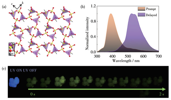
Single-crystal X-ray diffraction (SCXRD) analysis reveals that compound 1 crystallizes in the monoclinic space group of P21/c and features a 2D layered framework (Fig. 1a). Each Zn2+ ions has a tetrahedral configuration {O4} donor sets and coordinate to three oxygen atoms from three different sulfate groups and an oxygen atom in DMSO. Thus, Zn2+ ions can be treated as 3‑ connected nodes. The Zn—O bond lengths and O—Zn—O bond angle fall in the ranges of 0.193 10(16)-0.193 81(15) nm and 102.71(6)°-167.82(7)°, respectively. Two adjacent Zn2+ ions are bridged by one sulfate with the Zn…Zn separation being 0.446 2 nm. The sulfate moiety in the structure acts as a bridging group with a μ3-κO1∶κO1∶κO1 coordination mode for linking the three Zn2+ ions. Thus, the sulfate moiety can also be treated as a 3-connected node. The combination of these two nodes finally forms a 2D layered framework extending along the b- and c-axes, which has a fes topology (Fig.S1a).
图 1
Figure 1.
(a) Structure of 1 viewed along the a-axis; (b) Solid-state phosphorescence spectrum (purple) and fluorescence spectrum (yellow) of P@1; (c) Corresponding luminescence photographs under irradiation and upon removal of a 365 nm UV lamp
2.2
Characterization of compound 1 and composite P@1
The phase purity of the crystal structure of compound 1 was verified by powder X-ray diffraction (PXRD) experiments. PXRD analysis revealed that the experimental and simulated patterns were consistent, indicating the phase purity of the sample (Fig.S2a). The PXRD patterns of composite P@1 showed almost no change compared to 1. In addition, the solid UV-Vis diffuse reflectance spectra of 1 and P@1 were further measured and the results are shown in Fig.S2b. The spectrum of P@1 has apparent shoulders in the 340 nm region compared to 1, which can be attributed to the new absorption generated by the packing of 2, 6-naphthalic acid (P). The above experiments showed that P was successfully trace-loaded into the compound 1.
The solid-state luminescent properties of compound 1 were investigated at room temperature (Fig.S3). Upon excitation at 365 nm, a strong emission peak at 410 nm could be observed in the fluorescence spectrum of 1. Comparatively, the phosphorescence spectrum was silent, meaning 1 itself has no phosphorescence, which is very favorable for exploring phosphorescent emission based on guest P. As expected, P@1 could produce a green RTP visible to the naked eye, lasting for about 2 s (Fig. 1c). The result shows that 1 can limit the vibration of guest P and suppress the non-radiative energy loss of the triplet exciton, which is conducive to the increase of the RTP lifetime. The solid-state luminescence properties of P@1 were tested at room temperature. As shown in Fig. 1b, the fluorescence emission peak of P@1 was located at 390 nm (λex=365 nm), while the emission band of the phosphorescence spectrum was in a range of 520-545 nm with a lifetime of 284.88 ms (Fig.S4). P@1 has such excellent long-lived luminescence properties that it is promising as an ideal phosphorescent probe material.
2.3
Detecting of Pb2+ ion
The DMF suspension of P@1 was used to detect various metal ions (M(NO3)x, Mx+=Al3+, Zn2+, Ba2+, Bi3+, Ni2+, Mg2+, Mn2+, Ca2+, Cr3+, Pb2+, Sn2+, Na+, Fe3+, K+, Cd2+) via luminescent sensing. The phosphorescence spectra of P@1 dispersed in DMF at room temperature were recorded. As shown in Fig.S5, P@1 dispersed in DMF has a strong fluorescence emission, but their phosphorescence spectra are almost silent, prompting us to explore its application as phosphorescent recovery probes.
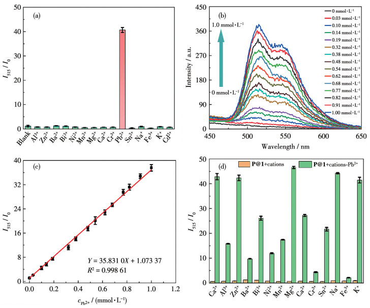
The phosphorescence spectra were measured after adding the above metal salts to a suspension of P@1 (Fig.S6). The concentrations of metal salts were all maintained at 1.0 mmol·L-1 in suspensions. When in contact with Pb2+ ions, the phosphorescence intensity of P@1 was enhanced by a maximum of about 40 times compared with the original (Fig. 2a). In contrast, the introduction of other metal ions has a negligible effect on emission intensity apart from Pb2+ ions. All the above results indicate that P@1 can selectively detect Pb2+ ions based on phosphorescence recovery. So far, some probes for detecting Pb2+ have been reported (Table S3). However, most of them are fluorescence probes, phosphorescent probes are rare among them.
图 2
Figure 2.
(a) Phosphorescence luminescence intensity of P@1 in DMF suspensions of different metal ions (1 mmol•L-1); (b) Phosphorescence spectra of P@1′s suspension at different Pb2+ concentrations; (c) Linear relationship between the phosphorescence intensity ratio (I515/I0) of P@1′s suspension and the Pb2+ concentration in a range of 0-1 mmol• L-1; (d) Phosphorescence intensities of P@1′s suspension in the presence of various interferents when with or without 1 mmol•L-1 Pb2+
I0 and I515 are the phosphorescence intensities of P@1′s suspension at 515 nm before and after the addition of metal ions.
To better understand the luminescence response of P@1 to Pb2+ ions, phosphorescent titration experiments were conducted by adding Pb2+ ions to the suspension of compound P@1. As shown in Fig. 2b, the intensity of phosphorescent emission at 515 nm also increased with the rising concentration of Pb2+ concentration. In a range of 0-1 mmol·L-1, the phosphorescent intensity of the P@1′s suspension at 515 nm showed a good linear relationship with the Pb2+ concentration, and the corresponding correlation coefficient (R2) was 0.996 (Fig. 2c). According to the detection limit formula, LOD=3σ/k (where σ is the standard deviation of 15 blank measurements and k is the slope of the fitting line), the limit of detection (LOD) of Pb2+ ions was calculated to be 2.52 μmol·L-1. Such results indicate that P@1 can serve as an excellent probe for detecting Pb2+ ions at low concentrations.
Further interference experiments were conducted to investigate P@1 selectivity towards Pb2+ ions. The phosphorescence spectra of P@1 dispersed in a mixed solution of equal amounts of Pb2+ ions and above other interfering metal ions (1 mmol·L-1) were measured. As shown in Fig. 2d, when metal ions such as Al3+, Zn2+, Ba2+, Bi3+, Ni2+, Mg2+, Mn2+, Ca2+, Cr3+, Sn2+, Na+, Fe3+, K+, Cd2+ were added to the suspension of P@1, the change in phosphorescent emission intensity could be ignored. However, the subsequent addition of an equal amount of Pb2+ ions significantly increased the intensity. Although the presence of Cr3+ and Fe3+ had a certain impact on the detection of Pb2+ ions, significantly enhanced phosphorescent emission could still be observed. These results indicate that P@1 has good anti-interference ability and can selectively detect Pb2+ ions.
The response time is an important factor in evaluating probe performance. To confirm the response time between P@1 and Pb2+ ions, the phosphorescence spectrum of the P@1′s suspension was monitored over time in the presence of Pb2+ ions (1 mmol·L-1). As shown in Fig.S7a, its phosphorescence intensity ratio (I515/I0) rapidly increased within 30 s after adding Pb2+ ions to the suspension of P@1, stabilized after 10 min, and remained almost unchanged within 30 min, indicating that P@1 can serve as a rapid responsive phosphorescent probe for Pb2+ ions.
2.4
Detection mechanism
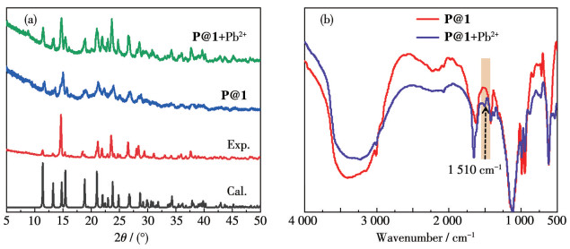
To understand the performance of P@1 detection of Pb2+ ions, its detection mechanism was deeply explored. A solid sample of the P@1+Pb2+ was prepared by adding 1 mmol·L-1 Pb2+ to P@1′s DMF suspension. PXRD test was conducted, and the results are shown in Fig. 3a. The pattern of P@1+Pb2+ was almost the same as that of P@1, indicating that the sharp increase in luminescence is not caused by the collapse of the framework. Therefore, we will further investigate the reasons for the sharp increase in the phosphorescence emission peak.
图 3
Figure 3.
(a) PXRD patterns of calculated compound 1 (black), as-synthesized 1 (red), as-synthesized P@1 (blue), and P@1+Pb2+ (green); (b) Comparison of FTIR spectra of P@1 and P@1+Pb2+
The two O atoms on the carboxyl group of P have strong coordination with Pb2+[15, 30, 43]. During the detection process, the carboxyl groups on P can anchor more Pb2+, thereby expanding the linear range. In addition, Pb2+ significantly enhanced ISC and hindered nonradiative loss of triplet excitons, thus P@1+Pb2+ exhibited significant phosphorescence enhancement. To test this hypothesis, we measured the decay time of P@1′s suspension before and after the addition of Pb2+ ions (Fig.S7b and S7c). As the concentration of Pb2+ increased, the average decay time of P@1 decreased gradually (5.53 μs for P@1, 0.42 μs for P@1+Pb2+). The shortened lifetime indicates that the carboxyl group on P anchor Pb2+, which is affected by the heavy atom effect of Pb2+, shortens the decay time of P@1+Pb2+.
A comparison of the FTIR spectra between P@1 and P@1+Pb2+ was compared to further elucidate the detecting mechanism. As depicted in Fig. 3b, it can be seen that the two spectra were the same except for the following differences. Compared with P@1, the C—O (carboxylate group) antisymmetric stretching vibration of P@1+Pb2+ at 1 682 cm-1 shifted to 1 510 cm-1. Such results, together with the above results, indicate that a new Pb—O bond is formed between the carboxyl oxygen atom on P and the Pb2+ ions. According to the literature, the frequency difference (D) between asymmetric and symmetric stretching vibrations of the carboxylate group can be used to determine its coordination mode[44]. In the IR spectra of 1@P+Fe3+, D was 90 cm-1
[Fig. 3b, νas(COO-)=1 510 cm-1, νs(COO-)=1 420 cm-1], indicating that the carboxylate group of P and Pb2+ ion form a bidentate coordination mode. Raman spectroscopy shows that a signal at 1 120 cm-1 appeared in the spectra of P@1+Pb2+, which can be attributed to the Pb—O stretching vibration of Pb-COO moiety (Fig.S8), further confirming the existence of Pb—O coordination bonds in the P@1+Pb2+[45-46].
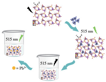
Based on the results summarized above, the mechanism for P@1 detecting Pb2+ was proposed. As shown in Fig. 4, a trace doping of P into the framework of 1. In the presence of Pb2+, carboxylate group oxygen atoms from different P binds with Pb2+ ions to form a Pb—O coordination bond. Such coordination locks and solidifies the molecular conformation of P, thereby reducing the nonradiative loss of triplet excitons and enhancing phosphorescent emission. In addition, Pb2+ exhibits a pronounced heavy-atom effect, which minimizes the nonradiative loss of triplet excitons and improves the ISC efficiency. Therefore, phosphorescent emission can be activated by introducing Pb2+ into the P@1 framework.
图 4
Figure 4.
Diagram of the synthetic routes for P@1 and proposed mechanism to detect Pb2+
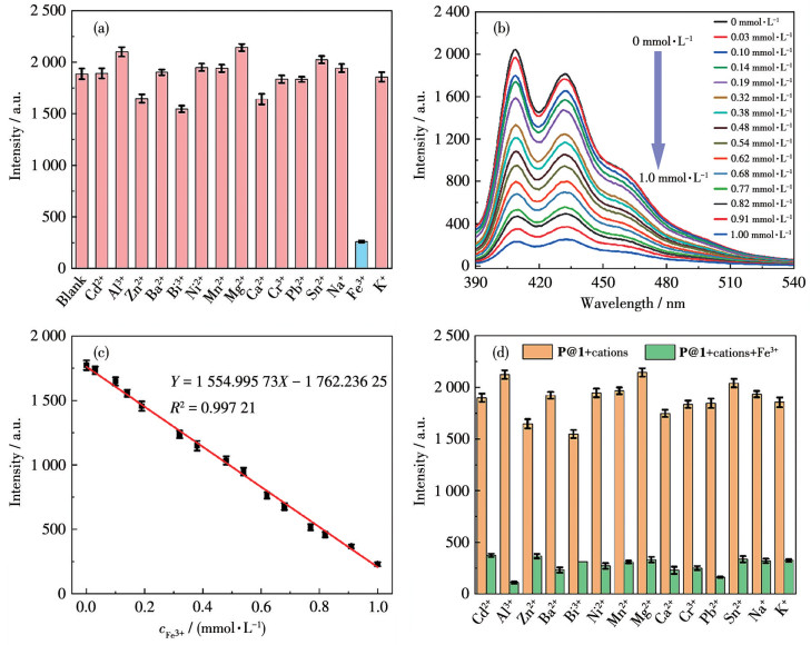
Given the strong fluorescence emission performance of P@1, we were prompted to explore its application in fluorescence detection. To explore the potential of P@1 for sensing metal ions, luminescence experiments were conducted by adding different metal ions to P@1 suspension. After the addition of 1.0 mmol·L-1 metal nitrate solution, the fluorescence spectra of the suspensions were tested. As shown in Fig. 5a, only Fe3+ exhibited a significant quenching effect on the luminescence of DMF suspension of P@1, with a quenching rate of over 86%, while the influence of other metal ions on the fluorescence intensity of P@1 suspension could be ignored. This indicates that P@1 can selectively detect Fe3+ ions.
图 5
Figure 5.
(a) Fluorescence intensity of the DMF suspension of P@1 in the presence of different metal ions (1 mmol•L-1); (b) Emission spectra of P@1′s suspension upon an incremental addition of Fe3+ (λex=365 nm); (c) Fluorescence intensity of P@1 at 430 nm as a function of Fe3+ concentration; (d) Fluorescence intensities of P@1′s suspension in the presence of various interferents when with or without 1 mmol•L-1 Fe3+
To investigate the sensitivity of P@1 in detecting Fe3+, different concentrations of Fe3+ were added to the suspension of P@1 for fluorescence titration experiments. As shown in Fig. 5b, with the continuous increase of Fe3+ concentration, the emission intensity at 430 nm decreased progressively. There was a good linear relationship between the concentration of Fe3+ and the fluorescence intensity in a range of 0-1.0 mmol·L-1 (Fig. 5c). The calculated LOD was 0.038 μmol·L-1. The above experimental results indicate that P@1 can be used for quantitative detection of Fe3+ ions in the concentration range of 0‑0.1 mmol·L-1. The anti‑ interference experiments show that the quenching effect of Fe3+ ions was not affected by the co-existing metal ions (Fig. 5d), implying that P@1 has good anti-interference ability, and can selectively detect Fe3+ ions.
To confirm the response time between P@1 and Fe3+ ions, the fluorescence spectrum of the suspension of P@1 was measured over time in the presence of Fe3+ (1 mmol·L-1). As shown in Fig.S10a, after adding Fe3+ ions to the suspension of P@1, its fluorescence intensity rapidly decreased within 30 s, stabilized after 1 min, and remained almost unchanged within 30 min, indicating that P@1 can serve as a rapid responsive fluorescent probe for Fe3+ ions.
To further explore the detection mechanism of P@1 towards Fe3+ ions, the 465 nm emission lifetime of P@1 and P@1+Fe3+ suspensions were determined and found to be 6.55 and 5.52 ns, respectively (Fig.S11). The UV-Vis absorption spectrum of the Fe3+ ions and the emission spectrum of P@1 were measured. As illustrated in Fig.S10b, the UV-Vis absorption curve of Fe3+ ions overlapped significantly with the excitation curve of P@1, indicating the existence of competitive absorption. Therefore, we speculate that the quenching may be caused by the fluorescence resonance energy transfer mechanism[47-48].
So far, multiple reports of fluorescence detection of Pb2+ have been reported, however, almost all detections are achieved through changes in fluorescence intensity. As we know, only a “turn-on” phosphorescent probe was developed to detect Pb2+. In contrast, P@1 can act as a phosphorescence recovery probe for Pb2+ detection, which can effectively reduce background luminescence and interference. Undoubtedly, such probes have better application prospects.
The above results indicate that a versatile strategy for the efficient induction of organic-based RTP via [Zn(SO)4(DMSO)] matrices encapsulation is proposed. The molecular vibration and rotation of RTP materials in solvents can open nonradiative channels. Dissolved oxygen can also act as a quencher for triplet excitons, suppressing phosphorescence. Furthermore, by introducing heavy metal Pb2+ into a rigid matrix and coordinating with a lock, the efficiency of ISC can be improved, while limiting nonradiative transitions, thus achieving phosphorescent recovery detection of Pb2+.
3.
Conclusions
In summary, a composite material P@1 with an efficient RTP effect was synthesized by doping 2, 6-naphthalic acid (P) phosphors into matrix 1in situ. P@1 exhibited a green RTP visible to the naked eye, lasting for approximately 2 s. Upon contact with only Pb2+ ions among 15 metal ions, the phosphorescence emission of P@1 in DMF suspensions was awakened, which can be used as a phosphorescent recovery probe for selective detection of Pb2+ ion with an LOD down to 2.52 μmol·L-1. The detection mechanism can be attributed to the fact that Pb2+, as a coordinating ion, not only enhances spin-orbit coupling to promote ISC but also enhances the rigidity of ligand P to prevent nonradiative loss of triplet excitons, thereby triggering phosphorescence emission. In addition, P@1 can also be used to detect Fe3+ by fluorescence quenching, with an LOD of 0.038 μmol·L-1. Therefore, these results open up a new avenue for developing probes with dual-channel high-detection performance.
Supporting information is available at http://www.wjhxxb.cn
Acknowledgments:
This work is financially supported by the Natural Science Foundation of Shandong of China (No.ZR2024QB308), Guangxi Key Laboratory of Electrochemical and Magnetochemical Functional Materials (No.EMFM20241110), the National Natural Science Foundation of China (No.42067041).
[1]
TANG Q, LI L Y, ZHANG S, ZHENG L, MIAO C H. Characterization of heavy metals in coal gangue-reclaimed soils from a coal mining area[J]. J. Geochem. Explor.,
2018, 186:
1-11.
doi: 10.1016/j.gexplo.2017.11.018
[2]
LIU X Y, BAI Z K, SHI H D, ZHOU W, LIU X C. Heavy metal pollu-tion of soils from coal mines in China[J]. Nat. Hazards,
2019, 99(2):
1163-1177.
doi: 10.1007/s11069-019-03771-5
[3]
USMAN K, ABU-DIEYEH M H, ZOUARI N, AL-GHOUTI M A. Lead (Pb) bioaccumulation and antioxidative responses in Tetraena qataranse[J]. Sci. Rep.,
2020, 10(1):
17070.
doi: 10.1038/s41598-020-73621-z
[4]
YANG C Y, DU C Y, YUAN F Y, YU P T, WANG B X, SU C S, ZOU R Q, WANG J Y, YAN X, SUN C Y, LI H X. CRISPR/Cas12a-derived ratiometric fluorescence sensor for high-sensitive Pb2+ detec-tion based on CDs@ZIF-8 and DNAzyme[J]. Biosens. Bioelectron.,
2024, 251:
116089.
doi: 10.1016/j.bios.2024.116089
[5]
AHMED H E H, MOHAMMED A M A, SOYLAK M. A magnetic sol-id phase extraction procedure for Pb(Ⅱ) at trace levels on magnetic Luffa@TiO 2 in food and water samples[J]. Food Chem.,
2023, 428:
136794.
doi: 10.1016/j.foodchem.2023.136794
[6]
ZHANG N, SHEN K, YANG X M, LI Z X, ZHOU T K, ZHANG Y, SHENG Q L, ZHENG J B. Simultaneous determination of arsenic, cadmium and lead in plant foods by ICP-MS combined with automated focused infrared ashing and cold trap[J]. Food Chem.,
2018, 264:
462-470.
doi: 10.1016/j.foodchem.2018.05.058
[7]
TIGHE M, BIELSKI M, WILSON M, RUSCIO-ATKINSON G, PEASLEE G F, LIEBERMAN M. A sensitive XRF screening method for lead in drinking water[J]. Anal. Chem.,
2020, 92(7):
4949-4953.
doi: 10.1021/acs.analchem.9b05058
[8]
XU J M, LIU M B, ZHAO W H, WANG S Q, GUI M F, LI H B, YU R Q. DNAzyme-based cascade signal amplification strategy for highly sensitive detection of lead ions in the environment[J]. J. Hazard. Mater.,
2022, 429:
128347.
doi: 10.1016/j.jhazmat.2022.128347
[9]
DU X X, LIU Y J, WANG F, ZHAO D Y, GLEESON H F, LUO D. A fluorescence sensor for Pb2+ detection based on liquid crystals and aggregation-induced emission luminogens[J]. ACS Appl. Mater. Inter-faces,
2021, 13(19):
22361-22367.
doi: 10.1021/acsami.1c02585
[10]
YANG C Y, YU P T, LI Y, WANG J Y, MA X Y, LIU N, LV T, ZHENG H R, WU H, LI H X, SUN C Y. Platform formed from ZIF-8 and DNAzyme: "Turn-on" fluorescence assay for simple, high-sensi-tivity, and high-selectivity detection of Pb2+[J]. J. Agric. Food Chem.,
2022, 70(30):
9567-9576.
doi: 10.1021/acs.jafc.2c03503
[11]
NIU X F, ZHONG Y B, CHEN R, WANG F, LIU Y J, LUO D. A "turn-on" fluorescence sensor for Pb2+ detection based on graphene quantum dots and gold nanoparticles[J]. Sens. Actuator B -Chem.,
2018, 255:
1577-1581.
doi: 10.1016/j.snb.2017.08.167
[12]
LIU T Q, WAN X J, YAO Y W. Dual sensitive and selective sensor for Pb2+ and Al3+ with distinctive fluorescence response[J]. Sens. Ac-tuator B-Chem.,
2018, 254:
1094-1100.
doi: 10.1016/j.snb.2017.07.114
[13]
XUE T, SHI Y Y, GUO J, GUO M X, YAN Y. Preparation of AgInS2 quantum dots and their application for Pb2+ detection based on fluo-rescence quenching effect[J]. Vacuum,
2021, 193:
110514.
doi: 10.1016/j.vacuum.2021.110514
[14]
WANG H Y, MIAO L, ZHANG B L, SUN Y J, CHEN J, LIU S Q, ZHANG W Q, WANG T, ZHANG J J. Coordinated solvent mole-cules enable the excellent capabilities of two Zn2+-based complexes in detecting L-arginine via long-lived luminescence recovery[J]. Adv. Funct. Mater.,
2024, :
202403734.
[15]
ZHANG B L, ZHANG P P, NI A Y, ZHANG J J, WANG H Y, FENG K X, LIU S Q, ZHAO Z B, DUAN C Y. Efficient, multicolored, and stable room-temperature phosphorescence doped materials based on a lead halide matrix: A coordination-driven doping strategy[J]. Adv. Opt. Mater.,
2023, 11(21):
2300717.
doi: 10.1002/adom.202300717
[16]
JIN J B, JIANG H, YANG Q Q, TANG L I, TAO Y, LI Y Y, CHEN R F, ZHENG C, FAN Q L, ZHANG K Y, ZHAO Q, HUANG W. Thermally activated triplet exciton release for highly efficient tri-mode organic afterglow[J]. Nat. Commun.,
2020, 11(1):
842.
doi: 10.1038/s41467-020-14669-3
[17]
YAN Z A, LIN X H, SUN S Y, MA X, TIAN H. Activating room-tem-perature phosphorescence of organic luminophores via external heavy-atom effect and rigidity of ionic polymer matrix[J]. Angew. Chem.-Int. Edit.,
2021, 60(36):
19735-19739.
doi: 10.1002/anie.202108025
[18]
CHEN T H, MA Y J, YAN D P. Single-component 0D metal-organic halides with color-variable long-afterglow toward multi-level informa-tion security and white-light LED[J]. Adv. Funct. Mater.,
2023, 33(18):
2214962.
doi: 10.1002/adfm.202214962
[19]
LIU S Y, LIN Y H, YAN D P. Dynamic multi-color long-afterglow and cold-warm white light through phosphorescence resonance energy transfer in host-guest metal-organic frameworks[J]. Sci. Chin. Chem.,
2023, 66(12):
3532-3538.
doi: 10.1007/s11426-023-1656-y
[20]
XING C, ZHOU B, YAN D P, FANG W H. Integrating full-color 2D optical waveguide and heterojunction engineering in halide microsheets for multichannel photonic logical gates[J]. Adv. Sci.,
2024, 11(17):
202310262.
[21]
TIAN S A, MA H L, WANG X, LV A Q, SHI H, GENG Y, LI J, LIANG F S, SU Z M, AN Z F, HUANG W. Utilizing d-pπ bonds for ultralong organic phosphorescence[J]. Angew. Chem. -Int. Edit.,
2019, 58(20):
6645-6649.
doi: 10.1002/anie.201901546
[22]
YANG Z, XU C, LI W L, MAO Z, GE X Y, HUANG Q Y, DENG H J, ZHAO J, GU F L, ZHANG Y, CHI Z G. Boosting the quantum effi-ciency of ultralong organic phosphorescence up to 52% via intramo-lecular halogen bonding[J]. Angew. Chem. -Int. Edit.,
2020, 59(40):
17451-17455.
doi: 10.1002/anie.202007343
[23]
YANG Y S, WANG K Z, YAN D P. Ultralong persistent room tem-perature phosphorescence of metal coordination polymers exhibiting reversible pH-responsive emission[J]. ACS Appl. Mater. Interfaces,
2016, 8(24):
15489-15496.
doi: 10.1021/acsami.6b03956
[24]
WIBOWO A H, SURYANDARI Y, MASYKUR A, PÉREZ-YÁÑEZ S, RODRíGUEZ-DIÉGUEZ A, CEPEDA J. Zinc/itaconate coordina-tion polymers as first examples with long-lasting phosphorescence based on acyclic ligands[J]. J. Mater. Chem. C,
2018, 6(40):
10870-10880.
doi: 10.1039/C8TC03598A
[25]
YIN Y J, ZHAO H, ZHANG L W, HUANG J X, ZHANG J J, CHEN J, NI J, SONG B, LIU S Q, DUAN C Y. Color-tunable long-lived room-temperature phosphorescence in a coordination polymer based on a nonaromatic ligand and its phosphor/coordination polymer-doped systems[J]. Chem. Mater.,
2021, 33(18):
7272-7282.
doi: 10.1021/acs.chemmater.1c01514
[26]
LUCENTI E, FORNI A, BOTTA C, CARLUCCI L, GIANNINI C, MARINOTTO D, PAVANELLO A, PREVITALI A, RIGHETTO S, CARIATI E. Cyclic triimidazole derivatives: Intriguing examples of multiple emissions and ultralong phosphorescence at room tempera-ture[J]. Angew. Chem.-Int. Edit.,
2017, 56(51):
16302-16307.
doi: 10.1002/anie.201710279
[27]
LI M K, CAI X Y, CHEN Z J, LIU K K, QIU W D, XIE W T, WANG L Y, SU S J. Boosting purely organic room-temperature phosphores-cence performance through a host-guest strategy[J]. Chem. Sci.,
2021, 12(40):
13580-13587.
doi: 10.1039/D1SC03420K
[28]
SHI H F, SONG L L, MA H L, SUN C, HUANG K W, LV A Q, YE W P, WANG H, CAI S Z, YAO W, ZHANG Y J, ZHENG R L, AN Z F, HUANG W. Highly efficient ultralong organic phosphorescence through intramolecular-space heavy-atom effect[J]. J. Phys. Chem. Lett.,
2019, 10(3):
595-600.
doi: 10.1021/acs.jpclett.8b03712
[29]
ZHU C Y, WANG Z, MO J T, FAN Y N, PAN M. A long persistent phosphorescent metal-organic framework for multi-level sensing of oxygen[J]. J. Mater. Chem. C,
2020, 8(29):
9916-9922.
doi: 10.1039/D0TC02391D
[30]
XU S F, ZHAN L H, HONG C Y, CHEN X M, CHEN X, OYAMA M. Metal-organic framework-5 as a novel phosphorescent probe for the highly selective and sensitive detection of Pb(Ⅱ) in mussels[J]. Sens. Actuator B-Chem.,
2020, 308:
127733.
doi: 10.1016/j.snb.2020.127733
[31]
NIE Y J, CHEN X W, WANG Y Q, LAI W Q, ZHENG N, WENG W. Matrix-free nitrogen-doped carbon dots with room temperature phos-phorescence for information encryption and temperature detection[J]. Microchem. J.,
2022, 175:
107126.
doi: 10.1016/j.microc.2021.107126
[32]
ZHANG X P, LIU J K, CHEN B, HE X W, LI X Y, WEI P F, GAO P F, ZHANG G Q, LAM J W Y, TANG B Z. Highly efficient and persistent room temperature phosphorescence from cluster exciton enables ultrasensitive off-on VOC sensing[J]. Matter,
2022, 5(10):
3499-3512.
doi: 10.1016/j.matt.2022.07.010
[33]
ANDREWS N C. Iron metabolism: Iron deficiency and iron overload[J]. Annu. Rev. Genomics Hum.,
2000, 1:
75-98.
doi: 10.1146/annurev.genom.1.1.75
[34]
FLEMING ROBERT E, PONKA P. Iron overload in human disease[J]. N. Engl. J. Med.,
2012, 366(4):
348-359.
doi: 10.1056/NEJMra1004967
[35]
PASRICHA S R, TYE-DIN J, MUCKENTHALER M U, SWINKELS D W. Iron deficiency[J]. Lancet,
2021, 397(10270):
233-248.
doi: 10.1016/S0140-6736(20)32594-0
[36]
STOLTZFUS R J. Iron-deficiency anemia: Reexamining the nature and magnitude of the public health problem. Summary: Implications for research and programs[J]. J. Nutr.,
2001, 131(2S-2):
697S-700S.
[37]
HOU L L, SONG Y H, XIAO Y J, WU R, WANG L. ZnMOF-74 responsive fluorescence sensing platform for detection of Fe3+[J]. Microchem. J.,
2019, 150:
104154.
doi: 10.1016/j.microc.2019.104154
[38]
RUAN B, YANG J, ZHANG Y J, MA N, SHI D, JIANG T, TSAI F C. UiO-66 derivate as a fluorescent probe for Fe3+ detection[J]. Talanta,
2020, 218:
121207.
doi: 10.1016/j.talanta.2020.121207
[39]
ÜÇÜNCÜ M. A phenalenone-based fluorescent probe for the detec-tion of Fe3+ions[J]. 2022, 33(2): 707-712
[40]
LI Y J, ZHANG X J, WANG Z C, ZHAO L N, LI Y X. Highly sensi-tive Fe3+ luminescence detection via single-ion adsorption[J]. Chin. Chem. Lett.,
2024, 35(1):
108532.
doi: 10.1016/j.cclet.2023.108532
[41]
CHEN Z E, ZANG X F, ZHANG H. An ethyl thioglycolate-based chemosensor: Spectrophotometric detection of Fe3+ and fluorometric detection of Hg2+ with high selectivity[J]. Spectroc. Acta Pt. A -Molec. Biomolec. Spectr.,
2021, 260:
119955.
doi: 10.1016/j.saa.2021.119955
[42]
VADIA F Y, GHOSH S, MEHTA V N, JHA S, MALEK N I, PARK T J, KAILASA S K. Fluorescence"turn off-on"detection of Fe3+ and propiconazole pesticide using blue emissive carbon dots from lemon peel[J]. Food Chem.,
2023, 428:
136796.
doi: 10.1016/j.foodchem.2023.136796
[43]
ALQADAMI A A, NAUSHAD M, ALOTHMAN Z A, ALSUHYBANI M, ALGAMDI M. Excellent adsorptive performance of a new nano-composite for removal of toxic Pb(Ⅱ) from aqueous environment: Adsorption mechanism and modeling analysis[J]. J. Hazard. Mater.,
2020, 389:
121896.
doi: 10.1016/j.jhazmat.2019.121896
[44]
VERPOORT F, HAEMERS T, ROOSE P, MAES J P. Characteriza-tion of a surface coating formed from carboxylic acid-based coolants[J]. Appl. Spectrosc.,
1999, 53:
1528.
doi: 10.1366/0003702991946262
[45]
BROOKER M H, SUNDER S, TAYLOR P, LOPATA V J. Infrared and Raman spectra and X-ray diffraction studies of solid lead(Ⅱ) car-bonates[J]. Can. J. Chem.,
2011, 61(3):
494-502.
[46]
OTERO V, SANCHES D, MONTAGNER C, VILARIGUES M, CARLYLE L, LOPES J A, MELO M J. Characterisation of metal car-boxylates by Raman and infrared spectroscopy in works of art[J]. J. Raman Spectrosc.,
2015, 45(11/12):
1197-1206.
[47]
ZHANG X F, FENG L H, MA S Y, XIA T F, JIAO F F, KONG Z, DUAN X. A microporous Tb-based MOF for multifunctional detec-tion of the α-CHC, Cu2+ and Fe3+[J]. J. Solid State Chem.,
2022, 312:
123232.
doi: 10.1016/j.jssc.2022.123232
[48]
ZHAO F H, ZHAO Z H, LI Y S, FENG R, HAN T, HE Y C, LI Z L. Two diverse 3D 6-connected Cd(Ⅱ)/Co(Ⅱ) MOFs based on binuclear clusters as fluorescence sensors for detection of Fe3+, Cr2O72- and nitrobenzene[J]. J. Mol. Struct.,
2024, 1298:
137051.
doi: 10.1016/j.molstruc.2023.137051
Figure 1
(a) Structure of 1 viewed along the a-axis; (b) Solid-state phosphorescence spectrum (purple) and fluorescence spectrum (yellow) of P@1; (c) Corresponding luminescence photographs under irradiation and upon removal of a 365 nm UV lamp
Figure 2
(a) Phosphorescence luminescence intensity of P@1 in DMF suspensions of different metal ions (1 mmol•L-1); (b) Phosphorescence spectra of P@1′s suspension at different Pb2+ concentrations; (c) Linear relationship between the phosphorescence intensity ratio (I515/I0) of P@1′s suspension and the Pb2+ concentration in a range of 0-1 mmol• L-1; (d) Phosphorescence intensities of P@1′s suspension in the presence of various interferents when with or without 1 mmol•L-1 Pb2+
I0 and I515 are the phosphorescence intensities of P@1′s suspension at 515 nm before and after the addition of metal ions.
Figure 5
(a) Fluorescence intensity of the DMF suspension of P@1 in the presence of different metal ions (1 mmol•L-1); (b) Emission spectra of P@1′s suspension upon an incremental addition of Fe3+ (λex=365 nm); (c) Fluorescence intensity of P@1 at 430 nm as a function of Fe3+ concentration; (d) Fluorescence intensities of P@1′s suspension in the presence of various interferents when with or without 1 mmol•L-1 Fe3+

 下载:
下载:

 下载:
下载:


