Table 1.
Main crystallographic parameters of complexes 1 and 2
引用本文:
韩雅惠, 赵金金, 任宁, 张建军. 苯甲酸与4-羟基-2,2′∶6′,2″-三联吡啶镧系配合物的合成、晶体结构、热分解机理及荧光性能[J]. 无机化学学报,
2025, 41(5): 969-982.
doi:
10.11862/CJIC.20240395

Citation: Yahui HAN, Jinjin ZHAO, Ning REN, Jianjun ZHANG. Synthesis, crystal structure, thermal decomposition mechanism, and fluorescence properties of benzoic acid and 4-hydroxy-2, 2′: 6′, 2″-terpyridine lanthanide complexes[J]. Chinese Journal of Inorganic Chemistry, 2025, 41(5): 969-982. doi: 10.11862/CJIC.20240395
Citation: Yahui HAN, Jinjin ZHAO, Ning REN, Jianjun ZHANG. Synthesis, crystal structure, thermal decomposition mechanism, and fluorescence properties of benzoic acid and 4-hydroxy-2, 2′: 6′, 2″-terpyridine lanthanide complexes[J]. Chinese Journal of Inorganic Chemistry, 2025, 41(5): 969-982. doi: 10.11862/CJIC.20240395
苯甲酸与4-羟基-2,2′∶6′,2″-三联吡啶镧系配合物的合成、晶体结构、热分解机理及荧光性能
摘要:
用混合配体苯甲酸(HBA)和4-羟基-2,2'∶6',2″-三联吡啶(4-OH-terpy),与镧系硝酸盐通过超声溶解和常规溶液法成功合成了2种新型镧系元素配合物:[Sm2(BA)6(4-OH-terpy)2]·2H2O·2EtOH (1)和[Pr2(BA)6(4-OH-terpy)2(H2O)2]·HBA·H2O (2)。在合成过程中,4-羟基-2,2'∶6',2″-三联吡啶作为中性配体参与反应,而苯甲酸则以去质子化形式(BA-)作为酸性配体与镧系离子配位。这2种配合物的晶体结构通过单晶X衍射得到了精确解析。同时还采用了元素分析、红外和拉曼光谱以及粉末X射线衍射技术来深入探究这2种配合物的物理化学性质。单晶X射线衍射数据显示,尽管2种配合物的结构存在差异,但它们均属于三斜晶系$P \overline{1}$空间群,且中心镧系元素离子具有相同的配位数,但配位环境却有所不同。为了全面评估这2种配合物的热稳定性,进一步实施了包括热重分析-微分热重分析-差示扫描量热法、傅里叶变换红外光谱分析以及质谱联用(TG-DTG-DSC/IR/MS)技术在内的综合测试。同时对配合物逸出气体的三维红外堆积图和质谱图进行了深入探究。此外,对配合物1的荧光特性研究表明,它能够展现出与Sm3+特征跃迁相匹配的荧光发射。
English
Synthesis, crystal structure, thermal decomposition mechanism, and fluorescence properties of benzoic acid and 4-hydroxy-2, 2′: 6′, 2″-terpyridine lanthanide complexes
Abstract:
Two novel lanthanide complexes, [Sm2(BA)6(4-OH-terpy)2]·2H2O·2EtOH (1) and [Pr2(BA)6(4-OH-terpy)2 (H2O)2]·HBA·H2O (2), where HBA=benzoic acid, 4-OH-terpy=4-hydroxy-2, 2'∶6', 2″-terpyridine, were successfully synthesized using ultrasonic dissolution and the conventional solution method with two mixed ligands HBA and 4OH-terpy. During the synthesis, 4-OH-terpy was involved in the reaction as a neutral ligand, while HBA, in its deprotonated form (BA-), coordinated with the lanthanide ions as an acidic ligand. The crystal structures of these two complexes were precisely determined by single crystal X ray diffraction. Elemental analysis, infrared and Raman spectroscopy, and powder X-ray diffraction techniques were also employed to further explore the physicochemical properties of the two complexes. The single-crystal X-ray diffraction data indicate that, despite their structural differences, both complexes belong to the triclinic crystal system $P \overline{1}$ space group. The central lanthanide ions have the same coordination number but exhibit different coordination environments. To comprehensively evaluate the thermal stability of these two complexes, comprehensive tests including thermogravimetric analysis, differential thermogravimetric analysis, differential scanning calorimetry, Fourier transform infrared spectroscopy, and mass spectrometry were conducted. Meanwhile, an in depth investigation was conducted into the 3D infrared stacked images and mass spectra of the gases emitted from the complexes. In addition, studies of the fluorescence properties of complex 1 showed that it exhibited fluorescence emission matching the Sm3+ characteristic transition.
-
Key words:
- lanthanide complexes
- / crystal structure
- / thermochemistry
- / fluorescence spectrum
-
0. Introduction
In recent years, the synthesis and properties of lanthanide complexes[1] have been the focus of scientists. Due to their unique electronic structure and abundant physicochemical properties[2], these complexes have shown great application potential in many high-tech fields. The introduction of aromatic carboxylic acid ligands and nitrogen-containing ligands[3] has, in particular, ushered in new development opportunities for the synthesis and exploration of lanthanide complexes.
Aromatic carboxylic acid ligands, based on their stable aromatic structure, exhibit diverse coordination patterns and good solubility. These properties make aromatic carboxylic acid ligands an ideal choice for the design of lanthanide complexes. They can not only form stable complexes with lanthanide ions but also precisely regulate the properties of lanthanide complexes by adjusting the structure and coordination mode of ligands[4-6]. This regulatory capability provides abundant possibilities for the development of lanthanide complexes with specific functions.
At the same time, nitrogen-containing ligands also occupy an important position in the synthesis of lanthanide complexes with their unique charm. Their flexible coordination modes, good electron transport properties, and unique spatial configurations allow nitrogen‑ containing ligands to play a key role in regulating the functionalization of lanthanide complexes[7-9]. By introducing nitrogen-containing ligands, scientists can further expand the types and structures of lanthanide complexes while endowing them with richer functions and properties[10].
The introduction of these two types of ligands not only greatly enriches the types and structures of lanthanide complexes[11-13], but also provides a solid foundation for the application of lanthanide complexes in many high-tech fields. In the field of luminescent materials, lanthanide complexes have attracted much attention due to their unique luminescence properties[14-15]. In the field of magnetic materials, they exhibit excellent magnetic properties. In the field of catalysts, the efficient catalytic activity of lanthanide complexes provides a new solution for greening and efficient chemical reactions[16-19]. In the biomedical field, the biocompatibility and imaging performance of lanthanide complexes provide a new means for the diagnosis and treatment of diseases[20].
In the process of in-depth exploration of the synthesis and properties of lanthanide complexes, the simplest aromatic carboxylic acid, benzoic acid (HBA), was selected as the acidic ligand, and 4-hydroxy-2, 2′∶6′, 2″-terpyridine (4-OH-terpy), a neutral nitrogen-containing ligand, was introduced, aiming to construct a series of lanthanide complexes with rich structures and excellent performance. As an aromatic carboxylic acid without any substituents, HBA provides favorable conditions for the synthesis of complexes due to its stable aromatic structure and good solubility. More importantly, deprotonated benzoic acid (BA-) can form a variety of coordination modes with lanthanide atoms, which not only increases the structural diversity of the complexes but also provides more possibilities for regulating their properties. At the same time, the introduction of the 4-OH-terpy ligand has added new highlights to the design and synthesis of the complexes. The ligand not only satisfies the requirements for the properties of an electron donor but also has a high coordination number, enabling it to form stable complexes with lanthanide atoms[21]. This coordination not only enriches the structure of the complex but also significantly enhances the fluorescence intensity of the complex, making it potentially valuable in the field of luminescent materials.
In this work, two novel complexes based on HBA, 4-OH-terpy, and lanthanides (Sm and Pr) were successfully synthesized, specifically [Sm2(BA)6(4-OH-terpy)2]·2H2O·2EtOH (1) and [Pr2(BA)6(4-OH-terpy)2(H2O)2]·HBA·H2O (2). To comprehensively and deeply reveal the structural characteristics and physicochemical properties of these complexes, we have adopted a variety of advanced characterization techniques, including X‑ray crystallography to accurately determine their crystal structure, infrared spectroscopy, and Raman spectroscopy to explore their molecular vibrational patterns and chemical bond information. In addition, we systematically studied the thermal decomposition mechanism of these two complexes by thermogravimetric analysis-differential thermogravimetric analysis‑ differential scanning calorimetry (TG-DTG-DSC) combined with Fourier transform infrared spectroscopy/mass spectrometry (FTIR/MS) and explored the fluorescence properties of complex 1 to further expand its application potential in the field of optical materials.
1. Experimental
1.1 Experimental synthesis method and reagents utilized
The reagents used in the experiment are listed in Table S1 of the Supporting information.
0.6 mmol of HBA and 0.2 mmol of 4-OH-terpy were jointly dissolved in 6 mL of an ethanol solution with a concentration of 95%. Subsequently, 1 mol·L-1 sodium hydroxide solution was added dropwise to precisely adjust the pH value of the solution to 5.4, 5.8, 6.2, 6.4, 6.7, and 7.0, ensuring the accuracy and reproducibility of the experimental conditions. Next, 0.2 mmol of Sm(NO3)3·6H2O and an equal amount of Pr(NO3)3·6H2O were respectively dissolved in 3 mL of distilled water to form an aqueous solution of lanthanide nitrate. After that, the previously prepared ligand ethanol solutions were added to the aqueous solution of lanthanide nitrate to achieve the mixing of the ligands and metal ions. In order to promote the rate of the chemical reaction and ensure adequate and homogeneous mixing of the reactants, the entire mixed solution was transferred to an ultrasonic cleaner. Using ultrasound agitation, the solution was ultrasonicated for 2 h. After the treatment was completed, the solution was allowed to stand for 12 h. Subsequently, the solution was separated by filtration, and the filtrate was covered with a plastic film with micropores and allowed to volatilize and crystallize at room temperature. Elemental Anal. Calcd. for C72H52N6O14Sm2·2H2O·2C2H6O(%): C 55.19, H 4.14, N 5.08; Found(%): C 56.36, H 4.25, N 5.12. Elemental Anal. Calcd. for C72H56N6O16Pr2·H2O·C7H6O2(%): C 56.37, H 3.83, N 4.99; Found(%): C 57.12, H 3.90, N 5.12.
1.2 Instruments
Vario-EL cube elemental analyzer (Elementar, Germany): for analyzing the C, H, and N element content of complexes. Tensor 27 FTIR spectrometer (Bruker, Germany) was for infrared spectroscopy within the spectral range of 4 000-400 cm-1. LabRAMSoleil laser confocal Raman imaging spectrometer (HORIBA, France) was for performing Raman spectroscopy. The powder X-ray diffraction (PXRD) data were measured on a SmartLab X-ray diffractometer (Rigaku, Japan) at 298 K with a working voltage of 40 kV and a working current of 40 mA, using Cu Kα radiation with λ= 0.154 18 nm, and measured over a 2θ range of 5°-50°. TG‑DTG‑DSC/FTIR/MS integrated test system (PerkinElmer, United States) includes a synchronous thermal analyzer (model: STA800), an infrared spectrometer (model: Spectrum 3), and a mass spectrometer (model: Clarus SQ8T). It was used for evaluating the thermal stability of compounds. Measurements were conducted in air at a flow rate of 40 mL·min-1, with the temperature gradually increasing at a rate of 10 K·min-1 over a range from 298.15 to 1 305.15 K. Spectrofluorometer FS5 (Edinburgh Instruments, United Kingdom) was used to perform fluorescence analysis of complexes under xenon lamp irradiation.
1.3 Single-crystal X-ray diffraction
At 293(2) K, a Smart-1000 single-crystal diffractometer with a graphite monochromator (Mo Kα, λ=0.071 073 nm) was used to collect diffraction data from the crystals. Subsequently, the ShelXT program was used to preliminarily analyze the crystal structure, and in the OLEX2 interface[22], all non-hydrogen atoms were refined by the least squares method[23] to ensure the accuracy and precision of the structure. Table 1 outlines the key crystallographic parameters of complexes 1 and 2. Table 2 furnishes detailed information on the hydrogen bonds present within these two complexes. Additionally, the primary bond length and bond angle data for complexes 1 and 2 have been included in Table S2 and S3.
表 1

 下载:
导出CSV
下载:
导出CSV
Parameter 1 2 Empirical formula C72H52N6O14Sm2·2H2O·2C2H6O C72H56N6O16Pr2·H2O·C7H6O2 Formula weight 1 654.08 1 683.18 Crystal system Triclinic Triclinic Space group P1 P1 a / nm 1.132 94(11) 1.239 65(4) b / nm 1.308 25(12) 1.378 50(5) c / nm 1.414 41(13) 1.393 58(5) α / (°) 66.872(2) 115.901(4) β / (°) 78.982(3) 96.657(3) γ / (°) 79.865(4) 110.992(3) Volume / nm3 1.880 5(3) 5.439 1(9) Z 1 1 Dc / (Mg·m-3) 1.461 1.477 Absorption coefficient / mm-1 1.618 2.420 F(000) 834 850 Crystal size / mm 0.17×0.11×0.05 0.12×0.07×0.05 2θ range for data collection / (°) 3.680-50.040 6.580-50.100 Limiting indices -13 ≤ h ≤ 13, -15 ≤ k ≤ 13, -15 ≤ l ≤ 9 -14 ≤ h ≤ 14, -15 ≤ k ≤ 16, -16 ≤ l ≤ 16 Independent reflection 6 340 (Rint=0.073 3) 6 703 (Rint=0.035 9) Completeness to θ=25.02° / % 97.0 99.8 Max. and min. transmission 0.923 5 and 0.770 5 0.935 7 and 0.855 1 Data, restraint, number of parameters 6 340, 0, 461 6 703, 200, 512 Goodness-of-fit on F 2 1.091 1.069 Final R indices [I > 2σ(I)] R1=0.076 6, wR2=0.128 8 R1=0.046 1, wR2=0.112 8 Final R indices (all data) R1=0.131 3, wR2=0.152 5 R1=0.062 2, wR2=0.121 8 Largest diff. peak and hole / (e·nm-3) 1 500 and -2 090 1 343 and -512 表 2
Table 2. Hydrogen bonds of complexes 1 and 2下载:
导出CSV
D—H…A d(D—H) / nm d(H…A) / nm d(D…A) / nm ∠D—H…A / (°) 1 O8—H8D…O1#1 0.085 0.210 9 0.295 4 172.98 O8—H8C…O3 0.085 0.191 5 0.276 1 172.76 O7—H7…O8#2 0.082 0.191 9 0.269 1 156.67 O9—H9…O2#2 0.082 0.205 7 0.287 5 175.18 2 O7—H7…O4#3 0.082 0.184 9 0.265 0 164.87 O8—H8C…O4 0.085 0.203 9 0.285 0 159.22 O8—H8D…O2#2 0.085 0.200 7 0.281 8 159.11 O10—H10…O11 0.082 0.191 7 0.268 1 154.58 O11—H11C…O2 0.085 0.234 3 0.306 4 142.92 O11—H11D…O8#2 0.085 0.251 6 0.324 4 144.16 Symmetry codes: #1: -x+1, -y, -z+1; #2: -x+1, -y+1, -z+1 for 1; #2: -x+1, -y+1, -z+1; #3: -x+2, -y+2, -z+1 for 2 2. Results and discussion
2.1 Crystal structure
2.1.1 Structure of complex 1
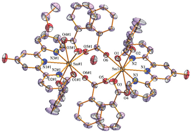
As shown in Fig. 1, complex 1 is a unique dinuclear structural unit with a core composed of two Sm3+ ions. In addition to the lanthanide ions in these two cores, the carboxylic acid and nitrogen‑containing ligands further enrich their structural composition. 1 also contains two free water molecules and ethanol solvent molecules, which may affect the physical and chemical properties of the complex to a certain extent.
图 1
In complex 1, six BA- ligands exhibit flexible coordination patterns that coordinate with Sm3+ ions in two distinct ways. Four of the BA- ligands are tightly bound to Sm3+ ions through double-dentate chelation, forming stable coordination bonds. The other two BA- ligands adopt a bridging coordination mode, connecting two Sm3+ ions via two oxygen atoms on their carboxyl groups. Thereby, they play the role of a bridge and support within the structure, enhancing the stability and connectivity of the dinuclear structural unit.
In addition to the carboxylic acid ligand, the 4-OH-terpy ligand is also an important component in complex 1. This ligand has a high coordination number, and the three nitrogen atoms in its structure are tightly bound to the Sm3+ ion in a tridentate-chelated manner. This tridentate chelation not only increases the interaction force between the ligand and Sm3+ ions but also contributes to the formation of more stable and complex structures. Therefore, in complex 1, the introduction of the 4-OH-terpy ligand further improves the stability and overall performance of the complex.
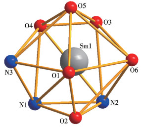
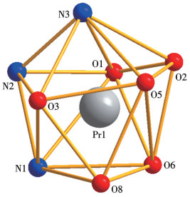
An Sm3+ metal ion is tightly and orderly surrounded by nine atoms, forming a stable coordination environment. Of these atoms, six are oxygen atoms from BA-, and the other three nitrogen atoms are part of the 4-OH-terpy ligand. These atoms work together through different coordination modes to construct a molecular structure that is both stable and functional. It is important to note that the coordination environment of each central Sm3+ ion is the same, and the coordination number of each central Sm3+ ion is nine, which ensures a high degree of consistency and stability throughout the molecular structure. The crystal data was input into the shape-win32 software, and the Sm3+ ions in complex 1 were determined to have the geometric configuration of the MFF-9 type after calculation and analysis (Fig. 2).
图 2
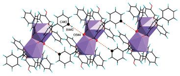
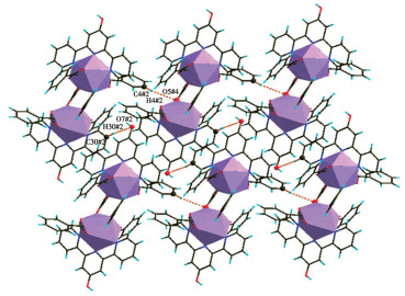
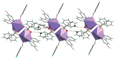
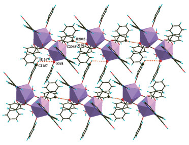
As shown in Fig. 3, the structural units of complex 1 are infinitely repeated along the a-axis of crystallography. Each structural unit involves C—H…O bonding, which forms a 1D chain structure. The distancebetween two adjacent structural units is 0.313 48 nm. At the same time, the structural units are also arranged along the c-axis, with a C…O distance of 0.343 26 nm formed between each adjacent unit. Therefore, parallel to the crystallographic ac plane, a 2D layered supramolecular planar structure is formed, as depicted in Fig. 4.
图 3
 Figure 3. One-dimensional chain structure of the structural unit of complex 1 connected by C—H…O hydrogen bonds
图 4
 Figure 4. Two dimensional layered supramolecular planar structure of the structural unit of complex 1 connected through C—H…O hydrogen bonds
2.1.2 Structure of complex 2
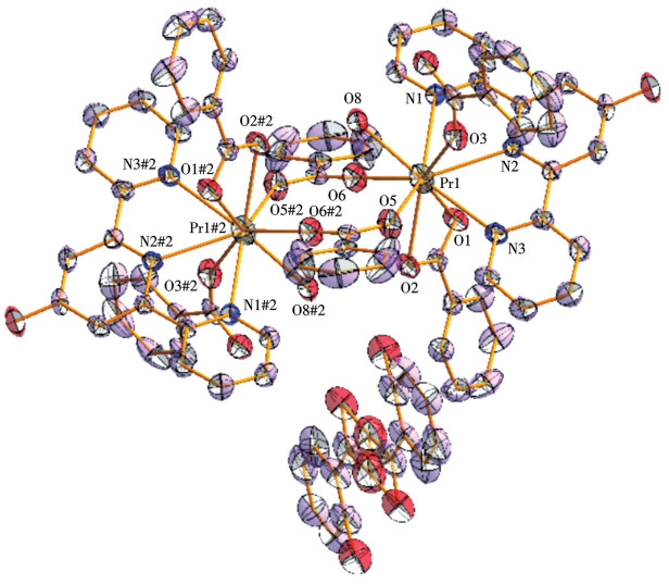
Complex 2 also has a dual-core structural unit, and its structure is both similar and different from complex 1. As shown in Fig. 5, the core of the complex contains two Pr3+ ions linked to six BA- ligands, two 4-OH-terpy ligands, and two coordination water molecules. In addition, there is a molecule of free HBA that binds to water. The six BA- ligands exhibit three different coordination modes with Pr3+ ions: two BA- ligands participate in coordination in a bidentate manner, two others are monodentate, and the remaining two BA- ligands are also bidentate, bridging two Pr3+ ions through two oxygen atoms on a carboxyl group. The 4-OH-terpy ligand is characterized by its high coordination number, the three nitrogen atoms on it are tightly bound to Pr3+ ions in a three-toothed chelation manner, forming a more stable complex structure. In addition, there are two oxygen atoms in the water molecule that also coordinate with Pr3+ ions.
图 5
Nine atoms together surround the central lanthanide ions, and Fig. 6 shows the geometry of Pr3+. The relevant data for complex 2 were input into the shape-win32 software program, and after the calculation and analysis, it was finally determined that the ion showed the geometric configuration of the MFF-9 type. As shown in Fig. 7, along the crystallographic a-axis, the structural units of the complex expand, and each structural unit is connected through some form of interaction involving C—H…O bonds, forming a 1D chain structure. The distance between two adjacent structural units is 0.290 27 nm. At the same time, the structural units expand along the b-axis, with a distance of 0.354 00 nm formed between each structural unit. Therefore, parallel the crystallographic ab plane, a 2D layered supramolecular structure is formed, as shown in Fig. 8.
图 6
图 7
 Figure 7. One-dimensional chain structure of the structural unit of complex 2 connected by C—H…O hydrogen bonds
图 8
 Figure 8. Two-dimensional layered supramolecular planar structure of the structural unit of complex 2 connected through C—H…O hydrogen bonds
2.2 Analysis of infrared and Raman spectroscopy data
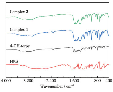
To further analyze the structure of the complexes, the ligands and their resulting complexes were characterized using infrared and Raman spectroscopy. The specific absorption peaks observed in the infrared spectrum can reflect changes in the ligand functional groups before and after complex formation, thereby allowing us to infer the coordination mode between the ligand and the central metal ion within the complex, the strength of the coordination bond, and possible molecular structure characteristics of the complex.
In the infrared spectrum of HBA (Fig. 9), the absorption peak corresponding to the carbonyl stretching vibration (νC=O) was located at 1 640 cm-1. However, when HBA combined with other molecules to form complexes, the original νC=O absorption peak was replaced by two new characteristic peaks. These peaks include the antisymmetric carboxyl stretching vibration [νas(COO-)] absorption peak appearing in a range of 1 577-1 550 cm-1 and the absorption peak appearing in a range of 1 462-1 456 cm-1. The emergence of these peaks marks the formation of the complex. In addition, the vibrational absorption peak of the Ln—O bond was observed at 424 cm-1, directly demonstrating that the O atom in the BA- ion has been coordinated with the central lanthanide ion[24]. It is also worth noting that the absorption peak of the 4-OH-terpy molecule, which originally exhibited a carbon-nitrogen stretching vibration (νC=N) at 1 552 cm-1, shifted to a higher wavenumber region after the formation of the complex. This shift indicates that 4-OH-terpy acts as a neutral ligand and that its N atom is also involved in the coordination process[25].
图 9
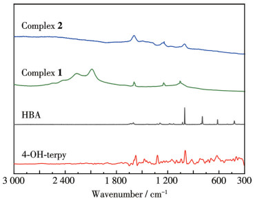
As shown in Fig. 10, Raman spectroscopy revealed changes in the HBA ligand before and after the formation of the complex: the characteristic absorption peak of νC=O, originally appearing at 1 603 cm-1, disappeared after the complex was formed, which may indicate that the C=O group in the HBA has undergone a chemical change or structural rearrangement[26]. At the same time, the δC—H absorption peak in 4-OH-terpy, originally located at 744 cm-1, shifted to the long wavelength direction after the complex formation; this shift indicated the effect of coordination with the lanthanide center atom on the vibration frequency of the C—H bond in 4-OH-terpy[27]. This further confirms the successful synthesis of the complex. In addition, despite their similar structures, the two complexes differ in subtle ways, as clearly shown by their Raman patterns.
图 10
2.3 PXRD data analysis
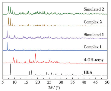
PXRD experiments were performed on powder samples of the two ligands and their complexes (Fig. 11). The experimental results show that there is a significant difference, between the diffraction peak shape of the complex and the peak shape of the two ligands, and that a material with a new crystal phase is successfully formed[28]. The formation of this new crystal phase is a key indicator indicating the success of the complex synthesis. In addition, we compared the experimental diffraction data with the single-crystal CIF data obtained from theoretical simulations. The comparison results show that the positions and shapes of the diffraction peaks in the experimental data are in complete agreement with those in the simulated data. This high degree of consistency not only further validates the reliability of the experimental data, but also fully demonstrates the extremely high chemical purity of the synthesized complexes.
图 11
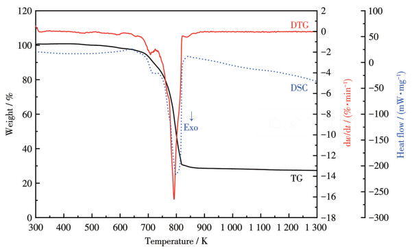
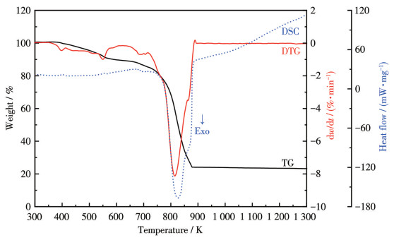
2.4 Thermal decomposition analysis
The thermal decomposition properties of the two complexes were studied and analyzed at a heating rate of 10 K·min-1 in the simulated air atmosphere. The TG curves, DTG curves, and DSC curves were successfully obtained, as shown in Fig. 12 and 13. These two diagrams indicate that these two complexes follow a very similar mechanism of thermal decomposition.
图 12
For complex 1, the DTG curve exhibited two downward peaks with peak temperatures of 708 and 791 K (Fig. 12), respectively, clearly indicating that 1 has undergone two stages of decomposition. The first stage of decomposition occurred in a temperature range of 300-727 K, accompanied by a certain weight loss of 11.68%, corresponding to the loss of free water, ethanol, and a small amount of neutral ligand. Within this temperature range, a slightly upward peak appeared on the DSC curve, suggesting that the first step of decomposition is an endothermic process. Immediately afterward, the second stage of decomposition occurred in a temperature range of 727-1 300 K, with a weight loss of 61.03% in this stage. Unlike the first step, this part of the DSC curve corresponded to a downward peak, indicating that the second step of decomposition is an exothermic reaction, during which a large number of neutral and acidic ligands are lost. Notably, the weight loss stabilized around 900 K with no significant further changes, suggesting that the decomposition process of 1 is nearly complete. Throughout the experiment, the total weight loss rate was 72.71%, indicating that most of the original complex had been converted into other forms after decomposition. The final residue product is mainly the lanthanide oxide Sm2O3[29] and also contains small amounts of carbonaceous compounds, which may be incompletely decomposed organic ligand residues or carbides formed during the reaction.
For complex 2, the DTG curve exhibited four obvious downward peaks, with peak temperatures of 398, 550, 687, and 814 K, respectively (Fig. 13), clearly indicating that 2 has undergone a four-stage decomposition process. The first stage of decomposition occurred in a temperature range from 300 to 430 K and was accompanied by a weight loss of 1.08%, corresponding to the loss of free water molecules (Calcd. 1.07%). The second stage of decomposition occurred in a temperature range from 430 to 570 K, accompanied by a weight loss of 8.18% (Calcd. 7.94% for the loss of free HBA molecules). Both of these first two decomposition steps were endothermic reactions.
图 13
The third stage of decomposition occurred in a temperature range from 570 to 719 K with a weight loss of 5.45%, during which coordination water and part of the neutral ligand are lost. In the fourth stage, the decomposition temperature ranged from 719 to 1 300 K, during which the neutral ligand and acid ligand are lost successively, resulting in a weight loss of 61.97%. It is important to note that each step of the decomposition of this complex is an exothermic reaction. The total experimental weight loss rate of 2 was 76.68%, slightly lower than the theoretical weight loss rate of 79.77% when completely converted to Pr6O11. This difference suggests that in addition to the expected lanthanide oxide Pr6O11 as the main residual product, there are still a small number of carbonaceous compounds that have not yet fully decomposed, which may account for the lower theoretical total weight loss rate.
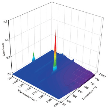
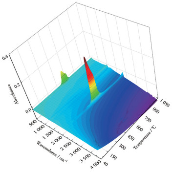
2.5 Evolved gases analyzed by FTIR/MS
The evolved gas of the complex thermal decomposition products was passed into the infrared spectrometer, and through continuous scanning and data processing, a 3D infrared accumulation map was obtained (Fig. 14 and 15). The infrared spectra of the evolved gases from complexes 1 and 2 at specific temperatures were also obtained (Fig. S1 and S2). Subsequently, these evolved gases were introduced into a mass spectrometer for further mass-to-charge analysis and identification of the molecular fragments contained therein[30].
图 14
图 15
Taking complex 1 as an example, Fig. 14 visualizes the changes in the vibration frequency and intensity of the chemical bonds as the complex evolved gases. These changes can be attributed to the breaking and formation of chemical bonds during the reaction process, thereby helping to identify the types of gases released by the complex. At a specific temperature of 516 K, the single infrared spectrum was visible: the CO₂ bending vibration absorption peak at 672 cm-1 and the CO₂ stretching vibration absorption peak in a range of 2 324-2 358 cm-1. The presence of these characteristic peaks directly proves the release of CO₂. In addition, the characteristic absorption peaks of several organic fragments were also visible, including νC=C at 1 542 and 1 360 cm-1, νC=N at 1 478 cm-1, and νC—H multiple absorption peaks from 3 590 to 3 768 cm-1. The presence of these characteristic peaks not only reveals the complex chemical reaction mechanism in the thermal decomposition of complexes but also indicates that the process involves the decomposition of both acidic and neutral ligands, which is consistent with our previous conclusions from TG curves. Through the observation of the 3D infrared patterns of the two complexes, it was found that their peak positions and peak shapes were approximately the same, indicating that even though there are slight differences in the structure of the two complexes, the final pyrolysis products are similar. Mass spectra of the evolved gases of the two complexes also provided key evidence.
The peaks in the mass spectrum with different mass‑to‑charge ratios (m/z) correspond to different molecular fragments or ions, and their relative abundances reflect the amount and stability of each product during thermal decomposition. By comparing the mass spectral data with the standard mass spectra of known compounds, we can further confirm the specific identity of the decomposition products and thus deepen our understanding of the mechanism of thermal decomposition of complexes[31]. Although the structures of complexes 1 and 2 are different, they utilize the same ligand, and thus the fragments of organic molecules produced through the thermal decomposition process are similar at higher temperatures. There is also some similarity in their mass spectra. Fig.S3 and S4 show the mass spectra of 1 and 2 as they evolve during thermal decomposition, respectively. By detailed attribution analysis of the molecular fragments in these spectra, we can observe the following key fragments: the m/z of about 252 corresponds to the fragments of 4-OH-terpy, m/z of 122 corresponds to the fragment of the BA- ion, m/z of 78 corresponds to the fragment of the benzene molecule after the removal of the carboxyl group, and m/z of 79 corresponds to the fragment of the pyridine molecule, and in a m/z range of 128-235, there was a series of molecular fragments belonging to the decomposition products of 4-OH-terpy. Of particular note is the fragmentation peak with a m/z of 44, suggesting that the decomposition products contain a large number of CO2 molecular fragments, which is in perfect agreement with our previous inferences based on the thermal decomposition mechanism, further validating our understanding of the thermal decomposition process of complexes.
2.6 Fluorescence properties of the complexes
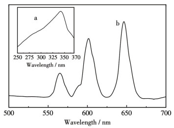
The emission spectra of complex 1 were measured at an excitation wavelength of 342 nm (Fig. 16). Three distinct energy level transition peaks were observed: 565 nm (corresponding to the transition of 4G5/2→6H5/2), 601 nm (4G5/2→6H7/2), and 646 nm (4G5/2→6H9/2)[32]. It is worth noting that among these transition peaks, the intensity of the 4G5/2→6H9/2 transition peak at 646 nm was the largest, which not only confirms the luminescence characteristics of the 1 but also directly correlates to its orange-red luminescence visual performance. Similarly, the CIE1931 color coordinate software was used to input the emission spectrum data of 1 into it for calculation and analysis. The chromatic coordinates of 1 were (0.582 2, 0.412 9), which were located at the edge of the orange light region on the CIE1931 chromaticity map (Fig.S5). It is further verified that the light emitted by complex 1 is indeed close to orange-red, and is consistent with the luminescence characteristics obtained by spectral analysis[33].
图 16
The fluorescence excitation and emission spectra of complex 2 are presented in Fig.S6. Upon comparison, it was observed that, despite the two complexes being highly similar in structure and utilizing the same ligand, the fluorescence emission characteristics of Pr⁺, when compared to those of Sm3+, exhibited a relatively weaker emission ability. Consequently, under the same external environment and experimental conditions, the fluorescence emission peak intensity of 2, upon excitation, was notably lower than that of 1. This disparity can be attributed to the intrinsic fluorescence properties of the two metal ions.
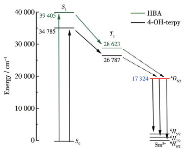
The combination of organic ligands with metal ions or metal clusters can significantly enhance the luminescent properties of the resulting complexes. The core of this sensitization effect resides in the ligands′ robust absorption of light and their efficient energy transfer mechanism with the metal center. Ligands frequently exhibit favorable absorption characteristics for ultraviolet or visible light. Upon excitation by light, they transition from the ground state to the excited state. If the excited state energy of the ligand aligns with a specific emission energy level of the metal ion, the excited energy can be seamlessly transferred to the metal ion, causing it to emit light of a particular wavelength. Consequently, the judicious selection of the ligand structure and metal center can optimize this energy transfer process, ensuring that the complex exhibits heightened luminous intensity and a narrower luminescence peak upon light excitation, thereby augmenting its potential applications as a luminescent material. We have calculated the energy levels of the two ligands using Gaussian 16, as presented in Table S4. Both ligands fulfill the criteria set by Reinhoudt′s rule of thumb and Latva's rule, thereby facilitating efficient energy transfer and substantially enhancing the luminescence efficiency of the complex[34]. The energy transfer mechanism of the two ligands and Sm3+ is shown in Fig. 17.
图 17
3. Conclusions
In summary, we successfully synthesized two novel lanthanide complexes using ultrasonic dissolution and traditional solution methods, with benzoic acid and 4-hydroxy-2, 2′∶6′, 2″-tripyridine as mixed ligands. The crystal structures of these two complexes were determined to belong to the triclinic P1 space group through single-crystal X-ray diffraction. The metal centers of both complexes have the same coordination number of nine, yet their molecular structures and coordination environments exhibit subtle differences. Furthermore, a comprehensive evaluation of the thermal stability of these two complexes was conducted using a combination of TG-DTG-DSC/FTIR/MS techniques, revealing their thermal reaction characteristics at high temperatures, with the main pyrolysis products being rare earth oxides. At the same time, quantum computational analysis further unveiled the complex energy transfer mechanism between the ligands and central ions, and it was found that complex 1 exhibited fluorescence emission that perfectly matches the characteristic transition of Sm⁺, suggesting a broad potential application value of 1 in the field of fluorescent materials.
Supporting information is available at
http://www.wjhxxb.cn
Acknowledgments: This work was supported by the National Natural Science Foundation of China (Grant No.22273015). -
-
[1]
YANG Y L, HU X M, YANG Z, HUANG W. Insights into molecular lanthanide complexes: Construction, properties and bioimaging and biosensing applications[J]. Adv. Funct. Mater., 2024, 21: 202412970.
-
[2]
ZHONG Y, WU Z N, ZHANG Y, DONG B, BAI X. Circularly polar-ized luminescence of lanthanide complexes: From isolated individu-als, discrete oligomers, to hierarchical assemblies[J]. InfoMat, 2023, 5: e12392. doi: 10.1002/inf2.12392
-
[3]
FAN M Y, YU H H, FU P, SU Z M, LI X, HU X L, GAO F W, PAN Q Q. Luminescent Cd(Ⅱ) metal-organic frameworks with anthracene ni-trogen-containing organic ligands as novel multifunctional chemosen-sors for the detection of picric acid, pesticides, and ferric ions[J]. Dyes Pigment., 2021, 185: 108834. doi: 10.1016/j.dyepig.2020.108834
-
[4]
TRUPP L, BRUTTOMESSO A C, VARDE M, ELISEEVA S V, RAMIREZ J A, PETOUD S, BARJA B C. Innovative multipodal ligands derived from Troger's bases for the sensitization of lanthanide(Ⅲ) luminescence[J]. Chem.-Eur. J., 2020, 26: 16900-16909. doi: 10.1002/chem.202003524
-
[5]
SHENG Y, JIANG Y J, CHENG Z H, LIU R C, GE J Y, GAO F. Syn-theses, structures, and magnetic properties of acetate-bridged lantha-nide complexes based on a tripodal oxygen ligand[J]. Front. Chem., 2022, 10: 1021358. doi: 10.3389/fchem.2022.1021358
-
[6]
KOSINSKA-PEZDA M, ZAPALA L, MACIOLEK U, BYCZYNSKI L, WOZNICKA E, ZAPALA W. Thermal study, temperature diffraction patterns and evolved gas analysis during pyrolysis and oxidative de-composition of novel ternary complexes of light lanthanides with mefe-namic acid and 1, 10-phenanthroline[J]. J. Anal. Appl. Pyrolysis, 2021, 159: 105293. doi: 10.1016/j.jaap.2021.105293
-
[7]
ROMANENKO G V, FOKIN S V, LETYAGIN G A, BOGOMYAKOV A S, OVCHARENKOV I. Structure of lanthanide semiquinolates with nitrogen-containing ligands[J]. J. Struct. Chem., 2020, 61: 1594-1598. doi: 10.1134/S002247662010011X
-
[8]
KHANAGWAL J, KHATKAR S P, DHANKHAR P, BALA M, KUMAR R, BOORA P, TAXAK V B. Synthesis and photolumines-cence analysis of europium(Ⅲ) complexes with pyrazole acid and nitro-gen containing auxiliary ligands[J]. Spectr. Lett., 2020, 53: 625-647. doi: 10.1080/00387010.2020.1817093
-
[9]
王晨璐, 宿素玲, 任宁, 张建军. 卤代芳香族羧酸与含氮配体合成镧系配合物的结构、热化学和荧光性质[J]. 物理化学学报, 2023,39,2206035. WANG C L, SU S L, REN N, ZHANG J J. Construction, thermochem-istry, and fluorescence properties of novel lanthanide complexes syn-thesized from halogenated aromatic carboxylic acids and nitrogen-con-taining ligands[J]. Acta Phys.-Chim. Sin., 2023, 39: 2206035.
-
[10]
NIELSEN L G, JUNKER A K R, SORENSEN T J. Composed in the f-block: Solution structure and function of kinetically inert lantha-nide(Ⅲ) complexes[J]. Dalton Trans., 2018, 47: 10360-10376. doi: 10.1039/C8DT01501E
-
[11]
YANG X P, JONES R A, HUANG S M. Luminescent 4f and d-4f polynuclear complexes and coordination polymers with flexible salen-type ligands[J]. Coord. Chem. Rev., 2014, 273: 63-75.
-
[12]
HAYASHI J, SHOJI S, KITAGAWA Y, HASEGAWA Y. Amor-phous lanthanide complexes for organic luminescent materials[J]. Coord. Chem. Rev., 2022, 467: 214607. doi: 10.1016/j.ccr.2022.214607
-
[13]
ZHANG L Y, DOU W, LIU W, XU C, JIANG H E, CHEN C Y, GUO L R, TANG X L, LIU W S. Lanthanide complexes with a biphospho-nate ester ligand and their fluorescent properties[J]. Inorg. Chem. Commun., 2015, 59: 53-56. doi: 10.1016/j.inoche.2015.06.024
-
[14]
MA J H, YANG D Q, SONG X F, WANG Y G. Luminescent materi-als of covalent grafting lanthanide complexes to the synthetic clays[J]. J. Lumin., 2019, 212: 126-132. doi: 10.1016/j.jlumin.2019.04.024
-
[15]
LI H R, CHENG W J, WANG Y, LIU B Y, ZHANG W J, ZHANG H J. Surface modification and functionalization of microporous hybrid material for luminescence sensing[J]. Chem.-Eur. J., 2010, 16: 2125-2130. doi: 10.1002/chem.200901687
-
[16]
LI X, LIU Y H, CHI X W, ZHU G Z, GAO F. Synthesis, structures, and magnetic properties of zigzag tetranuclear lanthanide complexes[J]. Z. Anorg. Allg. Chem., 2020, 646: 1292-1296. doi: 10.1002/zaac.202000235
-
[17]
SONG F, ZHANG Y, CHEN J H, HAN T, CHENG P. Syntheses, crystal structures and magnetic properties of three lanthanide-nitronyl nitroxide complexes[J]. J. Rare Earths, 2017, 35: 24-27. doi: 10.1016/S1002-0721(16)60168-0
-
[18]
FENG X, LIU L, WANG L Y, SONG H L, SHI Z Q, WU X H, NG S W. Lanthanide coordination polymers based on multi-donor ligand containing pyridine and phthalate moieties: Structures, lumines-cence and magnetic properties[J]. J. Solid State Chem., 2013, 206: 277-285. doi: 10.1016/j.jssc.2013.08.029
-
[19]
HUANG X D, WEN G H, BAO S S, JIA J G, ZHENG L M. Thermo-and light-triggered reversible interconversion of dysprosium-anthracene complexes and their responsive optical, magnetic and dielectric properties[J]. Chem. Sci., 2021, 12: 929-937. doi: 10.1039/D0SC04851H
-
[20]
ZHANG F H, WANG Y Y, LV C, LI Y C, ZHAO X Q. Luminescent complexes associated with isonicotinic acid[J]. J. Lumin., 2019, 207: 561-570. doi: 10.1016/j.jlumin.2018.11.051
-
[21]
SONG X Q, WANG L, ZHENG Q F, LIU W S. Synthesis, crystal structure and luminescence properties of lanthanide complexes with a new semirigid bridging furfurylsalicylamide ligand[J]. Inorg. Chim. Acta, 2012, 391: 171-178. doi: 10.1016/j.ica.2012.04.007
-
[22]
DOLOMANOV O V, BOURHIS L J, GILDEA R J, HOWARD J A K, PUSCHMANN H. OLEX2:A complete structure solution, refine-ment and analysis program[J]. J. Appl. Crystallogr., 2009, 42: 339-341. doi: 10.1107/S0021889808042726
-
[23]
SUNDARESWARAN T, JAGAN R, KARTHIKEYAN N, BOAZ B M. Rational analysis of hydrogen bonding interaction in phenazine, 2-hydroxynaphthalene (1:1) cocrystal: From molecular modeling to photophysical properties[J]. J. Mol. Model., 2024, 30: 351. doi: 10.1007/s00894-024-06128-3
-
[24]
杜丹丹, 郝娅帆, 王鑫鑫, 赵金金, 任宁, 张建军. 2-氯-4-氟苯甲酸与5, 5'-二甲基-2, 2'-联吡啶镧系配合物的晶体结构、光谱和热行为[J]. 无机化学学报, 2023,39,(9): 1807-1816. DU D D, HAO Y F, WANG X X, ZHAO J J, REN N, ZHANG J J. Crystal structure, spectra, and thermal behavior of lanthanide com-plexes with 2-chloro-4-fluorobenzoic acid and 5, 5'-dimethyl-2, 2'-bipyridine[J]. Chinese J. Inorg. Chem., 2023, 39(9): 1807-1816.
-
[25]
CEPEDA J, BALDA R, BEOBIDE G, CASTILLO O, FERNÁNDEZ J, LUQUE A, PÉREZ-YÁÑEZ S, ROMÁN P, VALLEJO-SÁNCHEZ D. Lanthanide(Ⅲ)/pyrimidine-4, 6-dicarboxylate/oxalate extended frameworks: A detailed study based on the lanthanide contraction and temperature effects[J]. Inorg. Chem., 2011, 50: 8437-8451. doi: 10.1021/ic201013v
-
[26]
LELLI M, DI BARI L. Solution structure and structural rearrange-ment in chiral dimeric ytterbium(Ⅲ) complexes determined by para-magnetic NMR and NIR-CD[J]. Dalton Trans., 2019, 48: 882-890. doi: 10.1039/C8DT03090A
-
[27]
HE S M, SUN S J, ZHENG J R, ZHANG J J. Molecular spectrum of lanthanide complexes with 2, 3-dichlorobenzoic acid and 2, 2-bipyri-dine[J]. Spectroc. Acta Pt. A -Molec. Biomolec. Spectr., 2014, 123: 211-215. doi: 10.1016/j.saa.2013.12.023
-
[28]
BABU S V, ESWARAMMA S, RAO K. Synthesis, characterization, luminescence and biological activities of lanthanide complexes with a hydrazone ligand[J]. Main Group Chem., 2018, 17: 99-110. doi: 10.3233/MGC-180251
-
[29]
HAO Y F, XU S L, ZHAO J J, GAO J, REN N, ZHANG J J. New nitrogen-containing lanthanide complexes with novel structural, ther-mal, and fluorescence properties[J]. J. Saudi Chem. Soc., 2024, 28: 101792. doi: 10.1016/j.jscs.2023.101792
-
[30]
CHEN M H, LIANG B, GUO Y H, LI C F, HE X, HU J H, LI R K, ZENG K, YANG G. Pyrolysis mechanism of polyimide containing bio-molecule adenine building block[J]. Polym. Degrad. Stabil., 2020, 175: 109124. doi: 10.1016/j.polymdegradstab.2020.109124
-
[31]
LIU B, LI Y M, WU S B, LI Y H, DENG S S, XIA Z L. Pyrolysis characteristic of tobacco stem studied by Py-GC/MS, TG-FTIR, and TG-MS[J]. BioResources, 2013, 8: 220-230.
-
[32]
SARIOGLU A O, KAHRAMAN D T, KÖSE A, SÖNMEZ M. Synthe-sis, characterization, photoluminescence properties and cytotoxic activities of Sm(Ⅲ) complexes of β-diketones[J]. J. Mol. Struct., 2022, 1260: 132786. doi: 10.1016/j.molstruc.2022.132786
-
[33]
邱深皓, 肖清泉, 唐华, 谢泉. 稀土元素X (X=Sc、Y、La、Ce、Eu)掺杂二维GaSe的电子结构、光学及磁学性质的第一性原理研究[J]. 无机化学学报, 2024,40,(11): 2250-2258. QIU S H, XIAO Q Q, TANG H, XIE Q. First-principles study on electronic structure, optical and magnetic properties of rare earth elements X (X=Sc, Y, La, Ce, Eu) doped with two-dimensional GaSe[J]. Chinese J. Inorg. Chem., 2024, 40(11): 2250-2258.
-
[34]
WANG C L, ZHANG J Y, LI X Y, REN N, ZHANG J J. Crystal structure, thermodynamic behavior, and luminescence properties of a new series of lanthanide halogenated aromatic carboxylic acid com-plexes[J]. Arab. J. Chem., 2022, 15: 104089.
-
[1]
-

Figure 1 Structural unit of complex 1
Ellipsoid probability: 50%; Symmetry code: #1:-x+1, -y, -z+1.
Figure 3 One-dimensional chain structure of the structural unit of complex 1 connected by C—H…O hydrogen bonds
Symmetry codes: #2:-x+1, -y+1, -z+1; #4: x, 1+y, z.
Figure 4 Two dimensional layered supramolecular planar structure of the structural unit of complex 1 connected through C—H…O hydrogen bonds
Symmetry codes: #2:-x+1, -y+1, -z+1; #4: x, 1+y, z.

Figure 5 Structural unit of complex 2
Ellipsoid probability: 50%; Symmetry code: #2:-x+1, -y+1, -z+1.
Figure 7 One-dimensional chain structure of the structural unit of complex 2 connected by C—H…O hydrogen bonds
Symmetry codes: #5: 1+x, 1+y, -1+z; #6: x, 1+y, -1+z.
Figure 8 Two-dimensional layered supramolecular planar structure of the structural unit of complex 2 connected through C—H…O hydrogen bonds
Symmetry codes: #5: 1+x, 1+y, -1+z; #6: x, 1+y, -1+z; #7: 1+x, y, -1+z; #8: 2-x, 2-y, -z.
Table 1. Main crystallographic parameters of complexes 1 and 2
Parameter 1 2 Empirical formula C72H52N6O14Sm2·2H2O·2C2H6O C72H56N6O16Pr2·H2O·C7H6O2 Formula weight 1 654.08 1 683.18 Crystal system Triclinic Triclinic Space group P1 P1 a / nm 1.132 94(11) 1.239 65(4) b / nm 1.308 25(12) 1.378 50(5) c / nm 1.414 41(13) 1.393 58(5) α / (°) 66.872(2) 115.901(4) β / (°) 78.982(3) 96.657(3) γ / (°) 79.865(4) 110.992(3) Volume / nm3 1.880 5(3) 5.439 1(9) Z 1 1 Dc / (Mg·m-3) 1.461 1.477 Absorption coefficient / mm-1 1.618 2.420 F(000) 834 850 Crystal size / mm 0.17×0.11×0.05 0.12×0.07×0.05 2θ range for data collection / (°) 3.680-50.040 6.580-50.100 Limiting indices -13 ≤ h ≤ 13, -15 ≤ k ≤ 13, -15 ≤ l ≤ 9 -14 ≤ h ≤ 14, -15 ≤ k ≤ 16, -16 ≤ l ≤ 16 Independent reflection 6 340 (Rint=0.073 3) 6 703 (Rint=0.035 9) Completeness to θ=25.02° / % 97.0 99.8 Max. and min. transmission 0.923 5 and 0.770 5 0.935 7 and 0.855 1 Data, restraint, number of parameters 6 340, 0, 461 6 703, 200, 512 Goodness-of-fit on F 2 1.091 1.069 Final R indices [I > 2σ(I)] R1=0.076 6, wR2=0.128 8 R1=0.046 1, wR2=0.112 8 Final R indices (all data) R1=0.131 3, wR2=0.152 5 R1=0.062 2, wR2=0.121 8 Largest diff. peak and hole / (e·nm-3) 1 500 and -2 090 1 343 and -512  下载: 导出CSV
Table 2. Hydrogen bonds of complexes 1 and 2
D—H…A d(D—H) / nm d(H…A) / nm d(D…A) / nm ∠D—H…A / (°) 1 O8—H8D…O1#1 0.085 0.210 9 0.295 4 172.98 O8—H8C…O3 0.085 0.191 5 0.276 1 172.76 O7—H7…O8#2 0.082 0.191 9 0.269 1 156.67 O9—H9…O2#2 0.082 0.205 7 0.287 5 175.18 2 O7—H7…O4#3 0.082 0.184 9 0.265 0 164.87 O8—H8C…O4 0.085 0.203 9 0.285 0 159.22 O8—H8D…O2#2 0.085 0.200 7 0.281 8 159.11 O10—H10…O11 0.082 0.191 7 0.268 1 154.58 O11—H11C…O2 0.085 0.234 3 0.306 4 142.92 O11—H11D…O8#2 0.085 0.251 6 0.324 4 144.16 Symmetry codes: #1: -x+1, -y, -z+1; #2: -x+1, -y+1, -z+1 for 1; #2: -x+1, -y+1, -z+1; #3: -x+2, -y+2, -z+1 for 2
下载: 导出CSV
-


 扫一扫看文章
计量
- PDF下载量: 0
- 文章访问数: 55
- HTML全文浏览量: 8
