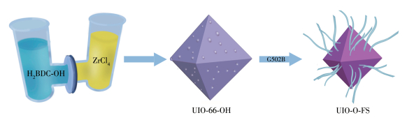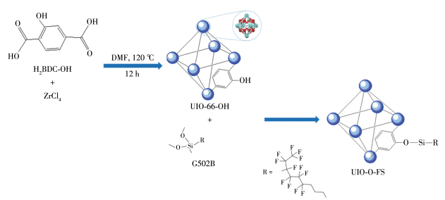
Figure 1.
Schematic illustration of the chemical reaction process of UIO-O-FS

A large number of stone cultural heritages exist in the world today, which are very precious but suffer from serious damage due to the long years of exposure to the natural environment and the resultant weathering and erosion[1-3]. In recent years, global warming and environmental pollution have accelerated the deterioration of stone cultural relics. Therefore, the protection of stone cultural relics has received increasing attention and has become an important research topic. At present, several conservation methods for rocks have been proposed by relevant researchers[4-15]. For example, Graziani, Tesser, and Shu et al.[4-8] coated different materials such as calcium oxalate, alicyclic epoxy resin, and rod-shaped TiO2 composite on the marble surface and investigated their protective properties respectively, and the test results showed that the materials had a certain protective effect. To protect sandstone, Shu, Tokarsky, and Aslanidou et al.[9-11] prepared modified sol-gel coating, nano-ZnO/poly (alkyl siloxane) coating, and nano-SiO2/alkoxysilane coating materials respectively, and applied them uniformly on the surface of rock samples, and the test results showed that the materials have good acid resistance and hydrophobicity. Al-Dosari et al.[12] prepared nano-Ca(OH)2/polymer composites for the protection of carbonate rocks using in-situ emulsion polymerization method and the hydrophobicity and consolidation properties of the materials were demonstrated by tests. Zhu et al.[16-17] prepared nanolime/ kaolin (N-K) nanocomposites and polydopamine (PDA) modified nanolime (PDA@NL) by in-situ growing Ca(OH)2 nanoparticles on the surface of kaolin nanosheets, respectively, which improved the stability, permeability, and consolidation of nanolime. PDA@NL also avoids the adverse effects of back migration and delayed carbonization on the conservation effect of nanolime in the protection of stone artifacts. Chai et al.[18] introduced the synergistic effect of ZnO nanoparticles and SiO2 nanoparticles, and showed experimentally that the nanocomposite coatings obtained after modification by fluorocarbon coupling agents have excellent hydrophobicity and weathering stability. Current protection materials can be categorized into three types, inorganic materials, organic materials, and new materials. Inorganic materials are resistant to weather but have low permeability. Organic materials are corrosion-resistant but have a shorter life span. New materials, such as nanomaterials and bionic, have unique functions and compatibility, indicating a new development prospect in the field of stone protection. Based on the above, this work aims to research the application of nanomaterials in the preservation of limestone-type stone relics of the Longmen Grottoes.
Metal-organic frameworks (MOFs) are nano- porous materials composed of inorganic metal nodes and organic linkers[19-22] and have attracted much attention in the research fields of sensors[23-25], catalysts[26-27], drug delivery[28-30], and gas adsorption and separation[31-32] because of their excellent properties such as high surface area, large pore volume, tunable pore size, favorable chemical and hydrothermal stability, and easy post-modification. According to the standard recommendations, tone cultural relic protection materials need to be hydrophobic, permeable, and stable and possess good stone adhesion and compatibility[33]. However, due to the high sensitivity of MOFs to moisture, their advantageous structural features are rapidly compromised, which restricts their applicability in stone heritage conservation[34-35]. There have been studies on the modification of MOFs materials and using them in the stone field. For example, Yoon et al. prepared semi-siloxane Zn-MOF liquid marbles using azobenzene-containing dicarboxylic acids as organic linkers and investigated their stability under acidic conditions[36]. Baah et al. summarized hydrophobic metal-organic frameworks and their applications, which included the use of MOF materials to prepare liquid marbles[37]. However, there are relatively few studies on the use of MOFs materials for the conservation of limestone-type stone heritage, so this paper addresses their application in the field of limestone-type stone heritage conservation.
Based on the available studies, we understand that special functional groups can be introduced into porous MOFs by post-synthetic modification methods[24-25]. In this work, porous fluorosilylated MOFs (UIO-O-FS) was synthesized from Zr-based UIO-66-OH containing 2-hydroxyterephthalic acid linkers by modification with dodecafluoro-heptyl-propyl-methyl-dimethoxy-silane(G502B). In UIO-O-FS, the interconnected discrete pores are covered with G502B, presenting gas channels, and the hydrophobicity induced by the fluorosilane surface makes the material ideal for exhibiting both good gas permeability and hydrophobicity. This work demonstrates an attempt at linker-oxo node engineering of hydrophobic MOFs for the protection of rock artifacts. The precise modification of the linker-oxo nodes of MOFs without blocking the windows of pores maximizes the preservation of the inherent accessibility of pore textures, ensuring the unique superiority of these MOFs in practical applications. According to the experimental results, it can be seen that UIO-O-FS has a good protective effect in the protection of stone heritage. Therefore, a new hydrophobic MOF material for stone heritage protection is proposed.
2-Hydroxyterephthalic acid (H2BDC-OH, 99%), terephthalic acid (H2BDC, 99%), and N,N-dimethylformamide (DMF, 99.9%) were provided by Aladdin Reagent Co., Ltd. Zirconium chloride (ZrCl4, 99.9%), acetic acid (CH3COOH, 99.5%), absolute ethanol (C2H5OH, 99.7%), sulfuric acid (H2SO4, 98.3%), hydrochloric acid (HCl, 36%-38%), sodium chloride (NaCl, 99.5%), dodecafluoroheptyl methacrylate (Actyflon-G04) and dodecafluoro-heptyl-propyl-methyl-dimethoxy-silane (G502B) were supplied by Sinopharm Chemical Reagent Co., Ltd. All reagents were used as received without further purification, and deionized water was used in all experiments.
The crystallographic structures of the prepared UIO-66, UIO-66-OH, and UIO-O-FS adsorbents were analyzed by X-ray diffraction (XRD) using a MAC-18XHF diffractometer (Rigaku, Japan) with Cu Kα radiation (λ=0.154 05 nm at 30 kV and 30 mA) in a 2θ range of 5°-60°. A Leo-Supra 55 field-emission scanning electron microscope (FESEM) (Carl Zeiss, Germany) was employed to survey the morphologies of samples. The Brunauer-Emmett-Teller (BET) specific surface areas of UIO-66, UIO-66-OH, and UIO-O-FS were determined with a JW-BK132F specific surface area ultrafine pore size analyzer (Beijing JWGB Sci. & Tech. Co., Ltd., China) using nitrogen sorption at 77 K. The functional groups of the adsorbents were assessed via FTIR) spectroscopy on a Tensor 27 instrument (Bruker, Germany). A JEM-2100F instrument (JEOL, Japan, 200 kV) was used to obtain high-resolution transmission electron microscopy (HRTEM) images and conduct energy-dispersive X-ray spectroscopy (EDS) for the mapping of elements of the materials. The static contact angle was measured with a DSA 100 instrument (Krüss, Germany) at room temperature. The thermogravimetric analysis (TGA) of the samples was performed by a TG209 instrument. The tests were performed under N2 protection with a N2 flow rate of 20 mL·min-1. The temperature was increased from 100 to 800 ℃ with a rate of 10 ℃·min-1.
To synthesize UIO-66-OH[19-20], 1 mmol each of ZrCl4 and H2BDC-OH were dissolved in 35 mL of DMF under magnetic stirring (1 500 r·min-1, 20 min). Then, the solution was transferred to a Teflon-lined autoclave and reacted at 120 ℃ for 12 h. After the reactor was cooled to room temperature, the resulting solid product was isolated by centrifugation (8 000 r·min-1, 10 min), washed with DMF and anhydrous ethanol, and finally dried under vacuum (12 h, 60 ℃) to obtain UIO-66-OH as a yellow product.
To perform a comparative test with UIO-66-OH, it is necessary to prepare UIO-66 to demonstrate the successful synthesis of UIO-66-OH. The synthesis process of UIO-66 employed H2BDC as the organic ligand and was otherwise the same as the synthesis of UIO-66-OH.
G502B was hydrolyzed by the following method. The fluorosilane was dropped into a mixture of deionized water and absolute ethanol (mass ratio of 1∶4, and the pH of this mixture was adjusted to three with glacial acetic acid). The hydrolysis reaction was continued under magnetic stirring until a clear solution was obtained.
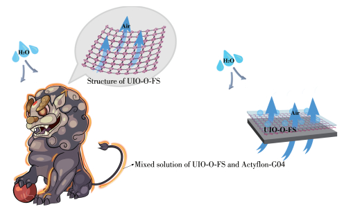
UIO-66-OH powder and the solution of hydrolyzed G502B were combined in a beaker and heated at 110 ℃ until dryness. The obtained crosslinked UIO-66-OH was named UIO-O-FS, as shown in Fig. 1. The flow chart of the entire experiment, as well as the structures of UIO-66-OH and UIO-O-FS, are shown in Fig. 2. Fig. 3 provides an artistic illustration of the UIO-O-FS protective material applied to the surface of a stone artifact, demonstrating the hydrophobicity and permeability of the protective material.
Because the prepared UIO-O-FS may be in the agglomerative state, to enable it to act directly on the surface of stone artifacts, it was ground before use to obtain UIO-O-FS powder. After that, UIO-O-FS powder was combined with Actyflon-G04 solution under ultrasonic treatment for 30 min to obtain a mixture with a mass fraction of 3%, and finally, the mixed solution of the protective material acting on the rock surface was obtained. The configured mixed solution was evenly applied to the surface of the rock specimen with a brush, followed by drying treatment (110 ℃, 2 h), and the above operation was repeated three times after drying. Actyflon-G04 contains unsaturated bonds that can undergo polymerization at high temperatures, forming a polymer film layer on the surface of rocks. Therefore, the above drying process was performed at 110 ℃. To verify whether Actyflon-G04 underwent the polymerization reaction., after dispersing the UIO-O-FS powder into the Actyflon-G04 solution in the experiment and found that the viscosity of the solution gradually increased as the temperature rose, indicating that polymerization occurred in Actyflon-G04.
To study the acid resistance of UIO-O-FS, twelve rock samples were divided into four groups. The first and third groups were unprotected samples, the second and fourth groups were protected samples, and the rock samples of the third and fourth groups were also immersed in sulfuric acid solution at pH=3 for 10 d. Similarly, to study the salt resistance of UIO-O-FS, the rock samples were prepared in the same way as the acid resistance test, with the difference that the third and fourth groups of rock samples needed to be immersed in a solution of sodium chloride at pH=3 and a concentration of 0.02 mol·L-1 for 7 d. A hydrochloric acid solution was used to adjust the pH of the NaCl solution.
The FTIR spectra of three MOF samples, UIO-66, UIO-66-OH, and UIO-O-FS, are provided in Fig. 4a. As can be seen in Fig. 4a, the peaks observed in a range of 400-600 cm-1 are attributed to the Zr—O stretching vibration in the three MOFs[38-40]. For the UIO-66 sample, the band in a range of 430-700 cm-1 is referred to as a combination of Zr—O modes with OH and CH bending vibrations. Moreover, the bands of C=O (1 708 cm-1), C=C (1 577 cm-1), and C—O (1 400 cm-1) stretching vibrations had a similar position for UIO-66, UIO-66-OH, and UIO-O-FS, hence a high degree of overlapping was observed in the spectra. In the spectrum of UIO-66-OH, the band in a range of 1 260-1 420 cm-1 is attributed to the deformation vibration peak of —OH, and this absorption band was wider than that in the UIO-66 spectrum because of the addition of hydroxyl groups to the MOF framework, indicates the successful synthesis of UIO-66-OH. In the spectrum of UIO-O-FS, characteristic bands at 1 313 and 867 cm-1 correspond to C—F and C—Si stretching vibrations, while these were not observed in the spectrum of UIO-66-OH, indicating that the modification experiment was completed, that is, the hydrophobic group grafted onto the UIO-66-OH surface, making the UIO-O-FS material hydrophobic.
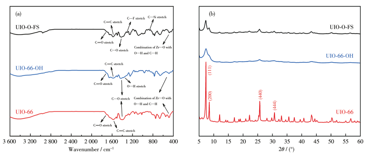
XRD analysis was used to determine the phase purity and crystalline structures of products, and their XRD patterns are shown in Fig. 4b. For the UIO-66 sample, the diffraction peaks were consistent with previously reported articles[39-40]. The analysis demonstrated that the characteristic reflection peaks of the prepared UIO-66-OH and UIO-O-FS samples matched those of the pattern of UIO-66, but the intensity of the UIO-O-FS peak was lower than that of UIO-66, the width of the UIO-O-FS peak was wider than that of UIO-66, and the intensity and width of the UIO-66-OH peak were in between. Therefore, from the Scherrer formula D=Kγ/(Bcos θ), the particle size of UIO-66-OH was smaller than that of UIO-66 due to that the OH groups embedded in the exo-ligand[41-42]. Similarly, the particle size of UIO-O-FS after hydrophobic modification was smaller than that of UIO-66 due to the fluorosilane groups attached to the UIO-66-OH surface, but because the volume of the fluorosilane groups was larger than that of the OH groups, it made the particle size of UIO-O-FS further reduced.
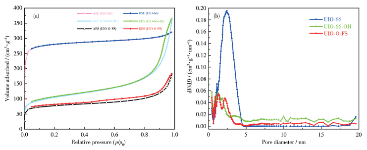
N2 adsorption-desorption isotherm at 77 K was used to determine the specific surface area and pore size, and distribution of the samples. Fig. 5a shows the measured N2 adsorption-desorption isotherm curves of UIO-66, UIO-66-OH, and UIO-O-FS. It can be seen that the N2 adsorption isotherm curve of UIO-66 belongs to the type Ⅰ adsorption isotherm curve, which indicates that UIO-66 is a microporous material. The adsorption trend of UIO-66 on N2 is as follows: at low relative pressure (p/p0 < 0.1), the adsorption amount increases rapidly and tends to saturate, which is caused by the rapid filling of the micropores in the material with N2; at higher relative pressure (p/p0 > 0.1), the adsorption amount increases slowly, and the adsorption and desorption lines coincide without desorption hysteresis[43-44]. Unlike the isotherms of UIO-66, pure UIO-66-OH, and UIO-O-FS had isotherms of type Ⅳ with H3-type adsorption hysteresis loops without an obvious saturated adsorption platform, proving the mesoporous structure of UIO-66-OH and UIO-O-FS[43]. At the same time, the same conclusion as the N2 adsorption-desorption isotherm can be concluded from Fig. 5b. Table 1 shows the BET-specific surface areas and average pore diameters of UIO-66, UIO-66-OH, and UIO-O-FS. From Table 1, we can see that the BET-specific surface area of UIO-66 (870 m2·g-1) is the largest, followed by UIO-66-OH (350 m2·g-1) and UIO-O-FS (249 m2·g-1). The average pore diameter of UIO-66 was about 2.279 0 nm, while those of UIO-66-OH and UIO-O-FS were about 6.481 2 and 4.548 6 nm, respectively. Since the average pore size of UIO-O-FS is larger than that of UIO-66, it indicates that the modified material can better provide channels for water vapor and air and reduce the resistance to mass transfer. However, the average pore size of UIO-O-FS was reduced compared with that of UIO-66-OH, which is caused by the successful grafting of the fluorosilane groups onto the surface of UIO-66-OH and the larger volume of the fluorosilane groups than that of the OH groups.
 下载:
导出CSV
下载:
导出CSV
| Sample | SBET/(m2·g-1) | Pore volume/(cm3·g-1) | Average pore diameter/nm |
| UIO-66 | 870 | 0.495 9 | 2.279 0 |
| UIO-66-OH | 350 | 0.567 8 | 6.481 2 |
| UIO-O-FS | 249 | 0.282 8 | 4.548 6 |
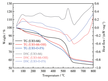
To obtain the composition and thermal stability of the materials and their intermediates, TGA was performed on UIO-66, UIO-66-OH, and UIO-O-FS, and the TG curves of the samples are shown in Fig. 6. As can be seen from Fig. 6, the weight of all three samples decreased continuously with the increase of temperature. At 100 ℃, the loss of sample weight is caused by the excluding of small molecules such as water, organic solvents, and ligands. This was followed by the maximum weight loss peak in the TG curve caused by the destruction of the UIO-66, UIO-66-OH, and UIO-O-FS structures. The differential scanning calorimetry (DSC) curve of UIO-66 shows that its thermal decomposition process is mainly based on heat absorption, for example, the weight loss of the material peaks at 550 ℃ due to the absorption of a large amount of heat by UIO-66, indicating that the main structure of the material is decomposed at this time, which also indicates that the sample has good thermal stability. The DSC curve of UIO-66-OH was the same as that of UIO-66, both of which were mainly endothermic. The difference was that the DSC curve of UIO-O-FS was mainly exothermic. The main weight loss peaks of UIO-66-OH and UIO-O-FS occurred at 500 to 650 ℃. In addition, we can also know that when the temperature rose from 100 to 800 ℃, the residual weight of UIO-O-FS, UIO-66-OH, and UIO-66 was about 56%, 57%, and 53%, respectively. This means that the carbon residual amount of UIO-66-OH and UIO-O-FS is larger than that of UIO-66 and larger than that reported in the literature[45-46]. Compared with UIO-66, both the weight loss rate and carbon residual amount after modification are increased due to the cross-linking within the MOF structure. In other words, the thermal stability of the modified UIO-O-FS is improved, which can extend the application of the material to relatively higher temperatures.
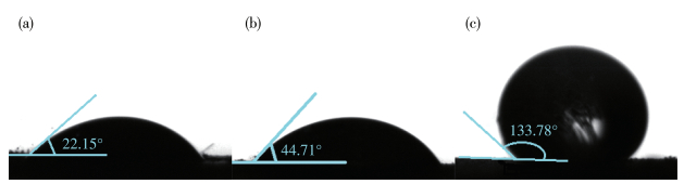
To examine the effect of the hydrophobic groups attached to the open metal sites of the framework on hydrophobicity, we measured the contact angles of water droplets dropped on the solid surface of each sample. It is known that when the contact angle of the material is less than 90°, the material is considered hydrophilic. At contact angles greater than 90° and less than 150°, the material is classified as hydrophobic, while materials with contact angles exceeding 150° are termed superhydrophobic. As shown in Fig. 7, the contact angles of UIO-66, UIO-66-OH, and UIO-O-FS were 22.15°, 44.71°, and 133.78°, respectively. The contact angle of UIO-66-OH was greater than that of UIO-66, indicating that the hydrophobicity of UIO-66-OH was stronger than UIO-66, but since the angle was still below 90°, the material did not exhibit hydrophobic properties and should rather be classified as hydrophilic. The contact angle of UIO-O-FS was 133.78°, which was much larger than that of UIO-66-OH, indicating that the modification experiment of UIO-66-OH was done successfully, and the hydrophobic groups have been linked on the surface of UIO-66-OH, that is, UIO-O-FS has a hydrophobic effect.
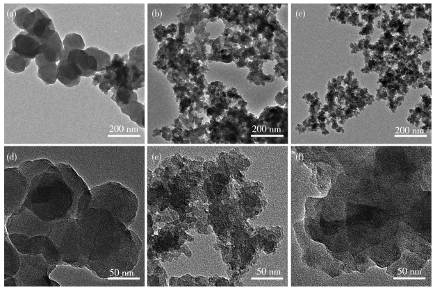
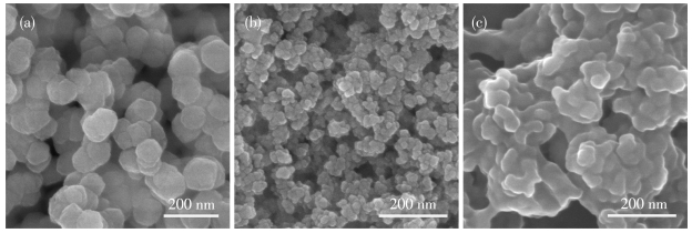
TEM and SEM images of the MOFs are shown in Fig. 8 and 9. As shown in Fig. 8, the pristine UIO-66 material had a typical octahedral morphology and uniform particle size, which was the same as previously reported[40]. As shown in Fig. 8c, the particles of UIO-O-FS obtained after the modification test had irregular morphology and decreased crystallinity, and there was a layer of adhesion on the surface of the particles, which were hydrophobic organic groups grafted on the surface of UIO-66-OH, indicating successful modification. This is consistent with the results of the FTIR analysis.
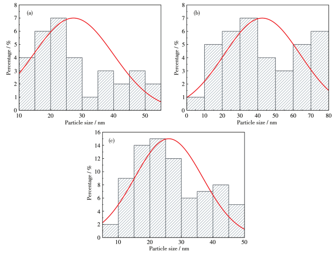
As can be seen in Fig. 9, the pure UIO-66 particles were large in size and uniformly octahedral[40], but the grain shapes of UIO-66-OH and UIO-O-FS were irregular due to the organic groups covered on their surfaces. Meanwhile, it is shown from the morphological analysis that UIO-O-FS had a pore structure, which can form permeable and breathable channels, effectively prevent the attachment of liquid water, the infiltration, and condensation of water vapor, avoid the damage caused by water on the surface and interior of cultural relics, which means that the material can effectively protect stone cultural relics. Fig. 10 shows the particle size distributions of UIO-66, UIO-66-OH, and UIO-O-FS. The particle size ranges of UIO-66, UIO-66-OH, and UIO-O-FS were 11-53 nm, 8-76 nm, and 9-46 nm, respectively, as can be obtained from Fig. 10.
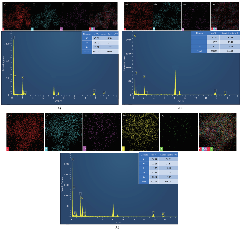
Fig. 11 shows the energy spectrum analysis diagram of UIO-66, UIO-66-OH, and UIO-O-FS. The information in the table in Fig. 11A and 11B revealed that the atomic ratios of Zr and O in the two synthesized materials were close to the theoretical ratios for UIO-66 and UIO-66-OH. Therefore, the prepared materials can be confirmed to be UIO-66 and UIO-66-OH. The table in Fig. 11C shows that the material contained F and Si elements, which were not present in UIO-66-OH, indicating that the hydrophobic groups have been successfully linked to the surface. This conclusion is consistent with the previous conclusions.
To test the actual effectiveness of the protective material, acid and salt resistance tests were conducted. The rock samples were processed according to Table 2, and the prepared rock samples are shown in Fig. 12.
下载:
导出CSV
| Group | Preparation method |
| 1 | Unprotected treatment, not immersed |
| 2 | Protected treatment, not immersed |
| 3 | Unprotected treatment, immersed |
| 4 | Protected treatment, immersed |
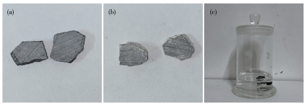
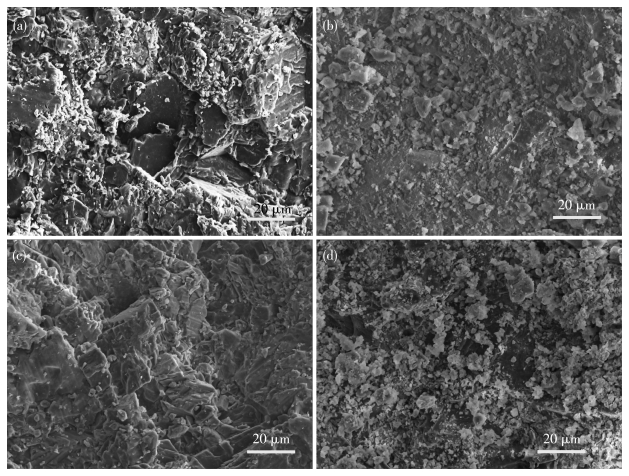
Table 3 shows the acid resistance test results. It can be seen that the masses of the samples treated with the protective material were larger than the mass before treatment, indicating that the protective material has entered the rock samples. In addition, after the erosion resistance test, the mass loss rate of the fourth group of rock samples was smaller than that of the third group of rock samples, which also indicates that the material has some protective effect. The comparison of Fig. 13a and 13b revealed that there were obvious crystal connections on the surface of the sample after treatment with the protective material, and the pores of the rock were filled, indicating that the tiny nanoparticles can densify the surface of the rock and have a cementing effect. In addition, many channels were present on the surface of the sample, which ensures that the sample is permeable to water vapor, reflecting the permeability of the protective material. When comparing Fig. 13c with 13d, it could be observed that the surface of rock sample 3 was uneven with obvious erosion, there was no cementation between mineral particles, and the structure was relatively loose. However, there was no obvious trace of corrosion on the surface of rock sample 4 treated with the protective material, again indicating that the increased association between particles of the rock sample treated with UIO-O-FS, and the rock sample structure is strengthened, thus improving the erosion resistance of the rock. In addition, according to Fig. 13b and 13d, SEM images of rock samples treated with UIO-O-FS protective material were different after acid resistance tests, but the difference between them was smaller than that of rock samples treated without UIO-O-FS protective material (Fig. 13a and 13c). The results confirm the effectiveness of the prepared material for rock protection.
下载:
导出CSV
| Group | Initial mass/g | Mass after treatment/g | Mass after soaking/g | Mass loss rate/% |
| 1 | 1.855 8 | |||
| 2 | 1.277 2 | 1.499 5 | ||
| 3 | 1.506 6 | 1.502 9 | 0.246 | |
| 4 | 1.752 0 | 1.810 3 | 1.806 2 | 0.226 |
For the salt resistance test, the data trends in Table 4 were the same as those in Table 3, both of which showed that the mass of rock samples treated with the protective material was greater than that before treatment, indicating that the protective material has entered the interior of the rock samples. And after the salt resistance test, the mass loss rate of the fourth group of rock samples was smaller than that of the third group of rock samples, which also indicates that the material has salt resistance.
下载:
导出CSV
| Group | Initial mass/g | Mass after treatment/g | Mass after soaking/g | Mass loss rate/% |
| 1 | 8.241 6 | |||
| 2 | 7.078 3 | 7.085 1 | ||
| 3 | 7.476 8 | 7.404 2 | 0.971 | |
| 4 | 8.179 4 | 8.182 5 | 8.108 1 | 0.909 |
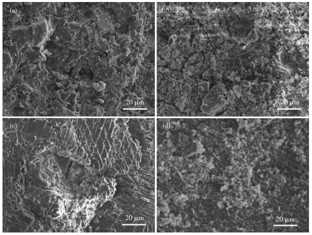
Comparing Fig. 14a and 14b, the same conclusion can be obtained as Fig. 13. By comparing Fig. 14c and 14d, it can be inferred that the third group of rock samples, which were not subjected to protective treatment, exhibited significant erosion similar to the results of the acid resistance test. The surface of the fourth group of rock samples, which underwent protective treatment, showed an obvious connection between the particles, making it possible to prevent salt erosion from the surroundings. The test results show that the prepared material can provide an excellent anti-salt effect for the rock.
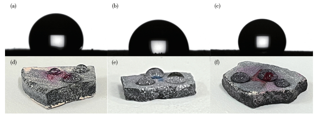
To prove that acid or salt solution immersion has little effect on the coating, we measured the contact angles of water droplets for the rock samples before the erosion test (Fig. 15a), after the acid resistance test (Fig. 15b), and after the salt resistance test (Fig. 15c). As shown in Fig. 15a and 15b, the contact angle of the rock sample decreased after the acid resistance test but was still greater than 90°, indicating that the protective material UIO-O-FS is still hydrophobic and has good acid resistance after the acid resistance test. Simultaneously, the validity of this conclusion is also corroborated by Fig. 15d and 15e. Similarly, as is evident from Fig. 15a and 15c, the contact angles of rock samples after the salt resistance test had little change compared with that before the erosion resistance test and were both greater than 90°, which shows that the protective material UIO-O-FS has good salt resistance. Also, the same conclusion can be drawn according to Fig. 15d and 15f.
In conclusion, we have established a simplified strategy to prepare a novel class of homogenous, stable, MOFs-based, hydrophobic materials by modifying linker-oxo nodes with fluorosilane. As expected, the BET-specific surface area, average pore size, and particle size of UIO-O-FS were 249 m2·g-1, 4.548 6 nm, and 9-46 nm, respectively, which were all decreased compared to UIO-66-OH. Moreover, the contact angle of the water droplet for UIO-O-FS was 133.78°, higher than that of UIO-66-OH by a factor of 2, indicating that UIO-O-FS has a porous molecular structure and hydrophobic functional groups, which enable the material to form air-permeable channels to facilitate the internal and external flow of air and water in stone cultural relics, greatly reducing the erosion probability of the relics without compromising their ornamental effect and mechanical properties, indicating that the material has a potential application prospect in the field of limestone cultural relics protection.
Castro N F, Becerra J E, Bellopede R, Marini P, Dino G A. Introduction to 'natural stones and cultural heritage promotion and preservation'[J]. Resour. Policy, 2022, 78: 102775. doi: 10.1016/j.resourpol.2022.102775
Orlowsky J, Groh M, Braun F. About the effectiveness of a hydrophobic surface treatment of Baumberger Sandstones[J]. Environ. Earth Sci., 2022, 81: 90. doi: 10.1007/s12665-022-10186-2
刘妍, 吕新妍, 杨富巍, 张坤, 杨璐, 孙满利, 王丽琴. 无机材料在骨质文物加固保护中的应用[J]. 无机化学学报, 2022,38,(5): 777-786. LIU Y, LV X Y, YANG F W, ZHANG K, YANG L, SUN M L, WANG L Q. Application of inorganic materials in consolidation of bone relics[J]. Chinese J. Inorg. Chem., 2022, 38(5): 777-786.
Graziani G, Sassoni E, Scherer G W, Franzoni E. Resistance to simulated rain of hydroxyapatite- and calcium oxalate-based coatings for protection of marble against corrosion[J]. Corros. Sci., 2017, 127: 168-174. doi: 10.1016/j.corsci.2017.08.020
Tesser E, Lazzarini L, Bracci S. Investigation on the chemical structure and ageing transformations of the cycloaliphaticepoxy resin EP2101 used as stone consolidant[J]. J. Cult. Herit., 2017, 31: 72-82.
Shu H, Song Y J, Liu Q, Luo M B. The study of rod-shaped TiO2 composite material in the protection of stone cultural relics[J]. Green Process. Synth., 2020, 9: 359-365. doi: 10.1515/gps-2020-0034
Liu Q, Zhu Z, Zhang J, Zhang B. Application of TiO2 photocatalyst to the stone conservation[J]. Mater. Res. Innov., 2015, 19: 51-54. doi: 10.1179/1433075X14Y.0000000209
Aldoasri M A, Darwish S S, Adam M A, Elmarzugi N A, Ahmed S M. Protecting of marble stone facades of historic buildings using multifunctional TiO2 nanocoatings[J]. Sustainability, 2017, 9: 2002. doi: 10.3390/su9112002
Shu H, Yang M, Liu Q, Luo M B. Study of TiO2-modified sol coating material in the protection of stone-built cultural heritage[J]. Coatings, 2020, 10: 179-190. doi: 10.3390/coatings10020179
Tokarsky J, Martinec P, Kutláková K M, Ovcacikova H, Studentova S, Scucka J. Photoactive and hydrophobic nano-ZnO/poly(alkyl siloxane) coating for the protection of sand-stone[J]. Constr. Build. Mater., 2019, 199: 549-559. doi: 10.1016/j.conbuildmat.2018.12.045
Aslanidou D, Karapanagiotis I, Lampakis D. Waterborne superhydrophobic and superoleophobic coatings for the protection of marble and sandstone[J]. Materials, 2018, 11: 585. doi: 10.3390/ma11040585
Al-Dosari M A, Darwish S S, Adam M A, Elmarzugi N A, Al-Mouallimi N, Ahmed S M. Ca(OH)2 nanoparticles based on acrylic copolymers for the consolidation and protection of ancient Egypt calcareous stone monuments[J]. Journal of Physics Conference Series, 2017, 829: 012009. doi: 10.1088/1742-6596/829/1/012009
Fadwa J, Mara S, Encarnacionón R, Kerstin E, Inés M, María T G, Carlos R. Protection and consolidation of stone heritage by self-inoculation with indigenous carbonatogenic bacterial communities[J]. Nat. Commun., 2017, 8: 279. doi: 10.1038/s41467-017-00372-3
董讨玲, 杨富巍, 刘妍. 烷氧基钙材料在石质文物保护中的应用研究进展[J]. 化工新型材料, 2022,50,(10): 27-30. DONG T L, YANG F W, LIU Y. Research progress on calcium alkoxide material for the conservation of stone cultural relic[J]. New Chemical Materials, 2022, 50(10): 27-30.
杨雯, 王晨仰, 刘军民, 王尧宇, 杨国平. 无机纳米材料在文物修复与保护中的应用研究[J]. 无机化学学报, 2021,37,(8): 1345-1352. YANG W, WANG C Y, LIU J M, WANG Y Y, YANG G P. Studies on inorganic nanomaterials for restoration and protection of cultural heritages[J]. Chinese J. Inorg. Chem., 2021, 37(8): 1345-1352.
Zhu J M, Jia C, Li Y K, Zhang P Y, Ding J H, Xu G, Zhao X C, Li X H. Polydopamine-modified nanolime with high kinetic stability in water for the consolidation of stone relics[J]. ACS Appl. Mater. Interfaces, 2022, 14: 13622-13630. doi: 10.1021/acsami.1c24699
Zhu J M, Ding J H, Zhang P Y, Dong W Q, Zhao X C, Camaiti M, Li X H. In-situ growth synthesis of nanolime/kaolin nanocomposite for strongly consolidating highly porous dinosaur fossil[J]. Constr. Build. Mater., 2021, 300: 124312. doi: 10.1016/j.conbuildmat.2021.124312
Chai Y M, Wang G, Shi P, Luo H J, Zhao X C, Zhang B, Zhu J F. Nanosized ZnO/SiO2-based amphiphobic coatings for stone heritage protection[J]. ACS Appl. Nano Mater., 2022, 5: 18708-18717. doi: 10.1021/acsanm.2c04463
Sun D G, Adiyala P R, Yim S J, Kim D P. Pore-surface engineering by decorating metal-oxo nodes with phenylsilane to give versatile super-hydrophobic metal-organic frameworks (MOFs)[J]. Angew. Chem., 2019, 131: 7483-7487. doi: 10.1002/ange.201902961
Fakhri H, Bagheri H. Two novel sets of UIO-66@metal oxide/graphene oxide Z-scheme heterojunction: Insight into tetracycline and malathion photodegradation[J]. J. Environ. Sci., 2020, 91: 222-236. doi: 10.1016/j.jes.2020.01.013
徐承极, 汪城, 黄玉明. Cu-MOFs衍生的片状CuO活化过一硫酸盐降解左氧氟沙星抗生素污染物研究[J]. 西南大学学报(自然科学版), 2023,45,(4): 178-188. doi: 10.13718/j.cnki.xdzk.2023.04.017XU C J, WANG C, HUANG Y M. Cu-MOFs-derived flake-like CuO to activate PMS for degrading levofloxacin antibiotics pollutant[J]. Journal of Southwest University (Natural Science Edition), 2023, 45(4): 178-188. doi: 10.13718/j.cnki.xdzk.2023.04.017
刘厚亭, 丁利, 周传聪, 邹会琪, 卢静, 王素娜, 李允伍. 一个基于3-(3',5'-二羧基苯基)-6-羧基吡啶的Co-MOF的合成、结构及质子传导性能[J]. 无机化学学报, 2023,39,(4): 596-606. LIU H T, DING L, ZHOU C C, ZOU H Q, LU J, WANG S N, LI Y W. Synthesis, structure, and proton conductivity of a Co-MOF based on 3-(3',5'-dicarboxyphenyl)-6-carboxylic pyridine[J]. Chinese J. Inorg. Chem., 2023, 39(4): 596-606.
Wang H, Wang X L, Kong R M, Xia L, Qu F L. Metal-organic framework as a multi-component sensor for detection of Fe3+, ascorbic acid and acid phosphatase[J]. Chin. Chem. Lett., 2020, 32: 198-202.
Shi L H, Zou X, Wang T F, Wang D M, Fan M K. Sunlight photocatalytic degradation of ofloxacin using UIO-66/wood composite photocatalysts[J]. Chin. Chem. Lett., 2021, 33: 442-446.
秦聪, 王兵, 王应德. 金属有机框架及其衍生的金属氧化物在电阻式气体传感器中的应用[J]. 无机化学学报, 2022,38,(3): 377-398. QIN C, WANG B, WANG Y D. Applications of metal-organic frameworks and their derived metal oxides in resistive gas sensors[J]. Chinese J. Inorg. Chem., 2022, 38(3): 377-398.
白亚峰, 杨子恒, 冯勇, 刘富亮, 陈晓涛, 常继莹. MOF衍生的Zn/N共掺杂碳催化剂的制备及其电催化性能[J]. 无机化学学报, 2021,37,(6): 1055-1061. BAI Y F, YANG Z H, FENG Y, LIU F L, CHEN X T, CHANG J Y. Preparation and electrocatalytic performance of Zn/N co-doped carbon catalyst derived from MOF[J]. Chinese J. Inorg. Chem., 2021, 37(6): 1055-1061.
李石雄, 黄凤兰, 宾月景, 韦玉彩, 唐雪丽, 廖蓓玲. 离子对UIO-66-2OH光催化性能的影响[J]. 无机化学学报, 2021,37,(8): 1465-1474. LI S X, HUANG F L, BIN Y J, WEI Y C, TANG X L, LIAO B L. Effect of ions on photocatalytic performance of UIO-66-2OH[J]. Chinese J. Inorg. Chem., 2021, 37(8): 1465-1474.
Wang L, Zheng M, Xie Z G. Nanoscale metal-organic frameworks for drug delivery: A conventional platform with new promise[J]. J. Mater. Chem. B, 2018, 6: 707-717. doi: 10.1039/C7TB02970E
于佳玉, 蔺泽之, 曹威, 张建军, 魏元锋, 高缘, 钱帅. 生物金属有机框架在药物递送系统中的研究进展[J]. 中国药科大学学报, 2023,54,(1): 23-33. YU J Y, HE Z Z, CAO W, ZHANG J J, WEI Y F, GAO Y, QIAN S. Research progress of bio-metal organic frameworks in drug delivery system[J]. J. China Pharm. Univ., 2023, 54(1): 23-33.
郭弘, 李霞, 瞿鼎, 陈彦. Fe基金属-有机框架在抗肿瘤药物递送方面的研究进展[J]. 药学学报, 2022,57,(5): 1252-1262. GUO H, LI X, QU D, CHEN Y. Research progress on Fe-based metal-organic frameworks in antitumor drug delivery[J]. Acta Pharmaceutica Sinica, 2022, 57(5): 1252-1262.
Vo T K, Nguyen V C, Quang D T, Park B J, Kim J. Formation of structural defects within UIO-66(Zr)-(OH)2 framework for MOF-74 CO2 adsorption using a microwave assisted continuous-flow tubular reactor[J]. Microporous Mesoporous Mat., 2021, 312: 110746-1107556.
李志华, 刘鸿, 宋凌勇, 黄天辉. 双金属功能化的MOF-74合成及气体吸附性能[J]. 无机化学学报, 2017,33,(2): 237-242. LI Z H, LIU H, SONG L Y, HUANG T H. Synthesis of dual-metal functionalized MOF-74 and its adsorption properties[J]. Chinese J. Inorg. Chem., 2017, 33(2): 237-242.
董伟, 周士安, 唐刚, 项腾飞, 龙红明, 丁磊, 张奎, 钱付平, 李刚. 超疏水Ti-MOF涂覆PET复合滤料用于细颗粒物高效去除[J]. 中国环境科学, 2023,43,(5): 2171-2181. DONG W, ZHOU S A, TANG G, XIANG T F, LONG H M, DING L, ZHANG K, QIAN F P, LI G. Ultra-superhydrophobic Ti-MOF coated PET composite filter media for efficient removal of fine particulate matter[J]. China Environmental Science, 2023, 43(5): 2171-2181.
张倬铭, 杨江峰, 王勇, 李晋平. 水分子对金属有机骨架材料结构及性能的影响[J]. 无机化学学报, 2015,31,(4): 637-634. ZHANG Z M, YANG J F, WANG Y, LI J P. Effect of water molecules on structure and properties of metal-organic frameworks[J]. Chinese J. Inorg. Chem., 2015, 31(4): 637-634.
Jayaramulu K, Geyer F, Schneemann A, Otyepka S K M, Zboril R, Vollmer D, Fischer R A. Hydrophobic metal-organic frameworks[J]. Adv. Mater., 2019, 31: e1900820.
Yoon Y, Lee T S. Preparation of liquid marbles using an azobenzene-based metal-organic framework particles[J]. Mol. Cryst. Liquid Cryst., 2018, 660: 90-97.
Antwi-Baah R, Liu H Y. Recent hydrophobic metal-organic frameworks and their applications[J]. Materials, 2018, 11: 2225.
Fakhri H, Bagheri H. Highly efficient Zr-MOF@WO3/graphene oxide photocatalyst: Synthesis, characterization and photodegradation of tetracycline and malathion[J]. Mater. Sci. Semicond. Process, 2020, 107: 104815.
Zhang X W, Yang Y X, Qin P G, Han L Z, Zhu W L, Duan S F, Lu M H, Cai Z W. Facile preparation of Nano-g-C3N4/UIO-66-NH2 composite as sorbent for high-efficient extraction and preconcentration of food colorants prior to HPLC analysis[J]. Chin. Chem. Lett., 2021, 33: 903-906.
Xue R X, Liu N, Bao L Y, Lai C, Su Y F, Lu Y, Dong J Y, Chen S, Wu F. UIO-66 type metal-organic framework as a multifunctional additive to enhance the interfacial stability of Ni-rich layered cathode material[J]. J. Energy. Chem., 2020, 50: 378-386.
Ichrafa C, Ounisb D Y, Slimc S. X-ray diffraction analysis by modified Scherrer, Williamson-Hall and size-strain plot methods of ZnO nanocrystals synthesized by oxalate route: A potential antimicrobial candidate against foodborne pathogens[J]. J. Clust. Sci., 2022, 34: 623-638.
Xu T T, Shehzad M A, Wang X, Wu B, Ge L, Xu T W. Engineering leaf-like UIO-66-SO3H membranes for selective transport of cations[J]. Nano-Micro Lett., 2020, 12: 65-75.
张江华, 李子轩, 葛艳艳, 唐晶晶, 周庄, 孙乐乐, 张华薇, 周大勇. HPMo@UiO-66-SO3H用于中碳链结构磷脂的高效合成[J]. 精细化工, 2023. doi: 10.13550/j.jxhg.20221093ZHANG J H, LI Z X, GE Y Y, TANG J J, ZHOU Z, SUN L L, ZHANG H W, ZHOU D Y. Highly efficient synthesis of medium-chain structured phospholipids using HPMo@UiO-66-SO3H[J]. Fine Chemicals, 2023, : . doi: 10.13550/j.jxhg.20221093
何云鹏, 金雪阳, 李文卓, 杨水金, 吕宝兰. Bi2WO6/UiO-66复合材料的制备及其光催化性能[J]. 无机化学学报, 2019,35,(6): 996-1004. HE Y P, JIN X Y, LI W Z, YANG S J, LÜ B L. Synthesis and photocatalytic properties of Bi2WO6/UiO-66 composite[J]. Chinese J. Inorg. Chem., 2019, 35(6): 996-1004.
Hu Z G, Peng Y W, Kang Z X, Qian Y H, Zhao D. A modulated hydrothermal (MHT) approach for the facile synthesis of UIO-66-type MOFs[J]. Inorg. Chem., 2015, 54: 4862-4868.
李涵乐, 武晋海, 刘明珠, 彭媛, 赵嘉美, 邱玉, 吴建虎, 高志贤. 磁性金属有机框架材料对水中林可霉素的去除研究[J]. 食品安全质量检测学报, 2022,13,(17): 5656-5663. LI H L, WU J H, LIU M Z, PENG Y, ZHAO J M, QIU Y, WU J H, GAO Z X. Removal of lincomycin from water by magnetic metalorganic framework materials[J]. Journal of Food Safety & Quality, 2022, 13(17): 5656-5663.

Figure 5 (a) N2 adsorption-desorption isotherms and (b) pore size distributions of UIO-66, UIO-66-OH, and UIO-O-FS

Figure 7 Contact angles of water droplets for (a) UIO-66, (b) UIO-66-OH, and (c) UIO-O-FS

Figure 12 (a) Unprotected rock sample; (b) Protected rock sample; (c) Schematic diagram of erosion resistance test

Figure 13 SEM images for acid resistance tests: (a) Group 1, (b) Group 2, (c) Group 3, and (d) Group 4

Figure 14 SEM images for salt resistance tests: (a) Group 1, (b) Group 2, (c) Group 3, and (d) Group 4

Figure 15 Contact angles of water droplets for the rock samples: (a) before erosion resistance test; (b) after acid resistance test; (c) after salt resistance test; Hydrophobicity of the rock samples: (d) before erosion resistance test; (e) after acid resistance test; (f) after salt resistance test
Table 1. Physical structural properties of UIO-66, UIO-66-OH, and UIO-O-FS
| Sample | SBET/(m2·g-1) | Pore volume/(cm3·g-1) | Average pore diameter/nm |
| UIO-66 | 870 | 0.495 9 | 2.279 0 |
| UIO-66-OH | 350 | 0.567 8 | 6.481 2 |
| UIO-O-FS | 249 | 0.282 8 | 4.548 6 |
 下载: 导出CSV
Table 2. Rock sample preparation
| Group | Preparation method |
| 1 | Unprotected treatment, not immersed |
| 2 | Protected treatment, not immersed |
| 3 | Unprotected treatment, immersed |
| 4 | Protected treatment, immersed |
下载: 导出CSV
Table 3. Acid resistance test results
| Group | Initial mass/g | Mass after treatment/g | Mass after soaking/g | Mass loss rate/% |
| 1 | 1.855 8 | |||
| 2 | 1.277 2 | 1.499 5 | ||
| 3 | 1.506 6 | 1.502 9 | 0.246 | |
| 4 | 1.752 0 | 1.810 3 | 1.806 2 | 0.226 |
下载: 导出CSV
Table 4. Salt resistance test results
| Group | Initial mass/g | Mass after treatment/g | Mass after soaking/g | Mass loss rate/% |
| 1 | 8.241 6 | |||
| 2 | 7.078 3 | 7.085 1 | ||
| 3 | 7.476 8 | 7.404 2 | 0.971 | |
| 4 | 8.179 4 | 8.182 5 | 8.108 1 | 0.909 |
下载: 导出CSV

 扫一扫看文章

扫一扫关注我们