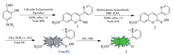
Scheme 1.
Synthesis of Cou-Py and proposed response mechanism of Cou-Py towards ClO-
A Fas-Responsive Mitochondria-Targeting Fluorescent Probe Detecting Hypochlorite in Living Cells and Zebrafish
Chang-Li ZHANG , Jing-Jing ZHANG , You SHEN , Jia-Cheng LU , Fang HUANG , Li XU

Hypochlorite (ClO-), as one of the most important and highly reactive oxygen species (ROS), plays a crucial role in a variety of physiological processes[1-2]. It not only contributes to the defense of bacterial and inflammatory but also acts as a natural adjuvant of adaptive immunity[3-5]. However, the excessive level of this strong oxidant in living systems can lead to oxidative stress, cellular damage, and tissue destruction, which will cause a series of related diseases, such as inflammatory diseases, cardiovascular diseases, lung injury, cardiopathy, angiocardiopathy, arthropathy, renal function damage, and even cancers[5-7]. The mitochondria, as the main source of hypochlorite production, play an important role in life-sustaining activities, however, the build-up of hypochlorite in mitochondria can result in cell death and various diseases[8-10]. Therefore, the development of mitochondria-targetable probes for ClO- detection in live cells and animals will help further our knowledge of the chemical and biological properties of ClO- in mitochondria.
As a classic noninvasive method, the fluorescence technique has been widely used as an effective tool for the detection of biomolecules in living cells and animals, because of its high selectivity, good sensitivity, ready operation, and rapid response rate[11-13]. Recently, a variety of mitochondria-targetable fluorescent probes for specific detection of ClO- have been designed and realized to trace ClO- in vitro and in vivo[14-29].
It is known that the basal ClO- level in cells is quite low. It is reported that the average ClO- generation from neutrophils is 0.47 nmol·min-1 per 106 cells[30]. Furthermore, Yu et al. estimated the average basal ClO- concentrations in the mitochondria of HeLa and A549 cells to be 46.3 and 49.7 nmol·L-1, respectively[21]. Besides, ClO- is very reactive and tends to be short-lived in cells[31]. However, at present, the limit of detection of most mitochondria-targeted fluorescent probes is micromolar level[13-16, 23-25, 27], and the response time of some probes is more than 1 min[14-15]. Some probes are easy to be disturbed by other reactive oxygen species such as peroxynitrite[32-33]. Therefore, the design of a fast responsive and highly sensitive probe for ClO- is still highly desired[34].
In organic chemistry, ClO- is often used to oxidize oximes to release carbonyl groups, thereby realizing the protection and deprotection of carbonyl groups. Notably, this reaction can be completed within 10 s[35]. Taking advantage of the fast and selective response, the reaction between ClO- and oxime has been employed as one of the common strategies to construct highly sensitive ClO- probes. Lin et al. reported the first deoximation-based fluorescent probe for ClO- detection[36]. After that, varied fluorescent ClO- probes based on the same response mechanism were obtained by employing different fluorophores or regulating the oxidation of oxime groups[37-39]. However, it should be noted that oxime groups are usually oxidized to aldehyde or ketone groups. These groups are easy to further react with sulfur-containing species (such as glutathione, cysteine) in the body, resulting in the change of the fluorescence signals and interfering with the detection of ClO- [40-41]. To overcome the interference of sulfur- containing species and meet the needs for real-time and rapid detection of ClO- in the mitochondria, herein, we report a new probe, Cou-Py (Scheme 1), which shows a highly sensitive and selective response to ClO-over other ROS, metal ions, and bio-thiols in mitochondria. Moreover, the probe has been successfully employed for visualizing the mitochondria ClO- in living MCF-7 cells and zebrafish larvae.
All chemicals and solvents were of analytical grade or spectroscopic grade and were used without further purification. Dichloromethane (DCM) was refluxed with calcium hydride and distilled at ambient pressure. 1H NMR and 13C NMR spectra were recorded on a Bruker DRX - 300 spectrometer (Germany) or Bruker DRX-500 spectrometer (Germany) with TMS as internal reference in CDCl3. Mass spectrometric data were determined with an LCQ ESI-MS Thermo Finnigan mass spectrometer.
The cells were obtained from the American Type Culture Collection (ATCC). Zebrafish larvae were purchased from EzeRinka company (Nanjing, Jiangsu). Confocal fluorescence imaging was carried out on a Zeiss LSM710 confocal microscope (Germany) with a 63×oil immersion objective for cells imaging and a 20×objective for zebrafish imaging, respectively.
Compound 1 was synthesized according to the literature[42]. Compound 1 (0.5 g, 2 mmol) was dissolved with 3 mL N, N-dimethylformamide (DMF), then a MeOH solution (3 mL) containing NH2OH·HCl (0.21 g, 3 mmol) and H2SO4 (10%, 100 μL) was added into the mixture. The mixture was heated to 90 ℃, stirred and refluxed for 3 h. After filtrating and washing with EtOH, compound 2 was obtained as a yellow solid (Yield: 61%). 1H NMR (600 MHz, CDCl3): δ 8.66 (s, 1H), 7.98 (d, J=7.86 Hz, 1H), 7.71 (t, J=7.50 Hz, 1H), 7.58 (s, 1H), 7.40 (s, 1H), 7.24 (t, J=8.88 Hz, 2H), 6.83 (s, 1H), 6.69 (d, J=8.28 Hz, 1H), 3.84 (s, 3H). 13C NMR (150 MHz, CDCl3): δ 161.9, 153.5, 152.7, 150.4, 149.3, 136.2, 131.7, 129.0, 124.3, 123.9, 123.0, 113.4, 111.3, 100.5, 55.7.
To a DCM solution (4 mL) of compound 2 (200 mg, 0.75 mmol) was added CH3I (140 μL, 1.5 mmol), and the resulted solution was stirred at room tempera- ture. After the disappearance of compound 2, the pre- cipitate was filtrated and washed with ether (3×10 mL). Pure product of Cou-Py was obtained as a yellow solid (Yield: 66%). 1H NMR (600 MHz, DMSO-d6): δ 9.88 (s, 1H), 6.88 (d, J=7.2 Hz, 1H), 6.49 (s, 3H), 6.39 (t, J= 7.8 Hz, 2H), 6.23 (d, J=7.8 Hz, 1H), 6.18 (d, J=7.8 Hz, 1H), 3.32 (s, 3H), 3.17 (s, 3H). 13C NMR (150 MHz, DMSO-d6): δ 163.0, 154.4, 150.9, 147.3, 147.2, 146.6, 134.4, 130.9, 130.5, 128.0, 117.6, 112.4, 111.6, 101.5, 56.5, 46.4. HRMS (ESI+) m/z Calcd. for C16H15N2O3: [M]+ 283.107 8; Found: [M]+ 283.107 1.
Cou-Py was dissolved in DMF to give a stock solution with a concentration of 1 mmol·L-1. For the photophysical properties measurement, the test solution with a concentration of 5 μmol·L-1 (VPBS∶VDMF= 200∶1) was prepared by diluting the stock solution with phosphate buffer solution (PBS, 10 mmol·L-1, pH= 7.4). ClO- was obtained from NaClO. Other ROS for selectivity response measurement were prepared based on the literature[43]. The absorption and fluorescence spectra were measured using a Shimadzu UV-1750 spectrophotometer (Japan) and FLUOROMAX-4 spectrofluorometer (Horiba, America), respectively.
MCF-7 cells were cultured by using standard cell culture procedures. The solution of Cou-Py was 5 μmol·L-1 in PBS (10 mmol·L-1, pH=7.4, containing 0.5% DMF). Imaging experiments were divided into the following five groups: (1) MCF-7 cells were stained with Cou-Py for 30 min before imaging. (2) MCF-7 cells were incubated with NaClO (20 μmol·L-1) for 4 h and subsequently incubated with Cou-Py for 30 min. (3) MCF-7 cells were incubated with 4-aminobenzoic acid hydrazide (ABH, 250 μmol·L-1) for 4 h and subsequently incubated with Cou-Py for 30 min. (4) MCF-7 cells were treated with lipopolysaccharide (LPS, 5 μg· mL-1) and phorbol 12-myristate 13-acetate (PMA, 5 μg·mL-1) for 12 h and stained with Cou-Py for 30 min. (5) ABH pretreated MCF-7 cells were stimulated with LPS and PMA for 12 h, treated with ABH for 4 h, and then stained with Cou-Py before imaging. Confocal fluorescence imaging was carried out on the Zeiss LSM710 confocal microscope with the excitation wave- length of 405 nm The excitation wavelength was 405 nm. The emission signals were collected in a range of 420-540 nm.
MCF-7 cells were stained with 50 nmol·L-1 MTDR (MitoTracker Deep Red) at 37 ℃ for 15 min, then the cells were washed with PBS (10 mmol·L-1, pH=7.4) two times. After rinsing with PBS, the cells were incubated with Cou-Py solution for 30 min at 37 ℃. Then, the cells were washed twice with PBS and imaged using the Zeiss LSM-710 microscope. For colocalization experiments after ClO- stimulation, after pretreated with NaClO solution (20 μmol·L-1) for 4 h at 37 ℃, MCF-7 cells were stained and imaged in the same way as before. The MTDR marked images were obtained upon irradiation at 633 nm with a band path from 650 to 740 nm, while the Cou-Py stained images were obtained upon irradiation at 405 nm with a band path of 420-540 nm.
To assess the cytotoxicity of Cou-Py, MCF-7 and HepG2 cells were incubated in Dulbecco´s modified Eagle´s medium (DMEM) at 37 ℃ with φCO2 of 5% and grown for 24 h. Subsequently, the medium was cast off, added Cou-Py with different concentrations (5, 10, 20, 30, 40, and 50 μmol·L-1), and co-incubated for an additional 48 h. MTT assay was used for testing the cytotoxicity of Cou-Py in MCF-7 and HepG2 cells and repeated three times for each experiment.
Zebrafish larvae were incubated with 5 μmol·L-1 Cou-Py at 28.5 ℃ for 30 min and washed with PBS twice. For exogenous ClO- imaging, the larvae were pretreated with NaClO solution (20 μmol·L-1) for 4 h and were then washed with PBS. Then, the larvae were incubated in Cou-Py solution for 30 min and were washed again with PBS. For endogenous ClO- removal imaging, the larvae were pretreated with ABH solution (250 μmol·L-1) for 4 h and were washed with PBS then the larvae were incubated in Cou-Py solution for 30 min and washed with PBS.
As a family of classical fluorophores, coumarin derivates are widely employed in bioimaging because of their high fluorescence quantum yields, excellent photo-stabilities, and biocompatibility. In the coumarin core, the oxime group can be conveniently introduced to the 2-position of the coumarin core via the reaction of carbonyl or imine groups with hydroxylamine hydrochloride[44]. Notably, our recent research found that once the oxime group is oxidized by ClO-, the resulting lactone carbonyl is not sensitive to biological thiols, so the interference of biological thiols can be effectively avoided[39]. Based on the above results, we designed Cou-Py as the new probe for detecting mitochondria ClO-, in which oxime was introduced to the 2-position of 3-pyridyl-7-methoxy-coumarin as the ClO- acceptor and pyridium cation as the mitochondria targeting group[16].
As shown in Scheme 1, compound 2 was obtained by reacting compound 1 with hydroxylamine hydrochloride in a methanol solution containing sulfuric acid and DMF. After that, compound 2 was further with CH3I in DCM solution to give Cou-Py. Because of the isomerization of C=N of oxime in the excited state, Cou-Py is expected to show weak fluorescence. After the reaction with ClO-, the oxime group was oxidized and hydrolyzed, and Cou-Py was transformed into compound 3 with good luminescence properties. Therefore, the response of Cou-Py to ClO- will show obvious fluorescence enhancement behavior. In addition, because the stability of lactone carbonyl is much higher than that of the aldehyde group, it is difficult that compound 3 can be reacted with sulfur-containing species such as glutathione. Therefore, in a living system, the fluorescence signal of ClO- response of Cou-Py is expected to be unaffected by these species. Cou-Py was fully charac- terized by 1H NMR, 13C NMR, HRMS, and FT-IR.
According to the molecular design, we speculate that probe Cou-Py was transformed into compound 3 and restored the structure of coumarin. To verify the recognition mechanism of probe Cou-Py for ClO-, the reaction product of Cou-Py with ClO- was characterized by the ESI-MS spectrometry. As shown in Fig.S1 (Supporting information), there were two prominent peaks, 283.11 assigned to [Cou-Py+H]+, and 268.99 assigned to [3+H]+, respectively. This result shows that Cou-Py was oxidized to compound 3 by ClO-, which further confirmed the above-mentioned mechanism in Scheme 1.
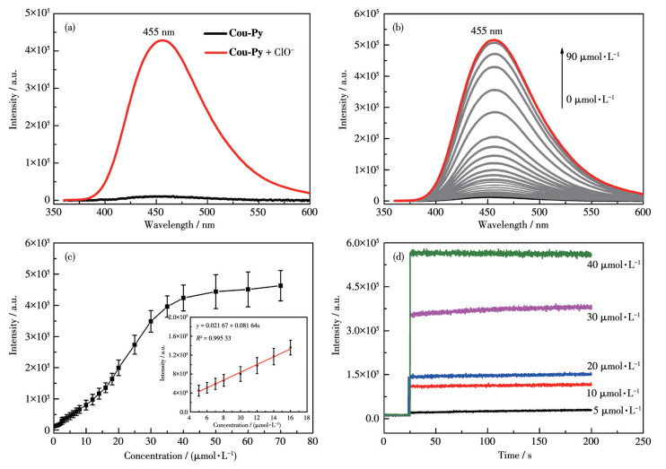
λex=350nm; Inset: plot of emission intensity at 455 nm as a function of cClO- in a linear range
We investigated the photophysical properties of Cou-Py towards ClO- in PBS (Fig. 1). Cou-Py showed one absorption band with an absorption maximum (λmax) centered at 350 nm (ε=82 000 L·mol-1·cm-1, Fig. S2). Due to the C=N isomerization of the oxime group in the excited state, Cou-Py displayed a very weak emission with an emission maximum centered at 455 nm, and the quantum yield (Φf) was calculated to be around 0.2%. After the addition of ClO- to the solution of the probe, the fluorescence intensity at 455 nm was increased linearly with the concentration of ClO- (cClO-) increasing from 0 to 20 μmol·L-1, indicating the generation of compound 3 (Fig. 1a). The Φf of the final titration system was calculated to be around 5.1%, which was 26-fold that of Cou-Py (Fig. 1b). Moreover, the detection limit of the probe was determined to be 6.87 nmol·L-1, demonstrating its highly sensitive responsi- bility (Fig. 1c and S3).
We investigated the kinetics of the reaction of Cou-Py with ClO-. As shown in Fig. 1d, Cou-Py can react with ClO- immediately to convert to compound 3 in the presence of different concentrations of ClO-, and the fluorescence was significantly enhanced. Moreover, no matter whether Cou-Py reacted with ClO- at a high or low concentration, it could react immediately and the fluorescence emission reached the maximum intensity within 5 s. These results indicate that Cou-Py can be used for the real-time ultrafast monitoring of ClO-.
To investigate the selective fluorescence response of Cou-Py to ClO-, we measured the fluorescence spectra of the probe in the presence of different biologically related species. Further, the fluorescence response of Cou-Py to ClO- was studied in the presence of various potential interfering agents. As shown in Fig. 2, the probe showed negligible fluorescence response to all the other ROS, metal ions, anions, as well as amino acids. In addition, the fluorescence intensity of Cou-Py solutions containing 10 Equiv. of interfering agent changed obviously after adding ClO- (Fig. S4). These results demonstrate the high selectivity of Cou-Py for ClO-. Furthermore, the influence of pH on the assay was investigated. As shown in Fig.S5, the free Cou-Py exhibited negligible fluorescent changes in a wide range of pH values. In the presence of NaClO, Cou-Py displayed remarkable performance during a pH range of 4-9 which was very close to the physiological conditions. The above studies indicated that Cou-Py could detect ClO- well in the complex biosystems.
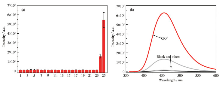
1: Blank, 2: Na+, 3: K+, 4: Mg2+, 5: Ca2+, 6: Cu2+, 7: Fe3+, 8: Fe2+, 9: Zn2+, 10: CO32-, 11: SO42-, 12: SO32-, 13: HSO3-, 14: S2-, 15: HS-, 16: homocysteine, 17: cysteine, 18: glutathione, 19: ROO·, 20: H2O2, 21: 1O2, 22: HO·, 23: NO·, 24: ONOO-, 25: ClO-; λex=350nm, Slit width: dex=dem=0.5nm, PMT voltage=950V
To extend the endogenous HOCl determination of Cou-Py in living cells, the MTT assay was applied to evaluate the cytotoxicity of Cou-Py against the HepG2 and MCF7 cells, and the results are illustrated in Fig. S6. The results show that the probe had no obvious toxicity to the cells at 0-50 μmol·L-1 with 48 h. The concentration of the probe used was 5 μmol·L-1, hence, it could be concluded that the concentration of the probe applied here is negligible to the cell toxicity.
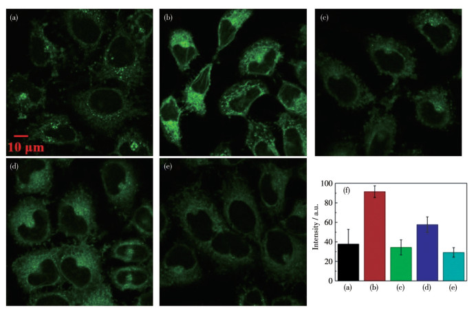
The excellent performance of the probe under physiological conditions encouraged us to evaluate the intracellular ClO- imaging ability of Cou-Py on MCF-7 cells. After incubation of MCF-7 cells with Cou-Py, very weak fluorescence was observed inside the cells (Fig. 3a). However, after staining the NaClO-treated cells with the probe, bright green fluorescence could be observed within the cells (Fig. 3b). The averaged intensity in Fig. 3b was about 2.4-fold more than that observed in Fig. 3a, indicating the increased intracellular cClO-. A slight decrease in fluorescence was observed when the cells were pretreated with ABH, a ClO- scavenger, followed by incubation with Cou-Py, suggesting the presence of endogenous ClO- in MCF-7 cells (Fig. 3c). Next, MCF-7 cells were stimulated with an endogenous ClO- inducer, LPS/PMA, and then incubated with Cou-Py, following which an approximately 1.5-fold fluorescence intensity enhancement was observed, as shown in Fig. 3d, in comparison with that in Fig. 3a. Moreover, when the cells were stimulated with LPS/ PMA and then treated with ABH, the incubation of Cou-Py showed almost the same fluorescence intensity as that observed in Fig. 3c. Therefore, the increased intracellular ClO- level should be responsible for the fluorescence enhancement observed in Fig. 3b and 3d.
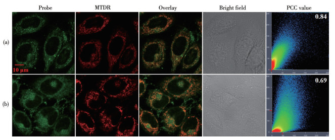
To further prove the mitochondria-targeting ability of Cou-Py, we carried out subcellular localization experiments for Cou-Py with MTDR in MCF-7 cells for 20 min at 37 ℃. As shown in Fig. 4a, Cou-Py could effectively target mitochondria in MCF-7 cells with a high Pearson´s correlation coefficient (PCC) value of 0.84 with MTDR. Further, the MCF-7 cells were costained with Cou-Py and MTDR and then incubated with NaClO. After 4 h, the green fluorescence generated from Cou-Py was found to overlap with the red fluorescence of MTDR, resulting in a high PCC value of 0.69. These results suggest that Cou-Py can target the mitochondria during the response to ClO-.
Probe: 420‐540 nm, λex=405 nm; MTDR: 650‐740 nm, λex=633 nm
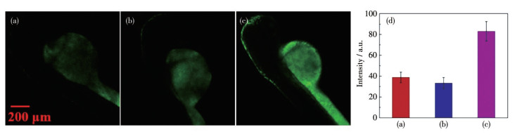
We further used the probe to image the dynamics of ClO- in zebrafish to confirm its in vivo imaging capability. We divided the zebrafish larvae into three portions. One portion was stained with Cou-Py at room temperature for 0.5 h, the second portion was incubated with ABH for 4 h and then stained with the probe, and the third portion was pretreated with NaClO for 4 h and then incubated with Cou-Py. As shown in Fig. 5a and 5b, the larvae in the first and second portions showed weak green fluorescence throughout the whole body, indicating the low cClO- in the larvae. Differently, the larvae in the third portion showed distinct enhanced green fluorescence with the emission enhancement factor of 2.2, suggesting the increment of the cClO- in the larvae (Fig. 5c). All the above imaging results demon- strate that Cou-Py can be used as an efficient probe for imaging mitochondria ClO- both in vitro and in vivo.
In summary, a novel mitochondria-targeted fluo- rescent probe Cou-Py was successfully developed. The fast response (within 5 s), high selectivity, and sensitivity of Cou-Py make it an effective tool for imaging ClO-. In addition, the colocalization coefficient suggests that Cou-Py can effectively target mitochondria. Moreover, Cou-Py had good permeability and low toxicity and was effectively employed in imaging ClO- in living cells as well as the zebrafish larvae. We expect that the probe could provide possibilities for studying and discovering specific physiological and pathological functions of ClO-.
Supporting information is available at http://www.wjhxxb.cn
D′Autréaux B, Toledano M B. ROS as Signalling Molecules: Mechanisms that Generate Specificity in ROS Homeostasis[J]. Nat. Rev. Mol. Cell Biol., 2017, 8(10): 813-824.
Winterbourn C C. Reconciling the Chemistry and Biology of Reactive Oxygen Species[J]. Nat. Chem. Biol., 2008, 4(5): 278-286. doi: 10.1038/nchembio.85
Harrison J E, Schultz J. Studies on the Chlorinating Activity of Myeloperoxidase[J]. J. Biol. Chem., 1976, 251(5): 1371-1374. doi: 10.1016/S0021-9258(17)33749-3
Prütz W A, Kissner R, Nauser T, Koppenol W H. On the Oxidation of Cytochrome c by Hypohalous Acids[J]. Arch. Biochem. Biophys., 2001, 389(1): 110-122. doi: 10.1006/abbi.2001.2321
Sam C H, Lu H K. The Role of Hypochlorous Acid as One of the Reactive Oxygen Species in Periodontal Disease[J]. J. Dent. Sci., 2009, 4(2): 45-54. doi: 10.1016/S1991-7902(09)60008-8
Khatib S, Musa R, Vaya J. An Exogenous Marker: A Novel Approach for the Characterization of Oxidative Stress[J]. Bioorg. Med. Chem., 2007, 15(11): 3661-3666. doi: 10.1016/j.bmc.2007.03.052
Steinbeck M J, Nesti L J, Sharkey P F, Parvizi J. Myeloperoxidase and Chlorinated Peptides in Osteoarthritis: Potential Biomarkers of the Disease[J]. J. Orthop. Res., 2007, 25(9): 1128-1135. doi: 10.1002/jor.20400
Murphy M P, Smith R A J. Targeting Antioxidants to Mitochondria by Conjugation to Lipophilic Cations[J]. Annu. Rev. Pharmacol. Toxicol., 2007, 47(1): 629-656. doi: 10.1146/annurev.pharmtox.47.120505.105110
Chalmers S, Caldwell S T, Quin C, Prime T A, James A M, Cairns A G, Murphy M P, McCarron J G, Hartley R C. Selective Uncoupling of Individual Mitochondria within a Cell Using a Mitochondria-Targeted Photoactivated Protonophore[J]. J. Am. Chem. Soc., 2012, 134(2): 758-761. doi: 10.1021/ja2077922
Niu W F, Guo L, Li Y H, Shuang S M, Dong C, Wong M S. HighlySelective Two-Photon Fluorescent Probe for Ratiometric Sensing and Imaging Cysteine in Mitochondria[J]. Anal. Chem., 2016, 88(3): 1908-1914. doi: 10.1021/acs.analchem.5b04329
Chen Y C, Bai Y, Han Z, He W J, Guo Z J. Photoluminescence Imaging of Zn2+ in Living Systems[J]. Chem. Soc. Rev., 2015, 44(14): 4517-4546. doi: 10.1039/C5CS00005J
Qian F, Zhang C L, Zhang Y M, He W J, Gao X, Hu P, Guo Z J. Visible Light Excitable Zn2+ Fluorescent Sensor Derived from an Intra-molecular Charge Transfer Fluorophore and Its In Vitro and In Vivo Application[J]. J. Am. Chem. Soc., 2009, 131(4): 1460-1468. doi: 10.1021/ja806489y
Wu D, Chen L Y, Xu Q L, Chen X Q, Yoon J. Design Principles, Sensing Mechanisms, and Applications of Highly Specific Fluorescent Probes for HOCl/OCl-[J]. Acc. Chem. Res., 2019, 52(8): 2158-2168. doi: 10.1021/acs.accounts.9b00307
Xu J C, Yuan H Q, Qin C Q, Zeng L T, Bao G M. A Mitochondria-Targeted Near-Infrared Probe for Colorimetric and Ratiometric Fluorescence Detection of Hypochlorite in Living Cells[J]. RSC Adv., 2016, 6(109): 107525-107532. doi: 10.1039/C6RA22868B
Xu Q L, Heo C H, Kim J A, Lee H S, Hu Y, Kim D, Swamy K M K, Kim G, Nam S J, Kim H M, Yoon J. A Selective Imidazoline-2-thione-Bearing Two-Photon Fluorescent Probe for Hypochlorous Acid in Mitochondria[J]. Anal. Chem., 2016, 88(12): 6615-6620. doi: 10.1021/acs.analchem.6b01738
Li K, Hou J T, Yang J, Yu X Q. A Tumor-Specific and Mitochondria-Targeted Fluorescent Probe for Real-Time Sensing of Hypochlorite in Living Cells[J]. Chem. Commun., 2017, 53(40): 5539-5541. doi: 10.1039/C7CC01679D
Ren M G, Zhou K, He L W, Lin W Y. Mitochondria and Lysosome-Targetable Fluorescent Probes for HOCl: Recent Advances and Perspectives[J]. J. Mater. Chem. B, 2018, 6(12): 1716-1733. doi: 10.1039/C7TB03337K
Zhong X L, Yang Q, Chen Y S, Jiang Y L, Wang B X, Shen J. A Mitochondria-Targeted Fluorescent Probe Based on Coumarin-Pyridine Derivatives for Hypochlorite Imaging in Living Cells and Zebrafish[J]. J. Mater. Chem. B, 2019, 7(46): 7332-7337. doi: 10.1039/C9TB01948K
Li M Y, Li K, Liu Y H, Zhang H, Yu K K, Liu X, Yu X Q. Mitochondria-Immobilized Fluorescent Probe for the Detection of Hypochlorite in Living Cells, Tissues, and Zebrafishes[J]. Anal. Chem., 2020, 92(4): 3262-3269. doi: 10.1021/acs.analchem.9b05102
Zhong X L, Yang Q, Chen Y S, Jiang Y L, Dai Z H. Aggregation-Induced Fluorescence Probe for Hypochlorite Imaging in Mitochondria of Living Cells and Zebrafish[J]. J. Mater. Chem. B, 2020, 8(33): 7375-7381. doi: 10.1039/D0TB01496F
Hou J T, Li K, Yang J, Yu K K, Liao Y X, Ran Y Z, Liu Y H, Zhou X D, Yu X Q. A Ratiometric Fluorescent Probe for In Situ Quantification of Basal Mitochondrial Hypochlorite in Cancer Cells[J]. Chem. Commun., 2015, 51(31): 6781-6784. doi: 10.1039/C5CC01217A
Teng H, Tian J Y, Sun D H, Xiu M X, Zhang Y H, Qiang X Y, Tang H Y, Guo Y. A Mitochondria-Specific Fluorescent Probe Based on Triazolopyridine Formation for Visualizing Endogenous Hypochlorous Acid in Living Cells and Zebrafish[J]. Sens. Actuators B, 2020, 319: 128288. doi: 10.1016/j.snb.2020.128288
Yang Q, Zhong X L, Chen Y S, Yang J, Jin C, Jiang Y L. A Mitochondria-Targeted Fluorescent Probe for Hypochlorite Sensing and Its Application in Bioimaging[J]. Analyst, 2020, 145(8): 3100-3105. doi: 10.1039/D0AN00245C
Huang L, Su W T, Zhao Y P, Zhan J T, Lin W Y. Synthesis, Molecular Docking Calculation, Fluorescence and Bioimaging of Mitochondria-Targeted Ratiometric Fluorescent Probes for Sensing Hypochlorite In Vivo[J]. J. Mater. Chem. B, 2021, 9(11): 2666-2673. doi: 10.1039/D0TB02735A
Xu J H, Wang C Y, Ma Q J, Zhang H T, Tian M J, Sun J G, Wang B Y, Chen Y C. Novel Mitochondria-Targeting and Naphthalimidebased Fluorescent Probe for Detecting HClO in Living Cells[J]. ACS Omega, 2021, 6(22): 14399-14409. doi: 10.1021/acsomega.1c01271
Yuan L, Wang L, Agrawalla B K, Park S J, Zhu H, Sivaraman B, Peng J J, Xu Q H, Chang Y T. Development of Targetable Two-Photon Fluorescent Probes to Image Hypochlorous Acid in Mitochondria and Lysosome in Live Cell and Inflamed Mouse Model[J]. J. Am. Chem. Soc., 2015, 137(18): 5930-5938. doi: 10.1021/jacs.5b00042
Zheng A S, Liu H, Peng C H, Gao X N, Xu K H, Tang B. A Mitochondria-Targeting Near-Infrared Fluorescent Probe for Imaging Hypochlorous Acid in Cells[J]. Talanta, 2021, 226: 122152. doi: 10.1016/j.talanta.2021.122152
Shen B X, Qian Y, Qi Z Q, Lu C G, Sun Q, Xia X, Cui Y P. Near-Infrared BODIPY-Based Two-Photon ClO-Probe Based on Thiosemicarbazide Desulfurization Reaction: Naked-Eye Detection and Mitochondrial Imaging[J]. J. Mater. Chem. B, 2017, 5(29): 5854-5861. doi: 10.1039/C7TB01344B
Shi D L, Chen S Q, Dong B, Zhang Y H, Sheng C Q, James TD, Guo Y. Evaluation of HOCl-Generating Anticancer Agents by an Ultrasensitive Dual-Mode Fluorescent Probe[J]. Chem. Sci., 2019, 10(13): 3715-3722. doi: 10.1039/C9SC00180H
Kaufmann S H E, Aratani Y, Koyama H, Nyui S, Suzuki K, Kura F, Maeda N. Severe Impairment in Early Host Defense against Candida albicans in Mice Deficient in Myeloperoxidase[J]. Infect. Immun., 1999, 67(4): 1828-1836. doi: 10.1128/IAI.67.4.1828-1836.1999
Klebanoff S J. Myeloperoxidase: Friend and Foe[J]. J. Leukoc. Biol., 2005, 77(5): 598-625. doi: 10.1189/jlb.1204697
Li H Y, Li X H, Wu X F, Shi W, Ma H M. Observation of the Generation of ONOO-in Mitochondria under Various Stimuli with a Sensitive Fluorescence Probe[J]. Anal. Chem., 2017, 89(10): 5519-5525. doi: 10.1021/acs.analchem.7b00503
Zhu B C, Zhang M, Wu L, Zhao Z Y, Liu C Y, Wang Z K, Duan Q X, Wang Y W, Jia P. A Highly Specific Far-Red Fluorescent Probe for Imaging Endogenous Peroxynitrite in the Mitochondria of Living Cells[J]. Sens. Actuators B, 2018, 257: 436-441. doi: 10.1016/j.snb.2017.10.170
Yang J J, Zheng W B, Shen Y, Xu Y Z, Lv G L, Li C X. A Novel Near-Infrared Fluorescent Probe Based on Phenoxazine for the Specific Detection of HOCl[J]. J. Lumin., 2020, 226: 117460. doi: 10.1016/j.jlumin.2020.117460
Dong S Q, Zhang L J, Lin Y J, Ding C F, Lu C. Luminescent Probes for Hypochlorous Acid In Vitro and In Vivo[J]. Analyst, 2020, 145(15): 5068-5089. doi: 10.1039/D0AN00645A
Lin W Y, Long L L, Chen B B, Tan W. A Ratiometric Fluorescent Probe for Hypochlorite Based on a Deoximation Reaction[J]. Chem. Eur. J., 2009, 15(10): 2305-2309. doi: 10.1002/chem.200802054
Shi Y, Huo F J, Yin C X. Malononitrile as the'Double-Edged Sword'of Passivation-Activation Regulating Two ICT to Highly Sensitive and Accurate Ratiometric Fluorescent Detection for Hypochlorous Acid in Biological System[J]. Sens. Actuators B, 2020, 325: 128793. doi: 10.1016/j.snb.2020.128793
Yang X F, Shi W D, Dong X L, Cui C Y, Huang X, Wang X, Xie HX, Li Y X, Yan M, Cui Y, Sun G X. A Simple but Sensitive and Efficient Fluorescent Probe for"Turn-On"Sensing of ClO-[J]. Polyhedron, 2020, 185: 114563. doi: 10.1016/j.poly.2020.114563
张晶晶, 严鸣, 卢雯, 徐莉, 王小青. 基于香豆素-肟的次氯酸根探针的设计、合成及荧光成像应用[J]. 无机化学学报, 2021,37,(6): 1071-1079. ZHANG J J, YAN M, LU W, XU L, WANG X Q. Design, Synthesis and Fluorescence Imaging Application of Hypochlorite Probe Based on Coumarin-Oxime[J]. Chinese J. Inorg. Chem., 2021, 37(6): 1071-1079.
Wang B S, Li P, Yu F B, Song P, Sun X F, Yang S Q, Lou Z R, Han K L. A Reversible Fluorescence Probe Based on Se-BODIPY for the Redox Cycle between HClO Oxidative Stress and H2S Repair in Living Cells[J]. Chem. Commun., 2013, 49(10): 1014-1016. doi: 10.1039/C2CC37803E
Liu S R, Wu S P. Hypochlorous Acid Turn-On Fluorescent Probe Based on Oxidation of Diphenyl Selenide[J]. Org. Lett., 2013, 15(4): 878-881. doi: 10.1021/ol400011u
Qin Q P, Wang Z F, Huang X L, Tan M X, Zou B Q, Liang H. Strong In Vitro and Vivo Cytotoxicity of Novel Organoplatinum(Ⅱ) Complexes with Quinoline-Coumarin Derivatives[J]. Eur. J. Med. Chem., 2019, 184: 111751. doi: 10.1016/j.ejmech.2019.111751
Sun Q, Xu J J, Ji C L, Shaibani M S S, Li Z, Lim K, Zhang C W, Li L, Liu Z P. Ultrafast Detection of Peroxynitrite in Parkinson′s Disease Models Using a Near-Infrared Fluorescent Probe[J]. Anal. Chem., 2020, 92(5): 4038-4045. doi: 10.1021/acs.analchem.9b05599
Carta F, Vullo D, Maresca A, Scozzafava A, Supuran C T. New Chemotypes Acting as Isozyme-Selective Carbonic Anhydrase Inhibitors with Low Affinity for the Offtarget Cytosolic Isoform Ⅱ[J]. Bioorg. Med. Chem. Lett., 2012, 22(6): 2182-2185. doi: 10.1016/j.bmcl.2012.01.129

Figure 1 (a) Fluorescence spectra of Cou-Py (5 μmol·L-1) and Cou-Py reacting with 10 Equiv. of NaClO in PBS (10 mmol·L-1, pH=7.4, containing 0.5% DMF); (b) Fluorescence spectra changes of Cou-Py (5 μmol·L-1) with increasing cClO-; (c) Fluorescence intensity at 455 nm of Cou-Py versus cClO-; (d) Plots of fluorescence intensity changes of Cou-Py (5 μmol·L-1) with increasing cClO- for different times in PBS
λex=350nm; Inset: plot of emission intensity at 455 nm as a function of cClO- in a linear range

Figure 2 (a) Fluorescent intensity at 455 nm and (b) fluorescent spectra of Cou-Py (5 μmol·L-1) with 10 Equiv. of ClO- and the other analytes in PBS (10 mmol·L-1, pH=7.4, containing 0.5% DMF)
1: Blank, 2: Na+, 3: K+, 4: Mg2+, 5: Ca2+, 6: Cu2+, 7: Fe3+, 8: Fe2+, 9: Zn2+, 10: CO32-, 11: SO42-, 12: SO32-, 13: HSO3-, 14: S2-, 15: HS-, 16: homocysteine, 17: cysteine, 18: glutathione, 19: ROO·, 20: H2O2, 21: 1O2, 22: HO·, 23: NO·, 24: ONOO-, 25: ClO-; λex=350nm, Slit width: dex=dem=0.5nm, PMT voltage=950V

Figure 3 Fluorescence images of Cou-Py in MCF-7 cells under different conditions: (a) cells incubated with 5 μmol·L-1 Cou-Py (10 mmol·L-1 PBS, pH 7.4, containing 0.5% DMF) for 30 min; (b) cells pretreated with NaClO (20 μmol·L-1, 4 h), and incubated with Cou-Py (5 μmol·L-1, 30 min); (c) cells pretreated with ClO- scavenger ABH (250 μmol·L-1, 4 h), and incubated with Cou-Py (5 μmol·L-1, 30 min); (d) cells treated with LPS (5 μg·mL-1) and PMA (5 μg·mL-1) for 12 h, and then incubated with Cou-Py (5 μmol·L-1, 30 min); (e) cells pretreated with ABH (250 μmol·L-1, 4 h), and then treated with LPS (5 μg·mL-1) and PMA (5 μg·mL-1) for 12 h, and incubated with ABH (250 μmol·L-1, 4 h), then combined with Cou-Py (5 μmol·L-1, 30 min) incubate; (f) Average fluorescence intensity in Fig. 3a-3e, where the data are mean±S.E.M., n=3

Figure 4 Intracellular co-localization fluorescence imaging of Cou-Py in MCF-7 cells: (a) cells stained with MTDR (50 nmol·L-1, 15 min), and then incubated with Cou-Py (5 μmol·L-1, 30 min); (b) cells pretreated with ClO- (20 μmol·L-1, 4 h), and then incubated with MTDR (50 nmol·L-1, 15 min), and finally stained with Cou-Py (5 μmol·L-1, 30 min)
Probe: 420‐540 nm, λex=405 nm; MTDR: 650‐740 nm, λex=633 nm

Figure 5 Fluorescence images of Cou-Py in zebrafish larvae under different conditions: (a) larvae pretreated with ClO- scavenger ABH (250 μmol·L-1, 4 h), and incubated with Cou-Py (5 μmol·L-1, 30 min); (b) larvae incubated with Cou-Py (5 μmol·L-1, 30 min); (c) larvae pretreated with NaClO (20 μmol·L-1, 4 h), and incubated with Cou-Py (5 μmol·L-1, 30 min); (d) Average fluorescence intensity in Fig. 5a‐5c, where the data are mean±S.E.M., n=3

 扫一扫看文章

扫一扫关注我们
 下载:
下载:

 下载:
下载: