Citation:
Yue-Jiao JIA, Xiao-Yu LIANG, Ming HU. A Water Stable Luminescent Zn-Complex Sensor for Detection of PO43- Ion, Fe3+ Ion, and Nitroaromatic Explosives[J]. Chinese Journal of Inorganic Chemistry,
2022, 38(6): 1146-1158.
doi:
10.11862/CJIC.2022.108
Received Date:
11 December 2021 Revised Date:
31 March 2022 Available Online:
10 June 2022
Abstract:
A luminescent Zn-complex based on the 3-(2, 4-dicarboxylphenyl)-6-carboxylpyridine (H3dpcp), namely [Zn(H2dpcp)2(H2O)2]·H2O (1), was successfully fabricated by the solvothermal process. Complex 1 features a mono-nuclear butterfly-like structure, which further extends to the 3D supramolecular architecture via π…π interactions. It is found that complex 1 exhibited excellent luminescent stability in a pH range of 1-10 in an aqueous solution. It should be noted that complex 1 can not only detect PO43- ion based on the turn-on effect with high selectivity and recyclability but also serve as a remarkably selective sensing material with the fluorescence quenching for Fe3+ ion. The examination of nitroaromatic compounds demonstrated that complex 1 also behaved as a functional probe with high selectivity, sensitivity, and the low detection limit of 2, 4, 6-trinitrophenol (TNP). Furthermore, the luminescent sensing mechanisms for the above analytes were also investigated in detail. CCDC: 2010615.
The phosphate ion is a common inorganic anion that is widely found in groundwater and rivers. As an essential nutrient, it stimulates the growth of phyto-plankton and aquatic plants, and further causes eutro-phication of aqueous systems accompanied by red tide in the ecosystems[1-2]. Due to industrial wastes, agricul-tural fertilizers, and domestic sewages, it has become a pollutant that can not be ignored in various sub-strates[3]. Considering phosphate ion as a buffering agent and structural material for the bone and teeth in the human body, it plays an important role in physiological metabolism[4]. Iron is one of the essential trace elements for human metabolism, which is of great significance for human cell growth, proliferation, biological oxidation, and transformation[5]. It is involved in a variety of biological processes such as the synthesis of hemoglobin, the transport of oxygen, and the formation of enzymes and immune system compounds. It has been demonstrated that serious health problems or diseases like cancer, anemia, atherosclerosis, Parkinson′s disease, and neurodegenerative Alzheimer′s disease are caused by related disorders of iron[6-7]. As well known, the recognition of nitroaromatic compounds (NACs) is of practical significance, such as nitrobenzene, 4-nitrotoluene, 2, 4-dinitrotoluene, trinitrotoluene, and 2, 4, 6-trinitrophenol (TNP), can result in serious antiterrorism or environmental issue[8-9]. Hence, it is quite imperative to develop a kind of sensor that is efficient, convenient, and reliable for simultaneous detection of the above materials due to considerable concerns for people′s health and security.
Complexes with multifarious structures are independently assembled by metal centers and organic ligands in multiple binding sites. Due to their structural diversity and practical functionality, complexes have aroused great interest of researchers in many fields, such as gaseous separations[10-11], magnetism[12-13], catalysis[14-16], and fluorescence sensors[17-22]. Heretofore, the unique luminescent properties make some complexes competing candidates to recognize and sense inorganic anions, cations, organic molecules, pharmaceuticals, temperature, humidity, etc[23-32].
Hitherto, some references based on luminescent complexes have been reported on the discerning of Fe3+ ion and TNP molecules[33-37]. At the same time, only a few of them were published to detect phosphate ions up to now[38-40]. We previously designed a series of 3D Ln-MOFs to sense Fe3+, Cr2O72-, and TNP in dimethylformamide solution[41]. We also synthesized a new lumi-nescent Eu-MOF that not only distinguished Fe3+, Cr3+, and Al3+ ions with high selectivity and recyclability, but also served as an excellent selective sensing material for PO43- ions among some anions[42]. Not long ago, we presented a Zn coordination polymer that could identify Cr(Ⅲ), trace amounts of Cr(Ⅵ), and TNP with good anti-interference performance and recyclability[43]. Similar to luminescent probes with a certain degree of potential applications, they still have some restrictions of practicability, such as awful usability in aqueous samples. Therefore, it is necessary to design multifunctional luminescent probes with satisfactory selectivity, reproducibility, and reusability in an aqueous system.
Following the aforementioned points, a Zn(Ⅱ) complex, namely, [Zn(H2dpcp)2(H2O)2] ·H2O (1), was successfully prepared, where H3dpcp=3-(2, 4-dicarboxyl-phenyl)-6-carboxylpyridine. Because of its outstanding aqueous stability, complex 1 is a potential luminescent material for the detection of PO43-, Fe3+, and TNP in aqueous solutions with excellent selectivity and sensitivity. It is noteworthy that phosphate ions exhibited a fluorescent enhancement effect on complex 1 rather than quenching responses on most sensors. Importantly, the examination of simultaneous detection of PO43-, Fe3+, and TNP showed that complex 1 might also behave as a multifunctional probe with high selectivity, sensitivity, and recovery.
1.
Experimental
1.1
Materials and general methods
All chemicals were commercially purchased and used without further purification. Elemental analyses (C, H, and N) were performed on Perkin-Elmer 2400 analyzer. IR spectra were recorded as KBr pellets on a Nicolet Avatar-360 spectrometer in the 400-4 000 cm-1 region. Thermogravimetric analysis (TGA) was performed on a Perkin-Elmer TG-7 analyzer heated from 30 to 970 ℃ under a nitrogen atmosphere. Powder X-ray diffraction (PXRD) was performed on an Analytical Empyrean instrument by using Cu Kα radiation (λ= 0.154 06 nm, 45 kV, 200 mA, 2θ=5°-50°) at room temperature. The luminescence spectra were measured on an FLS920 spectrophotometer. The UV-Vis spectro-scopic studies were performed on a Hitachi U-3900 Spectrophotometer. An S-4800 Electron Microscope was used to perform energy dispersive X-ray spectroscopy (EDX) analyses. X-ray photoelectron spectroscopy (XPS) was performed on a Thermo Scientific ESCALAB 250Xi photoelectron spectrometer.
1.2
Synthesis of {[Zn(H2dpcp)2(H2O)2]·H2O}n (1)
A mixture of ZnSO4·7H2O (0.15 mmol, 43.1 mg), H3dpcp (0.15 mmol, 43.1 mg), H2O (12.0 mL), and HCl (1.0 mol·L-1, 1.0 mL) was stirred for 30 min at room temperature and then was sealed in a 23 mL Teflon-lined stainless steel vessel, heated at 160 ℃ for 72 h and then followed by slow cooling to the room temperature at a rate of 5 ℃ ·h-1. After filtration, the product was washed with H2O and air-dried. The bright yellow block crystals of 1 were obtained. Yield: 55.0% (based on Zn). Elemental analysis Calcd. for C28H22N2O15Zn (%): C 48.57, H 3.18, N 4.05; Found(%): C 48.67, H 3.14, N 3.97. IR (KBr pellet, cm-1): 1 696(s), 1 637(m), 1 372(m), 1 280(m), 1 242(m), 842(m), 770(m), 686(m).
1.3
X-ray crystallography
Crystallographic data of 1 were collected on a Bruker Smart 1000 diffractometer equipped with the graphite-monochromatic Mo Kα radiation (λ = 0.071 073 nm) using the ω-scan technique at room temperature. Semiempirical absorption corrections were applied using the SADABS program. The structure was solved by direct methods using SHELXS-2014 and refined by full-matrix least-squares on F2 using the SHELXTL-2014 program. All non-hydrogen atoms were refined anisotropically. The organic hydrogen atoms were geometrically generated, and the hydrogen atoms of water molecules were located from different Fourier maps and refined with the common isotropic thermal parameter. Details of the crystal parameters data collection and refinement for 1 are summarized in Table S1 (Supporting information). The selected bond lengths and angles of 1 are listed in Table S2.
CCDC: 2010615.
1.4
Fluorescence measurements
The suspension of complex 1 was prepared by adding 10.0 mg of powder sample to 20.0 mL of deionized water, ultrasonicated for 30 min, and placed for subsequent use. In a typical fluorescence experiment, 2.7 mL suspension solution of 1 (ca. 1.0 mmol·L-1) was added to a quartz cuvette (1 cm×1 cm), and the fluorescence-emission spectra with an excitation at 333 nm and a slit width of 3.0 nm were recorded in situ after each incremental addition of 1.0 mmol·L-1 of analytes at room temperature.
The investigation of sensing anions was carried out as following procedure: 1.5 mg sample of 1 was ground, dispersed in 2.7 mL aqueous solution. The mixture was ultrasonicated to form a suspension, and an individual of NanX solution (0.30 mL, 0.01 mol·L-1, Xn-=PO43-, CO32-, F-, Cl-, Br-, I-, OH-, NO3-, Ac-, NO2-, S2O32-, HCO3-) was slowly added to form 1-Xn- suspension (1.0 mmol·L-1), respectively. The fluorescence-emission spectra with an excitation at 333 nm and a slit width of 3.0 nm were recorded.
In order to explore the fluorescence response of complex 1 toward various metal ions, 1.5 mg sample of 1 was ground and dispersed in 3.0 mL aqueous solution of M(NO3)x (Mx+=Al3+, Na+, K+, Mg2+, Ca2+, Fe2+, Co2+, Ni2+, Cu2+, Zn2+, Mn2+, Ag+, Hg2+, Pb2+, Cd2+, Fe3+, Cr3+), then ultrasonically stirred for 30 min to form a 1-Mx+ suspension (1.0 mmol·L-1). The fluorescence-emission spectra with an excitation at 333 nm and a slit width of 3.0 nm were recorded.
Similarly, 1.5 mg of 1 was ground into the fine powder and dispersed into 3.0 mL aqueous solution to make up a suspension, and then 0.3 mL MeOH solution of NACs was added to the above suspension for further detection of the explosives, respectively (NACs=1, 3, 5-trinitrotoluene (1, 3, 5-TNT), 2, 4-dinitrotoluene (2, 4-DNT), 2, 6-dinitrotoluene (2, 6-DNT), 2, 4-dinitrophenol (2, 4-DNP), 1, 3-dinitrobenzene (1, 3-DNB), 1, 4-dinitrobenzene (1, 4-DNB), 1, 3, 5-trinitrobenzene (1, 3, 5-TNB), o-nitrotoluene (o-NT), TNP, p-nitrotoluene (p-NT), m-nitrotoluene (m-NT), and nitrobenzene (NB)).
2.
Results and discussion
2.1
Structural descriptions of 1
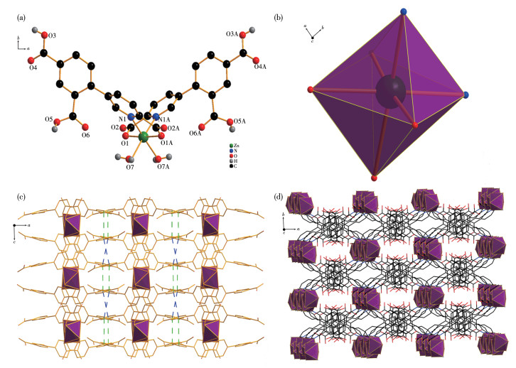
Complex 1 crystallizes in the orthorhombic Ccca space group. In an asymmetric structural unit of 1, a Zn (Ⅱ) ion, two H2dpcp- anions, and two coordinated water molecules are included. As shown in Fig. 1a, the six-coordinate Zn2+ is fulfilled by two nitrogen atoms (N1, N1A) from two pyridyl groups and two carboxylate oxygen atoms (O1, O1A) of the H2dpcp- anions, and two oxygen atoms (O7, O7A) from two coordinated water molecules, which generates a distorted octahedral structure (Fig. 1b). The bond lengths of Zn—O range from 0.208 1(2) to 0.234 6(10) nm and the bond length of Zn—N is 0.215 3(3) nm, which are well-matched to those observed in the reported compounds[44-46]. In complex 1, the adjacent coordination structural units form a three-dimensional supramolecular structure through two kinds of π…π interactions (Fig.S2), and the Cg…Cg distances for the weak π…π interactions among the benzene groups are 0.389 8 and 0.384 9 nm, respectively, which is similar to that reported value in the literature[47-48] (Fig. 1c and 1d).
Figure 1
Figure 1.
(a) Coordinated environment of Zn2+ ion in 1; (b) Distorted coordination geometry of Zn2+ ion; (c) π…π interactions of 1; (d) 3D supramolecular architecture formed by π…π interactions
The TGA curve of complex 1 is shown in Fig.S3. According to the TGA curve, a weight loss of 7.53% (Calcd. 7.81%) in a temperature range of 11-240 ℃ corresponds to the loss of one lattice water molecule and two coordinated water molecules. With the increasing temperature, the weight loss of 1 continued to rise rapidly, demonstrating that the structure of 1 successively decomposes. Meanwhile, the PXRD of complex 1 has been carried out to check its phase purity of crystal (Fig.S4). The experimental result of PXRD for 1 was in good agreement with the simulated one generated from the single crystal of 1, illuminating that the crystal structure of 1 is truly representative of the bulk crystal product.
2.3
Luminescent behaviors and stability in the solutions with different pH values
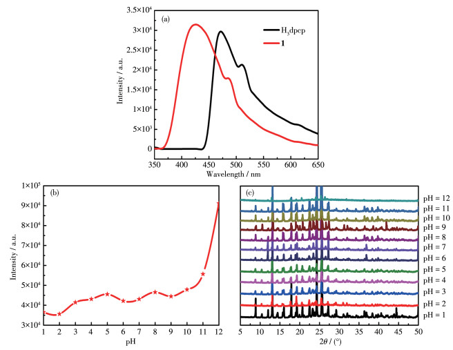
The luminescent properties of H3dpcp and 1 were investigated in the solid-state at room temperature. As shown in Fig. 2a, H3dpcp displayed a luminescent emission at 470 nm upon excitation at 357 nm, which may be assigned to the π*→π and π*→n electronic transitions of H3dpcp. Upon excitation at 333 nm, 1 showed the emission peaks centered at 422 nm, which indicates that the emission of 1 can still be assigned to the intra-ligand photoluminescence (PL). Compared with that of H3dpcp, the bathochromic shift of 1 is due to the bonding interactions between H2dpcp- ligands and zinc ions.
Figure 2
Figure 2.
(a) Emission spectra of H3dpcp and 1 at room temperature; (b) Fluorescence intensity of 1 dispersed in aqueous solutions with a pH range of 1-12 at the excitation of 333 nm; (c) PXRD patterns of 1 soaked in solutions with a pH range of 1-12
In addition, the luminescence spectra of 1 soaked in the solutions with different pH values exhibited a wide range of fluorescent stability. As shown in Fig. 2b, when the samples of 1 were immersed in acid or basic solution for a day with an extensive pH range of 1-12 (using 1.0 mol·L-1 HCl or 1.0 mol·L-1 NaOH solution), the luminescence intensities of 1 had no obvious changes in the pH range of 1-10. Subsequently, the fluorescence intensities increased with the increase of aqueous alkalinity in the general trend. The PXRD patterns of 1 elucidate that the framework of 1 retains integrity in the pH range of 1-11 (Fig. 2c). With the increase of pH value, H2dpcp- groups may be deprotonated, thereby changing the excited state energy of H2dpcp- ligands, which influences the energy transfer between H2dpcp- ligands and Zn2+ ions, resulting in the growth of fluorescence intensity[49-50]. It is worth pointing out that 1 is a promising sensor with fluorescent stability in an extensive pH range in an aqueous solution.
2.4
Effect of anion on the fluorescence of 1
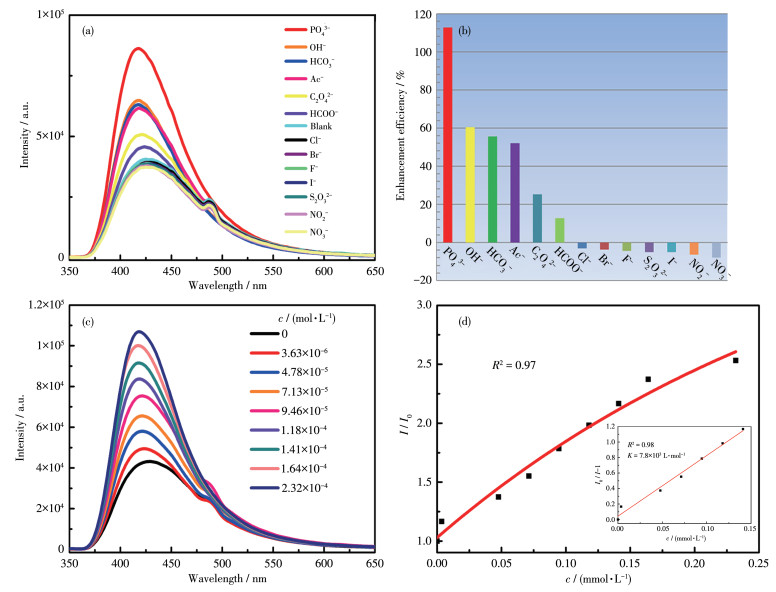
The emission spectra are shown in Fig. 3a and 3b. The luminescent intensities of 1 decreased to different extents caused by a few anions through the luminescence quenching, while some anions made the intensities of 1 rise conversely through the luminescence enhancement. It is noteworthy that the enhancement efficiency for 1.0 mmol·L-1 of PO43- was 112.73% cal- culated by the following formula: (I-I0)/I0×100%, where I represents the fluorescence intensity after the addition of PO43- ion to 1 and I0 is the initial fluorescence intensity of 1.
Figure 3
Figure 3.
(a) Luminescent spectra of 1 in the presence of various anions; (b) Comparison for enhancement efficiency of 1 detecting various anions; (c) Emission spectra of 1 with different concentrations of PO43- in aqueous solutions; (d) Nonlinear Stern-Volmer plot for 1 detecting PO43-
Inset: linear Stern-Volmer plot for 1 detecting PO43-
To further explore the sensitivity of 1 detecting PO43-, quantitative detection was also investigated. The PL spectra for 1 showed a certain degree of change after gradually adding a PO43- solution (Fig. 3c). It has been found that the intensity of 1 gradually enhanced with the increasing concentration of PO43- anion, indicating that the enhancement of PL intensity of 1 caused by the introduction of PO43- ions can be quantified.
A linear fit was performed by the Stern-Volmer equation I/I0=Kc+1 to further examine the relationship between the enhancement effect and PO43- concentration, where I and I0 are the fluorescence intensities in the presence and absence of PO43-, respectively; K is the Stern-Volmer constant, and c represents the concentration of PO43- (Fig. 3d). The K was found to be 7.8× 10³ L·mol-1 and the detection limit for PO43- ion is 0.162 mmol·L-1. Therefore, complex 1 can be considered as the sensor of PO43- ion with high sensitivity.
The experimental interference of mixed anions on the emission of 1 was also explored, and the PL spectra of 1 suspension with PO43- and other anions (1.0 mmol· L-1) are shown in Fig.S5a. The results suggest that the fluorescence enhancement effect of PO43- ion on the emission of 1 is not affected by introducing some anions, indicating that 1 can selectively sense PO43- ion among the above anions.
The recyclable performance of 1 as the sensor of PO43- was determined. 1.0 mmol·L-1 of PO43- was added dropwise into the suspension of 1 to form 1-PO43-, and then 1-PO43- was washed with deionized water. The fluorescent intensity could approximately be recovered to the initial value after five recycles (Fig.S6). The above results demonstrate the good recyclability of 1 for further detecting applications.
We also made a comparison of complex 1 with those of other reports for sensing PO43- ion (Table S3). It has been found that the detection limit of complex 1 was not in the lowest range, but its high sensitivity, selectivity, reversibility, and its extraordinary turn-on response make it a probable sensor in the determination of PO43- in an aqueous medium.
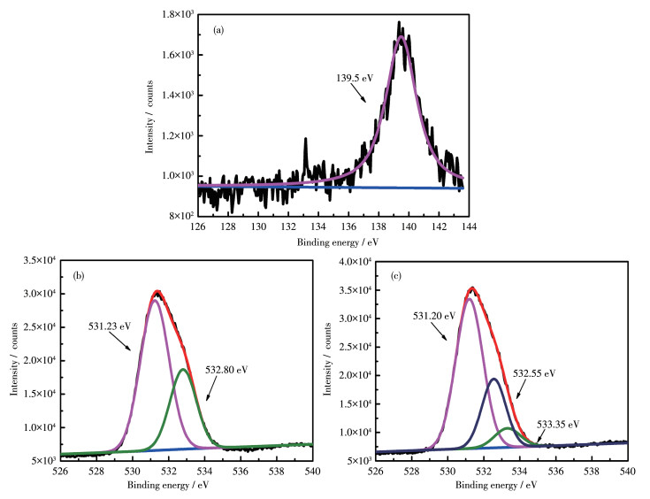
Furthermore, the mechanism for the enhancement of 1 after adding PO43- anion was also explored. Firstly, as shown in Fig. S7, the decay time of PO43- @1 was 0.090 5 ms, which was almost no change compared with 0.090 2 ms for 1, indicating the static mechanism[51]. Secondly, the PXRD pattern of 1 soaked in Na3PO4 solution (PO43-@1) excluded the collapse of the framework of 1, however, the peak at 7.86° disappeared (Fig. S8a). Meanwhile, the infrared spectrum result showed that the vibrational strength of carboxylate groups in PO43-@1 decreased in comparison to that of 1 (Fig.S8b). Thirdly, as shown in Fig.S5b, the UV-Vis absorption spectrum after the gradual addition of phos-phate ions in 1 was measured, and the results showed that the absorption peak at 292 nm moved slightly after the continuous addition of PO43-, and gradually changed into a wide absorption band of 265-287 nm. According to the structure of complex 1, it can be inferred that there may exist hydrogen bonding between PO43- ions and un-deprotonated carboxylate groups in complex 1. To deeply elucidate this hypothesis of fluorescence enhancement of 1 induced by the above anion, XPS of 1 and PO43- @1 were conducted. As shown in Fig.S9a and S9b, the high resolution of the C1s signal was deconvoluted into two peaks at 284.64 and 288.37 eV, corresponding to C=C/C—C, C=O of carboxylate groups, which was consistent with the FT-IR results[52]. The peaks at 1 021.57 and 1 044.61 eV in complex 1 can be distributed to Zn2p3/2 and 2p1/2 (Fig.S9c and S9d). As shown in Fig. 4a, a new peak was observed at 139.5 eV, which can be assigned to the P2p orbital of PO43-@1. The O1s peak was divided into two peaks at 531.23 and 532.80 eV, attributed to C=O and C—OH, respectively (Fig. 4b) [53-54]. Similarly, in Fig. 4c, it was observed that the O1s peak of PO43-@1 at 531.20 and 532.55 eV, indicating that its binding energy remains unchanged. Interestingly, a new peak at 533.35 eV appeared, which can be attributed to the weak bonding interaction between the Zn2+ ion and the O atom of the PO43- ion[55]. Because the phosphate ions have a competitive coordination relationship with the coordination waters of 1, the P—O bond interacts more strongly with the Zn2+ ion[56]. Thus, the Zn—O bond between the metal center and coordination waters could be partially broken and replaced by phosphate anions, which accelerates the energy transfer from H2dpcp- ligand to the Zn2+ center during excitation, leading to fluorescence enhancement[40].
Figure 4
Figure 4.
XPS spectra of complex 1 and PO43-@1: (a) P2p spectrum of PO43-@1; O1s spectra of (b) 1 and (c) PO43-@1
In a word, the enhanced fluorescence of complex 1 after the addition of PO43- ions may be caused by the formation of hydrogen bonds between phosphate ions and un-deprotonated carboxylate groups, and the weak bonding interactions of metal centers with O atoms of PO43- ions. Intriguingly, it was observed under natural light that the solution of complex 1 quickly became clear after the addition of PO43-, indicating that 1 can identify the phosphate ion with the naked eye.
2.5
Detection of metal cations
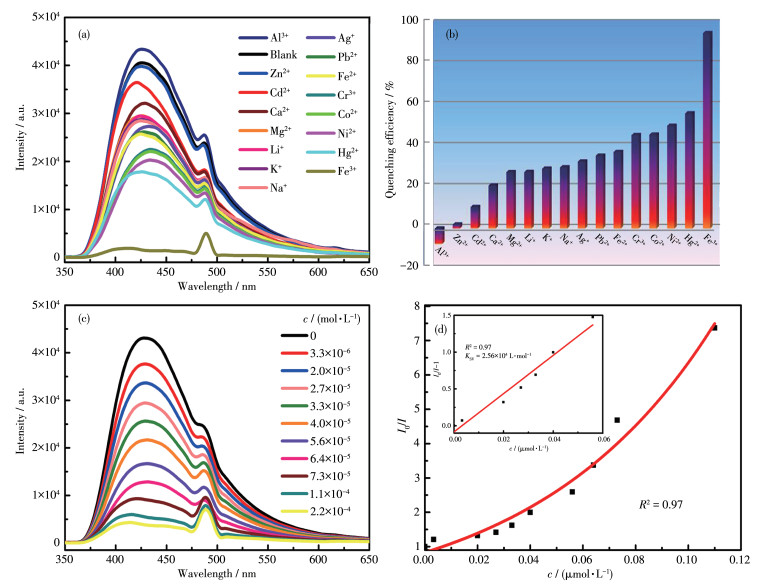
The samples of 1 were dispersed in the aqueous solutions of different metal cations and then the PL spectra were separately recorded (Fig. 5a). Notably, most cations exerted a relatively weak quenching effect on the emission of 1, while Fe3+ ions had the strongest quenching influence, indicating that complex 1 exhibits the selective fluorescent response toward Fe3+ ions (Fig. 5b). The quenching efficiency for 1.0 mmol·L-1 of Fe3+ was 95.33% calculated by the following formula: (I0-I)/I0×100%, where I represents the fluorescence intensity after the addition of Fe3+ ion and I0 is the initial fluorescence intensity of 1.
Figure 5
Figure 5.
(a) PL spectra of 1 dispersed in various metal ions solutions; (b) Comparison for quenching efficiency of 1 detecting various metal ions; (c) Emission spectra of 1 dispersed in the various concentrations of Fe3+ ions; (d) Nonlinear Stern-Volmer plot in a low concentration range of Fe3+ ions
Inset: linear Stern-Volmer plot for 1 detecting Fe3+
Homogeneously, the quantitative luminescence titration experiments were performed to better understand the sensing capabilities of complex 1 for Fe3+ ion. With the increase in the Fe3+ concentration, the fluorescence intensity of suspension of 1 gradually decreased (Fig. 5c). As observed from Fig. 5d, the curves of (I0/I-1) vs Fe3+ concentration showed a good linear correlation at low concentrations. The KSV of 1 was calculated to be 2.56×10⁴ L·mol-1 and the detection limit was 0.247 mmol·L-1, indicating that 1 can be considered as the sensor for Fe3+ ion.
The recyclable experiments exhibited that the initial luminescence intensity of complex 1 remained unchanged after five cycles, demonstrating that 1 has high recyclability and stability in the detectable application of Fe3+ ion (Fig.S10).
In previous research, the quenching mechanisms on fluorescent complexes by analytes can mainly arise from the collapse of the complexes framework, cation exchange between complexes and targeted cations, competition absorption, and resonance energy transfer caused by the strong interaction between incoming metal ions and luminophores in complexes[57-60]. The interaction between metal ions and luminophores is commonly related to two processes: fluorescence resonance energy transfer (FRET) and photoinduced electron transfer (PET) [5, 58, 61]. A clear indication of the FRET mechanism can be obtained when the electronic absorption band of the analytes has adequate overlap with the emission band of the luminophore[62-63]. While the PET process is mainly attributed to the weak interaction between the excited state of the luminophore and the species which can accept or donate electrons[64-65].
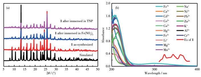
The mechanism experiments for complex 1 were further performed. The PXRD measurements were carried out to investigate the structures of complex 1 and 1 soaked in Fe(NO3)3 solutions with the concentration of 1.0 mmol·L-1 for three days (Fe3+ @1), respectively. As shown in Fig. 6a, the PXRD pattern of Fe3+@1 was similar to that of 1, which demonstrates that the framework of 1 remains intact after immersion of the iron ions. Consequently, the photoluminescence attenuations of 1 do not result from the collapse of the framework. The UV-Vis adsorptions spectrum of Fe(NO3)3 in the aqueous solution displayed a moderate overlap with the excitation spectrum of 1 (Fig. 6b). As given in Table S4, the EDX analysis of Fe3+@1 showed the presence of Fe3+ ions. This result can be explained that Zn (Ⅱ) ions can be partly replaced with Fe3+ ions, reducing the resonance energy transfer of 1. Based on the above results, it might be deduced that the luminescence quenching of 1 induced by Fe3+ ion can be attributed to the existence of competitive absorption of excitation energy between complex 1 and Fe3+ ions, and the replacement of Zn2+ ions with Fe3+ ions, which affects the energy transfer process[66-67].
Figure 6
Figure 6.
(a) PXRD patterns of 1 and 1 treated by different analytes solutions; (b) UV-Vis absorption spectra of various cations and the excitation spectrum of 1
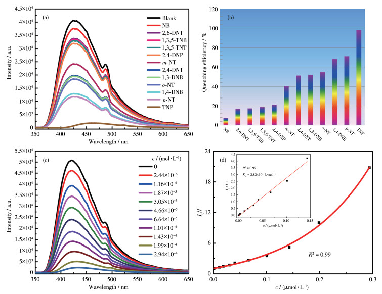
The structural feature associated with 1 implies this complex is the luminescent sensor for the detection of NACs. As shown in Fig. 7a and 7b, the highest quenching efficiency (99.12%) for TNP was found among the above NACs. The intensity of 1 decreased slowly with the increasing concentrations of TNP (Fig. 7c). According to Fig. 7d, the quenching constant KSV was 2.82×10⁴ L·mol-1 and the detection limit for TNP was 0.115 mmol·L-1. The initial luminescence intensity can almost be regained after five cycles (Fig. S11a). At the same time, the PXRD results after 1-5 cycles indicate that the framework of 1 remains unchanged (Fig. S11b). The above results demonstrate both reliable recyclability and structural stability of 1 in the detectable application of TNP.
Figure 7
Figure 7.
(a) Luminescent spectra of 1 with various NACs in MeOH solutions; (b) Comparison for quenching efficiency of 1 detecting various NACs; (c) Emission spectra of 1 with different concentrations of TNP in MeOH solutions; (d) Nonlinear Stern-Volmer plot in a low concentration range of TNP
Inset: linear Stern-Volmer plot for 1 detecting TNP
The sensing mechanisms for TNP were deeply investigated to better understand the highly selective detection. As shown in Fig. S11b, the framework of 1 treated in TNP/MeOH solutions remained intact. Furthermore, the UV-Vis absorption spectrum indicates that the absorption band of TNP overlapped with the emission spectrum of 1, which was far more than that of other nitroaromatic compounds, resulting in the most effective fluorescence quenching, and it is in accordance with the mechanism of resonance energy transfer (Fig. S12) [68-69]. Therefore, it can be deduced that the luminescent quenching of 1 for sensing TNP can be ascribed to the process of FRET.
3.
Conclusions
In summary, a new luminescent Zn-complex has been obtained based on the H3dpcp ligand under solvothermal conditions. Complex 1 displayed high fluorescence stability in a pH range of 1-10 in an aqueous solution. We have employed 1 as a fluorescent turn-on probe for selective detection of PO43- and also as a fluorescent turn-off sensor for the selective sensing of Fe3+, TNP in an aqueous medium. Especially complex 1 can identify the phosphate ion with naked eyes under natural light. The detection limits of complex 1 for PO43-, Fe3+, and TNP were 0.162, 0.247, and 0.115 mmol·L-1, respectively. Furthermore, the possible fluorescent sensing mechanisms of 1 for PO43-, Fe3+ and TNP have been elucidated, respectively. It is worth noting that the fluorescence turn-on response of complex 1 upon addition of the PO43- ions can be attributed to the formation of the hydrogen bonds and the weak bonding interactions between metal centers and O atoms of PO43- ions. This work displays that luminescent complexes could be rationally designed and explored as potential luminescence sensing materials in biological and environmental aspects.
Conflicts of interest: There are no conflicts to declare.
Acknowledgments: This work is financially supported by the National Natural Science Foundation of China (Grants No. 21761024, 22161032) and Inner Mongolia Natural Science Foundation (Grant No.2021MS02012).
Mainstone C P, Parr W. Phosphorus in Rivers-Ecology and Management[J]. Sci. Total Environ.,
2002, 282:
25-47.
[2]
Withers P J A, Jarvie H P. Delivery and Cycling of Phosphorus in Rivers: A Review[J]. Sci Total Environ.,
2008, 400:
379-395.
doi: 10.1016/j.scitotenv.2008.08.002
[3]
Lin K Y A, Chen S Y, Jochems A P. Zirconium-Based Metal Organic Frameworks: Highly Selective Adsorbents for Removal of Phosphate from Water and Urine[J]. Mater. Chem. Phys.,
2015, 160:
168-176.
doi: 10.1016/j.matchemphys.2015.04.021
[4]
Jia X L, Chen D J, Liu B, Hai L, Zhang R, Zheng Y D. Highly Selective and Sensitive Phosphate Anion Sensors Based on AlGaN/GaN High Electron Mobility Transistors Functionalized by Ion Imprinted Polymer[J]. Sci. Rep.,
2016, 6:
27728.
doi: 10.1038/srep27728
[5]
Farahani Y D, Safarifard V. Highly Selective Detection of Fe3+, Cd2+ and CH2Cl2 Based on a Fluorescent Zn-MOF with Azine-Decorated Pores[J]. J. Solid State Chem.,
2019, 275:
131-140.
doi: 10.1016/j.jssc.2019.04.018
[6]
Li L N, Shen S S, Ai W P, Song S Y, Bai Y, Liu H W. Facilely Synthe-sized Eu3+ Post-functionalized UiO-66-Type Metal-Organic Framework for Rapid and Highly Selective Detection of Fe3+ in Aqueous Solution[J]. Sens. Actuator B-Chem.,
2018, 267:
542-548.
doi: 10.1016/j.snb.2018.04.064
[7]
Hu X R, Cai X Y, Ma R S, Fu W W, Zhang C J, Du X J. Iron-Load Exacerbates the Severity of Atherosclerosis via Inducing Inflammation and Enhancing the Glycolysis in Macrophages[J]. J. Cell. Physiol.,
2019, 234:
18792-18800.
doi: 10.1002/jcp.28518
[8]
Gao E J, Liu D S, Xing J L, Feng Y H, Su J Q, Liu J X, Zhao H W, Wang N, Jia Z L, Zhang X Y, Fedin V P, Zhu M C. A Recyclable Bi-functional Luminescent Zinc(Ⅱ) Metal-Organic Framework as Highly Selective and Sensitive Sensing Probe for Nitroaromatic Explosives and Fe3+ Ions[J]. Appl. Organomet. Chem.,
2019, 33:
e5109.
[9]
Zhang Y Q, Blatov V A, Zheng T R, Yang C H, Qian L L, Li K, Li B L, Wu B. A Luminescent Zinc(Ⅱ) Coordination Polymer with Unusual (3, 4, 4)-Coordinated Self-Catenated 3D Network for Selective Detection of Nitroaromatics and Ferric and Chromate Ions: A Versatile Luminescent Sensor[J]. Dalton Trans.,
2018, 47:
6189-6198.
doi: 10.1039/C7DT04682K
[10]
Cai H, Xu L L, Lai H Y, Liu J Y, Ng S W, Li D. A Highly Emissive and Stable Zinc(Ⅱ) Metal-Organic Framework as a Host-guest Chemo-palette for Approaching White-Light-Emission[J]. Chem. Commun.,
2017, 53:
7917-7920.
doi: 10.1039/C7CC03350H
[11]
Li Y W, Yan H, Hu T L, Ma H Y, Li D C, Wang S N, Yao Q X, Dou J M, Xu J, Bu X H. Two Microporous Fe-Based MOFs with Multiple Active Sites for Selective Gas Adsorption[J]. Chem. Commun.,
2017, 53:
2394-2397.
doi: 10.1039/C6CC09923H
[12]
Wang J X, Li J, Wang Y N, Gao M X, Zhang X M, Yang P Y. Development of Versatile Metal-Organic Framework Functionalized Magnetic Graphene Core-Shell Biocomposite for Highly Specific Recognition of Glycopeptides[J]. ACS Appl. Mater. Interfaces,
2016, 8:
27482-27489.
doi: 10.1021/acsami.6b08218
[13]
Liu S J, Cao C, Yao S L, Zheng T F, Wang Z X, Liu C, Liao J S, Chen J L, Li Y W, Wen H R. Temperature- and Vapor-Induced Reversible Single-Crystal-to-Single-Crystal Transformations of Three 2D/3D GdⅢ-Organic Frameworks Exhibiting Significant Magnetocaloric Effects[J]. Dalton Trans.,
2016, 46:
64-70.
[14]
Jiao L, Wang Y, Jiang H L, Xu Q. Metal-Organic Frameworks as Platforms for Catalytic Applications[J]. Adv. Mater.,
2018, 30:
e1703663.
doi: 10.1002/adma.201703663
[15]
Zhao X X, Feng J R, Liu J W, Lu J, Shi W, Yang G M, Wang G C, Feng P Y, Cheng P. Metal-Organic Framework-Derived ZnO/ZnS Heteronanostructures for Efficient Visible-Light-Driven Photocatalytic Hydrogen Production[J]. Adv. Sci.,
2018, 5:
1700590.
doi: 10.1002/advs.201700590
[16]
Xiao J D, Jiang H L. Metal-Organic Frameworks for Photocatalysis and Photothermal Catalysis[J]. Acc. Chem. Res.,
2019, 52:
356-366.
doi: 10.1021/acs.accounts.8b00521
[17]
Eliseeva S V, Bunzli J C G. Lanthanide Luminescence for Functional Materials and Bio-sciences[J]. Chem. Soc. Rev.,
2010, 39:
189-227.
doi: 10.1039/B905604C
[18]
Hou L L, Song Y H, Xiao Y J, Wu R, Wang L. Ratiometric Fluorescence Detection of Dipicolinic Acid Based on Microporous Ln/Melamine-Terephthaladehyde Schiff Base Networks Complex[J]. Talanta,
2020, 209:
120534.
doi: 10.1016/j.talanta.2019.120534
[19]
He T, Zhang Y Z, Kong X J, Yu J M, Lv X L, Wu Y F, Guo Z J, Li J R. Zr(Ⅳ)-Based Metal-Organic Framework with T-Shaped Ligand: Unique Structure, High Stability, Selective Detection, and Rapid Adsorption of Cr2O72- in Water[J]. ACS Appl. Mater. Interfaces,
2018, 10:
16650-16659.
doi: 10.1021/acsami.8b03987
[20]
Wu J X, Yan B. Eu(Ⅲ)-Functionalized In-MOF (In(OH)bpydc) as Fluorescent Probe for Highly Selectively Sensing Organic Small Molecules and Anions Especially for CHCl3 and MnO4-[J]. J. Colloid Interface Sci.,
2017, 504:
197-205.
doi: 10.1016/j.jcis.2017.05.054
[21]
Zhang Q, Wang C F, Lv Y K. Luminescent Switch Sensors for the Detection of Biomolecules Based on Metal-Organic Frameworks[J]. Analyst,
2018, 143:
4221-4229.
doi: 10.1039/C8AN00816G
[22]
Gao Y X, Yu G, Liu K, Wang B. Luminescent Mixed-Crystal Ln-MOF Thin Film for the Recognition and Detection of Pharmaceuticals[J]. Sens. Actuator B-Chem.,
2018, 257:
931-935.
doi: 10.1016/j.snb.2017.10.180
[23]
Zhang Q S, Wang J, Kirillov A M, Dou W, Xu C, Xu C L, Yang L Z, Fang R, Liu W S. Multifunctional Ln-MOF Luminescent Probe for Efficient Sensing of Fe3+, Ce3+, and Acetone[J]. ACS Appl. Mater. Interfaces,
2018, 10:
23976-23986.
doi: 10.1021/acsami.8b06103
[24]
Li Y K, Wei Z H, Zhang Y, Guo Z F, Chen D S, Jia P Y, Chen P, Xing H Z. Multifunctional Ln-MOF Dual-Emitting EY@Zr-MOF Composite as Self-Calibrating Luminescent Sensor for Selective Detection of Inorganic Ions and Nitroaromatics[J]. ACS Sustainable Chem. Eng.,
2019, 7:
6196-6203.
doi: 10.1021/acssuschemeng.8b06500
[25]
Zhao J, Qu X L, Wang J M, Yan B. Photophysical Tuning of Viologen-Based Metal-Organic Framework Hybrids via Anion Exchange and Chemical Sensing on Persulfate (S2O82-)[J]. Ind. Eng. Chem. Res.,
2019, 58:
18533-18539.
doi: 10.1021/acs.iecr.9b04049
[26]
Cao Z, Chen L, Jiang F L, Zhou K, Yu M X, Jing T, Li S C, Li Z J, Hong M C. Incorporating Three Chiral Channels into an In-MOF for Excellent Gas Absorption and Preliminary Cu2+ Ion Detection[J]. Cryst. Growth Des.,
2019, 19:
3860-3868.
doi: 10.1021/acs.cgd.9b00295
[27]
Zhang Y, Yan B. A Portable Self-Calibrating Logic Detector for Gradient Detection of Formaldehyde Based on Luminescent Metal Organic Frameworks[J]. J. Mater. Chem. C,
2019, 7:
5652-5657.
doi: 10.1039/C9TC01288E
[28]
Li S D, Lu L P, Zhu M L, Yuan C X, Feng S S. A Bifunctional Chemosensor for Detection of Volatile Ketone or Hexavalent Chromate Anions in Aqueous Solution Based on a Cd(Ⅱ) Metal-Organic Framework[J]. Sens. Actuator B-Chem.,
2018, 258:
970-980.
doi: 10.1016/j.snb.2017.11.142
[29]
Yao W Q, Guo H, Liu H, Li Q, Xue R, Wu N, Li L, Wang M Y, Yang W. Simultaneous Electrochemical Determination of Acetaminophen and Dopamine Based on Metal-Organic Framework/Multiwalled Carbon Nanotubes-Au@Ag Nanocomposites[J]. J. Electrochem. Soc.,
2019, 166:
B1258-B1267.
doi: 10.1149/2.0101914jes
[30]
Rawool C R, Srivastava A K. A Dual Template Imprinted Polymer Modified Electrochemical Sensor Based on Cu Metal Organic Framework/Mesoporous Carbon for Highly Sensitive and Selective Recognition of Rifampicin and Isoniazid[J]. Sens. Actuator B-Chem.,
2019, 288:
493-506.
doi: 10.1016/j.snb.2019.03.032
[31]
Zhao D, Rao X T, Yu J C, Cui Y J, Yang Y, Qian G D. Design and Synthesis of an MOF Thermometer with High Sensitivity in the Physiological Temperature Range[J]. Inorg. Chem.,
2015, 54:
11193-11199.
doi: 10.1021/acs.inorgchem.5b01623
[32]
Zhou Z Q, Li M X, Wang L Y, He X, Chi T, Wang Z X. Antiferro-magnetic Copper(Ⅱ) Metal-Organic Framework Based Quartz Crystal Microbalance Sensor for Humidity[J]. Cryst. Growth Des.,
2017, 17:
6719-6724.
doi: 10.1021/acs.cgd.7b01318
[33]
Sun Y, Zhang N, Guan Q L, Liu C H, Li B, Zhang K Y, Li G H, Xing Y H, Bai F Y, Sun L X. Sensing of Fe3+ and Cr2O72- in Water and White Light: Synthesis, Characterization, and Fluorescence Properties of a Crystalline Bismuth-1, 3, 5-Benzenetricarboxylic Acid Framework[J]. Cryst. Growth Des.,
2019, 19:
7217-7229.
doi: 10.1021/acs.cgd.9b01098
[34]
Wang X Q, Feng D D, Tang J, Zhao Y D, Li J, Yang J, Kim C K, Su F. A Water-Stable Zinc(Ⅱ)-Organic Framework as a Multiresponsive Luminescent Sensor for Toxic Heavy Metal Cations, Oxyanions and Organochlorine Pesticides in Aqueous Solution[J]. Dalton Trans.,
2019, 48:
16776-16785.
doi: 10.1039/C9DT03195B
[35]
Shi X X, Qu X J, Chai J, Tong C X, Fan Y, Wang L. Stable Coordination Polymers with Linear Dependence Color Tuning and Luminescent Properties for Detection of Metal Ions and Explosives[J]. Dyes Pigment.,
2019, 170:
107583.
doi: 10.1016/j.dyepig.2019.107583
[36]
Wu K, Hu J S, Cheng X F, Li J X, Zhou C H. A Superior Luminescent Metal-Organic Framework Sensor for Sensing Trace Al3+ and Picric Acid via Disparate Charge Transfer Behaviors[J]. J. Lumin.,
2020, 219:
116908.
doi: 10.1016/j.jlumin.2019.116908
[37]
Wu K, Hu J S, Shi S N, Li J X, Cheng X F. A Thermal Stable Pincer-MOF with High Selective and Sensitive Nitro Explosive TNP, Metal Ion Fe3+ and pH Sensing in Aqueous Solution[J]. Dyes Pigment.,
2020, 173:
107993.
doi: 10.1016/j.dyepig.2019.107993
[38]
Zhao D, Wan X Y, Song H J, Hao L Y, Su Y Y, Lv Y. Metal-Organic Frameworks (MOFs) Combined with ZnO Quantum Dots as a Fluorescent Sensing Platform for Phosphate[J]. Sens. Actuator B-Chem.,
2014, 197:
50-57.
doi: 10.1016/j.snb.2014.02.070
[39]
Gao N, Huang J, Wang L Y, Feng J Y, Huang P C, Wu F Y. Ratiometric Fluorescence Detection of Phosphate in Human Serum with a Metal-Organic Frameworks-Based Nanocomposite and Its Immobilized Agarose Hydrogels[J]. Appl. Surf. Sci.,
2018, 459:
686-692.
doi: 10.1016/j.apsusc.2018.08.092
[40]
Das A, Das S, Trivedi V, Biswas S. A Dual Functional MOF-Based Fluorescent Sensor for Intracellular Phosphate and Extracellular 4-Nitrobenzaldehyde[J]. Dalton Trans.,
2019, 48:
1332-1343.
doi: 10.1039/C8DT03964J
[41]
Zhang X L, Zhan Z Y, Liang X Y, Chen C, Liu X L, Jia Y J, Hu M. Lanthanide-MOFs Constructed from Mixed Dicarboxylate Ligands as Selective Multi-responsive Luminescent Sensors[J]. Dalton Trans.,
2018, 47:
3272-3282.
doi: 10.1039/C7DT02966G
[42]
Zhan Z Y, Liang X Y, Zhang X L, Jia Y J, Hu M. A Water-Stable Europium-MOF as a Multifunctional Luminescent Sensor for Some Trivalent Metal Ions (Fe3+, Cr3+, Al3+), PO43- Ions, and Nitroaromatic Explosives[J]. Dalton Trans.,
2019, 48:
1786-1794.
doi: 10.1039/C8DT04653K
[43]
Liang X Y, Zhan Z Y, Jia Y J, Hu M. A Highly Selective Multifunctional Zn-MOF Probe for Sensing of Cr(Ⅲ), Cr(Ⅵ) Ions and TNP Molecule[J]. Appl. Organomet. Chem.,
2019, 33:
e4988.
[44]
Wang Y N, Zhu W C, Huo Q S, Yu J H, Xu J Q. A New Three-Dimensional Zn2+ Coordination Polymer Constructed from Oxalate and 1, 2, 4-Triazolate[J]. Spectrochim. Acta A,
2016, 161:
138-143.
doi: 10.1016/j.saa.2016.02.020
[45]
Pankajakshan A, Kuznetsov D, Mandal S. Ultrasensitive Detection of Hg(Ⅱ) Ions in Aqueous Medium Using Zinc-Based Metal-Organic Framework[J]. Inorg. Chem.,
2019, 58:
1377-1381.
doi: 10.1021/acs.inorgchem.8b02898
[46]
Das P, Mandal S K. A Highly Emissive Fluorescent Zn-MOF: Molecular Decoding Strategies for Solvents and Trace Detection of Dunnite in Water[J]. J. Mater. Chem. A,
2018, 6:
21274-21279.
doi: 10.1039/C8TA08546C
[47]
Gai Y L, Jiang F L, Chen L, Bu Y, Wu M Y, Zhou K, Pan J, Hong M C. A Series of Novel Zinc(Ⅱ) Entangled Coordination Polymers Based on Carboxyphenyl-Terpyridine Ligands[J]. Dalton Trans.,
2013, 42:
9954-9965.
doi: 10.1039/c3dt50532d
[48]
Guo D D, Zhu L N, Meng X X, Deng Z P, Huo L H, Gao S. Structural Variation from Linear, Layer to 3D Framework: Syntheses, Structures and Luminescence[J]. Appl. Organomet. Chem.,
2019, 33:
e5056.
[49]
Yu M, Zuo C S, Zhang N. An Experimental and Computational Study on Naphthylideneimine Based pH Sensitive Fluorescence Probe for Zinc[J]. Spectrochim. Acta A,
2020, 224:
117389.
doi: 10.1016/j.saa.2019.117389
[50]
Ambrosi G, Fanelli M, Paoli P, Formica M, Paderni D, Rossi P, Micheloni M, Giorgi L, Fusi V. Zn(Ⅱ) Detection and Biological Activity of a Macrocycle Containing a Bis(oxadiazole)pyridine Derivative as Fluorophore[J]. Dalton Trans.,
2020, 49:
7496-7506.
doi: 10.1039/C9DT03910D
[51]
Qu S M, Li Z, Jia Q. Detection of Purine Metabolite Uric Acid with Picolinic-Acid-Functionalized Metal-Organic Frameworks[J]. ACS Appl. Mater. Interfaces,
2019, 11:
34196-34202.
doi: 10.1021/acsami.9b07442
[52]
Ding H, Wei J S, Xiong H M. Nitrogen and Sulfur Co-doped Carbon Dots with Strong Blue Luminescence[J]. Nanoscale,
2014, 6:
13817-13823.
doi: 10.1039/C4NR04267K
[53]
Ming F L, Hou J Z, Huo D Q, Zhou J, Yang M, Shen C H, Zhang S Y, Hou C J. Copper-Based Metal-Organic Framework Nanoparticles for Sensitive Fluorescence Detection of Ferric Ions[J]. Anal. Methods,
2019, 11:
4382-4389.
doi: 10.1039/C9AY01093A
[54]
Ding H, Yu S B, Wei J S, Xiong H M. Full-Color Light-Emitting Carbon Dots with a Surface-State-Controlled Luminescence Mechanism[J]. ACS Nano,
2016, 10:
484-491.
doi: 10.1021/acsnano.5b05406
[55]
Onyiriuka E C. Zinc Phosphate Glass Surfaces Studied by XPS[J]. J. Non-Cryst. Solids,
1993, 163:
268-273.
doi: 10.1016/0022-3093(93)91304-L
[56]
Asha K S, Bhattacharjee R, Mandal S. Complete Transmetalation in a Metal-Organic Framework by Metal Ion Metathesis in a Single Crystal for Selective Sensing of Phosphate Ions in Aqueous Media[J]. Angew. Chem. Int. Ed.,
2016, 55:
11528-11532.
doi: 10.1002/anie.201606185
[57]
Besheli M E, Rahimi R, Farahani Y D, Safarifard V. A Porous Ni-Based Metal-Organic Framework as a Selective Luminescent Probe to Fe3+ Metal Ion and MeOH[J]. Inorg. Chim. Acta,
2019, 495:
118956.
doi: 10.1016/j.ica.2019.118956
[58]
Hu J S, Cheng T T, Dong S J, Zhou C H, Huang X H, Zhang L. Multi-functional Luminescent Cd(Ⅱ)-Based Metal-Organic Framework Material for Highly Selective and Sensitive Sensing 2, 4, 6-Trinitro-phenol (TNP) and Fe3+ Cation[J]. Microporous Mesoporous Mater.,
2018, 272:
177-183.
doi: 10.1016/j.micromeso.2018.06.013
[59]
Xu H, Gao J K, Qian X F, Wang J P, He H J, Cui Y J, Yang Y, Wang Z Y, Qian G D. Metal-Organic Framework Nanosheets for Fast-Response and Highly Sensitive Luminescent Sensing of Fe3+[J]. J. Mater. Chem. A,
2016, 4:
10900-10905.
doi: 10.1039/C6TA03065C
[60]
Wang J, Wu J, Lu L, Xu H J, Trivedi M, Kumar A, Liu J Q, Zheng M B. A New 3D 10-Connected Cd(Ⅱ) Based MOF with Mixed Ligands: A Dual Photoluminescent Sensor for Nitroaroamatics and Ferric Ion[J]. Front. Chem.,
2019, 7:
244.
doi: 10.3389/fchem.2019.00244
[61]
Gogoi C, Biswas S. A New Quinoline Based Luminescent Zr(Ⅳ) Metal-Organic Framework for the Ultrasensitive Recognition of 4-Nitrophenol and Fe(Ⅲ) Ions[J]. Dalton Trans.,
2018, 47:
14696-14705.
doi: 10.1039/C8DT03058H
[62]
Hussain S, Malik A H, Afroz M A, Iyer P K. Ultrasensitive Detection of Nitroexplosive-Picric Acid via a Conjugated Polyelectrolyte in Aqueous Media and Solid Support[J]. Chem. Commun.,
2015, 51:
7207-7210.
doi: 10.1039/C5CC02194D
[63]
Hou B L, Tian D, Liu J, Dong L Z, Li S L, Li D S, Lan Y Q. A Water-Stable Metal-Organic Framework for Highly Sensitive and Selective Sensing of Fe3+ Ion[J]. Inorg. Chem.,
2016, 55:
10580-10586.
doi: 10.1021/acs.inorgchem.6b01809
[64]
Gui B, Meng Y, Xie Y, Tian J W, Yu G, Zeng W X, Zhang G X, Gong S L, Yang C L, Zhang D Q, Wang C. Tuning the Photoinduced Electron Transfer in a Zr-MOF: Toward Solid-State Fluorescent Molecular Switch and Turn-On Sensor[J]. Adv. Mater.,
2018, 30:
1802329.
doi: 10.1002/adma.201802329
[65]
Cui Y J, Yue D, Huang Y K, Zhang J, Wang Z Y, Yang D R, Qian G D. Photo-Induced Electron Transfer in a Metal-Organic Framework: A New Approach towards a Highly Sensitive Luminescent Probe for Fe3+[J]. Chem. Commun.,
2019, 55:
11231-11234.
doi: 10.1039/C9CC05019A
[66]
Chen C H, Wang X S, Li L, Huang Y B, Cao R. Highly Selective Sensing of Fe3+ by an Anionic Metal-Organic Framework Containing Uncoordinated Nitrogen and Carboxylate Oxygen Sites[J]. Dalton Trans.,
2018, 47:
3452-3458.
doi: 10.1039/C8DT00088C
[67]
Arici M. Multifunctional Luminescent Coordination Polymers Based on Tricarboxylic Acid for the Detection of 2, 4-Dinitrophenol and Iron(Ⅲ) and Aluminum(Ⅲ) ions[J]. New J. Chem.,
2019, 43:
3690-3697.
doi: 10.1039/C8NJ04046J
[68]
Surya S G, Nagarkar S S, Ghosh S K, Sonar P, Rao V R. OFET Based Explosive Sensors Using Diketopyrrolopyrrole and Metal Organic Framework Composite Active Channel Material[J]. Sens. Actuator B-Chem.,
2016, 223:
114-122.
doi: 10.1016/j.snb.2015.09.076
[69]
Sharma A, Kim D, Park J H, Rakshit S, Seong J, Jeong G H, Kwon O H, Lah M S. Mechanistic Insight into the Sensing of Nitroaromatic Compounds by Metal-Organic Frameworks[J]. Comm. Chem.,
2019, 2:
39.
doi: 10.1038/s42004-019-0135-2
Figure 1
(a) Coordinated environment of Zn2+ ion in 1; (b) Distorted coordination geometry of Zn2+ ion; (c) π…π interactions of 1; (d) 3D supramolecular architecture formed by π…π interactions
Figure 2
(a) Emission spectra of H3dpcp and 1 at room temperature; (b) Fluorescence intensity of 1 dispersed in aqueous solutions with a pH range of 1-12 at the excitation of 333 nm; (c) PXRD patterns of 1 soaked in solutions with a pH range of 1-12
Figure 3
(a) Luminescent spectra of 1 in the presence of various anions; (b) Comparison for enhancement efficiency of 1 detecting various anions; (c) Emission spectra of 1 with different concentrations of PO43- in aqueous solutions; (d) Nonlinear Stern-Volmer plot for 1 detecting PO43-
Inset: linear Stern-Volmer plot for 1 detecting PO43-
Figure 5
(a) PL spectra of 1 dispersed in various metal ions solutions; (b) Comparison for quenching efficiency of 1 detecting various metal ions; (c) Emission spectra of 1 dispersed in the various concentrations of Fe3+ ions; (d) Nonlinear Stern-Volmer plot in a low concentration range of Fe3+ ions
Inset: linear Stern-Volmer plot for 1 detecting Fe3+
Figure 6
(a) PXRD patterns of 1 and 1 treated by different analytes solutions; (b) UV-Vis absorption spectra of various cations and the excitation spectrum of 1
Figure 7
(a) Luminescent spectra of 1 with various NACs in MeOH solutions; (b) Comparison for quenching efficiency of 1 detecting various NACs; (c) Emission spectra of 1 with different concentrations of TNP in MeOH solutions; (d) Nonlinear Stern-Volmer plot in a low concentration range of TNP
Inset: linear Stern-Volmer plot for 1 detecting TNP

 下载:
下载:

 下载:
下载:


