Figure 1.
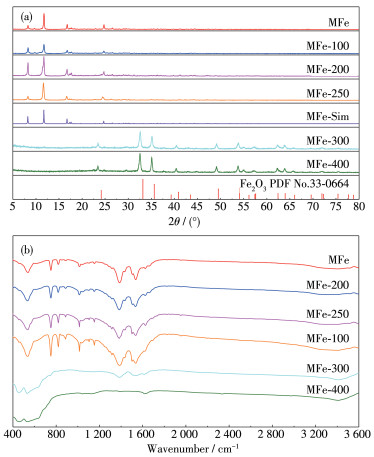
(a) PXRD patterns of MFe and MFe-T; (b) FTIR spectra of MFe and MFe-T
Synthesis of Quasi-MIL-53(Fe) Photocatalysts for Enhanced Visible Light Photocatalytic Degradation of Organic Dyes
Qi-Chao ZOU , Yan MA , Dian-Jun CHI , Hong-Bin QIAO , Jun-Ying ZHANG , Qian CHEN , Yu-Die SUN , Jian ZHANG , Kui ZHANG , Sheng-Jun LIU

The rapid development of modern industry has led to water pollution, including contamination with organic dyes. One harmful pollutant is methylene blue (MB). Although the adsorbent can effectively remove MB from water, its high cost limits practical applications[1]. In contrast, the solar photocatalytic degradation of organic pollutants has proven to be a clean technology that can address such environmental problems. Some successfully developed photocatalysts are metal oxides, metal sulfides and graphitic carbon nitride[1-2]. Howev- er, their low quantum and solar energy conversion effi- ciencies limit their practical applications. Therefore, the development of new and more efficient photocata- lysts is necessary.
Metal-organic frameworks (MOFs) are structures composed of organic ligands and metals (or metal clus- ters) and have a wide range of uses, such as in separa- tion, storage, detection, and catalysis[3-21]. In particular, MOFs featuring numerous active sites that generate numerous electron-hole pairs under adequate illumina- tion can be effectively used as photocatalysts. The first of such reported MOF catalysts used for photocatalytic degradation of phenol was MOF-5[22]. Thereafter, numerous other studies have focused on the develop- ment of MOF photocatalysts for CO2 reduction[23-24], water splitting[25-28] and organic pollutant degrada- tion[29-34].
The active sites of MOFs are important for the improvement of catalytic performance. Recent studies by Jiang's group revealed that structurally defective UiO-66-NH2 enhanced the efficiency of photocatalytic hydrogen production[35]. Huang et al. reported that the synthesis of MIL-53(Fe) (termed MFe hereafter) using hydrochloric acid as a modulator exposed more active sites than as-prepared MFe without hydrochloric acid, leading to the efficient photocatalytic degradation of tetracycline[36]. Further, Xu et al. reported that the use of quasi-MOFs obtained by thermal regulation enabled strong interactions between metal nanoparticles and the MOF, resulting in increased catalytic activity[37]. Furthermore, we reported the preparation of quasi-MFe at lower temperatures to improve their photocatalytic activity.
Herein, a series of MFe-based photocatalysts, col- lectively termed as MFe-T (T ℃ stands for calcination temperature), were activated at different temperatures in air and employed in the visible-light-assisted degra- dation of MB. Studying the photocatalytic performance of different samples allowed for the identification of the most suitable catalyst for optimized photodegradation. The resulting MFe-250 exhibited high photocatalytic activity for MB degradation, reaching a degradation rate of 99% within 90 min. Importantly, MFe-250 had excellent performance and chemical stability.
N, N-dimethyl formamide (DMF, GC, 99.5%), FeCl3·6H2O (AR, 99.0%), ethanol (EtOH, AR, 99.7%) and 2-propanol (AR, 99.7%) were purchased from General-Reagent. Methylene blue (MB, AR), ethylene- diamine tetraacetic acid disodium salt dihydrate (EDTA -2Na, AR) and hydrogen peroxide solution (30%, w/w) were purchased from Sinopharm Chemical Reagent Co., Ltd. (Shanghai, China). Terephthalic acid (AR, 99%) and ascorbic acid (VC, AR, 99.0%) were pur- chased from Aladdin. All chemicals were purchased from commercial sources and used without further treatments.
MFe was synthesized based on minor modifica- tions to the existing process[38], using the solvothermal method. In the process, ferric chloride hexahydrate (90 mg), terephthalic acid (55 mg) and DMF (8 mL) were mixed into a homogeneous solution. The solution was sealed in a 25 mL autoclave and heated at 180 ℃ for 12 h. After the autoclave cooled to room temperature, the product was washed with DMF and ethanol several times, and finally placed in a vacuum oven at 60 ℃ for 6 h for drying. Five samples were then taken from the dried product, following which each was successively placed in a muffle furnace and calcined in an air atmo- sphere for several hours at temperatures of 100, 200, 250, 300, and 400 ℃. The resultant samples were denoted as MFe-100, MFe-200, MFe-250, MFe-300, and MFe-400, respectively.
The powder X-ray diffraction (PXRD) patterns of samples were tested on a Bruker D8 Advance X-ray dif- fractometer with Cu Kα radiation (λ=0.154 07 nm, 40 kV, 40 mA, 5 (°)·min-1 from 5° to 80°). The FTIR spec- tra of the sample was tested on a Nicolet 6700 spec- trometer. The Brunauer-Emmett-Teller (BET) surface area was measured at 77 K with a Micromeritics ASAP2460 instrument (The air-dried sample was acti- vated in a vacuum at 120 ℃ for 4 h). UV-Vis diffuse reflectance spectra (UV-Vis DRS) were recorded on a UV-Vis-NIR spectrophotometer (Shimadzu 3600). Scanning electron microscopy (SEM, FEI NANO SEM430) was used to analyze the morphology of the samples. X-ray electron spectroscopy (XPS) measure- ments were performed with a Thermo Scientific K- Alpha+ XPS system using Mg Kα as an excitation source. Photodegradation solutions were analyzed by UV-Vis spectroscopic measurements (Persee TU-1810) in a range from 200 to 800 nm in ambient conditions. Thermogravimetric (TG) analyses were performed on a Shimadzu DTG-60H integration thermal analyzer from 25 to 600 ℃ at a heating rate of 10 ℃·min-1 under an air atmosphere.
The photocatalytic activity of each MFe-T was evaluated by its degradation rate of MB under visible light. The photocatalytic system consisted of photocata- lyst (20 mg), hydrogen peroxide (H2O2) (50 μL), and rhodamine B (RhB) (100 mL 20 mg·L-1) solution. After stirring for 60 min in the dark, the suspension was illu- minated with a 300 W Xe visible-light lamp (λ >420 nm, MC-XF300, Beijing Merry Change Co., Ltd.). Dur- ing the process, the solution was sampled every 10 min, filtered, and analyzed using a UV-Vis detector.
The photoelectrochemical properties of the materi- als were evaluated using an electrochemical worksta- tion (CHI-760E, Chenhua Instrument, Shanghai, China). The sample (10 mg) and mass fraction of 5% Nafion solution (10 μL) were added to ethanol (1 mL), and the mixture was sonicated for 1 h to form a uniform slurry. The slurry (50 μL) was added dropwise onto the conductive side of an indium tin oxide glass (1 cm×1 cm) and then dried at 100 ℃ for 5 h to obtain a work- ing electrode. The reference electrode was Ag/AgCl (saturated KCl solution) while the counter electrode was a Pt sheet. A three-electrode system with a Na2SO4 (0.5 mol·L-1) electrolyte was used to determine the pho- tocurrent and perform electrochemical impedance spec- troscopy (EIS). The light source was a 300 W Xe lamp (λ>420 nm).
PXRD patterns of pristine MFe and MFe-T are shown in Fig. 1a. The XRD patterns of MFe, MFe-100, MFe-200, and MFe-250 are in good agreement with simulated patterns (MFe-Sim), indicating that the MFe structure was stable only at low temperatures (Fig. 1a). From the patterns for MFe-300 and MFe-400, the absence of MFe peaks in the range of 5°-20° implied that the MFe framework was destroyed. Moreover, the diffraction patterns for MFe-300 and MFe-400 were consistent with that of Fe2O3 (PDF No.33-0664), indi- cating the complete decomposition of MFe at high tem- peratures.
At 100 ℃, the loss of adsorbed water molecules occurred, whereas at 200 ℃, the loss of coordinated water molecules commenced. At 250 ℃, Fe—O sites are exposed by the loss of organic ligands as CO2, while at temperatures above 300 ℃, MFe structure began to collapse. Hence, MFe structure is only retained below a certain temperature. To confirm this, TG analysis of MFe was carried out by heating the sample from 25 to 700 ℃ in the air (thermal calcination; Fig.S1, Support- ing information). When the temperature reached 427 ℃, the weight decreased sharply by 60%, indicat- ing that the original MOF structure was critically dam- aged. The removal of the organic ligand terephthalic acid occurs at such temperatures, resulting in the expo- sure of widely distributed Fe—O active sites. As the temperature approached 700 ℃, the weight dropped further by approximately 10%, suggesting the complete degradation of the MOF skeleton and the substantial oxidation of the iron to Fe2O3. These results show that TG analysis was a suitable method for sample analysis in this study.
To further confirm the presence of organic ligands in MFe-100, MFe-200, and MFe-250, FTIR spectra of MFe-T samples was compared to that of MFe sample (Fig. 1b). The FTIR spectra of MFe-100, MFe-200, and MFe-250 were very similar to that of MFe, suggesting that MFe structure was largely retained. Conversely, pyrolysis at 300 ℃ led to the disappearance of the peaks at 1 650-1 300 cm-1 (carboxylate groups), indi- cating the partial decomposition of MFe structure. The temperature at which most significant degradation of MFe structure occurred was 400 ℃.
Next, UV-Vis DRS was conducted. The spectra for MFe and MFe-T are shown in Fig.S2a. As the litera- ture described[39], MFe absorbs visible light in the red- edge region (approximately 650 nm). For MFe-T sam- ples, the initial increase in Fe2O3 (at temperatures below 300 ℃) led to a redshift (shift of the absorption edge towards higher wavelengths/lower energy), there- by enhancing light absorption. Consequently, the band gaps of MFe decreased from 2.50 to 2.15 eV (MFe- 200). Hence, thermally treated MFe-T potentially exhibits photocatalytic properties.
Fig.S3 shows the nitrogen (N2) adsorption-desorp- tion isotherms of MFe-T samples. The N2 adsorption- desorption isotherms of the samples pyrolyzed at 100 and 200 ℃ were the same, indicating that MFe porosity was preserved. MFe-300 and MFe-400 exhibited slight- ly increase in the surface area, whereas their porosity was more or less maintained. Furthermore, the Horvath- Kawazoe (H-K) micropore size distributions of MFe and MFe-T were very similar (Fig. S4), although MFe crystals shattered upon heat treatment (Fig. S5). These results indicate that the integrity of MFe structure was maintained in MFe-T, because it retained numerous Fe—O active sites.
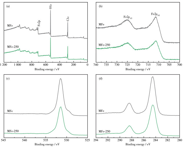
XPS was employed to determine the surface com- position and element valence of the samples (Fig. 2). The XPS survey spectra for both MFe and MFe-250 confirmed the presence of Fe, O, and C atoms, and did not contain peaks corresponding to any distinct impuri- ties (Fig. 2a). The results indicate a similarity in the composition and structure of MFe and MFe-250. The Fe2p3/2 and Fe2p1/2 XPS peaks for MFe were located at 711.39 and 725.09 eV, respectively[35], whereas those of MFe-250 were located at 711.34 and 724.84 eV, respectively (Fig. 2b). The close similarity between these values indicates that MFe-250 structure did not collapse. In the XPS O1s spectrum of MFe (Fig. 2c), the main peak corresponding to the carboxyl group of the terephthalic acid (H2BDC) linkers and the Fe—O bonds was located at 531.40 eV[40]. In contrast, the corresponding O1s peak for MFe-250 shifted to 531.48 eV because some Fe—O bonds remain after decarbox- ylation[40]. The high-resolution C1s XPS spectrum of MFe (Fig. 2d) exhibited two peaks with binding ener- gies of 288.43 and 284.50 eV, corresponding to the Fe- carboxylate moiety and benzoic acid linker, respective- ly[39]. The decrease in the intensity of the C1s peak in the spectrum for MFe-250 further indicates the heat- induced loss of organic ligands. Hence, the thermal treatment led to MFe decarboxylation and the corre- sponding generation of metal oxides, as revealed by the changes in the XPS peak intensity and position.
The MB degradation activities of MFe-T catalysts were evaluated at 25 ℃ in a jacketed glass reactor con- taining photocatalyst, H2O2, and MB in aqueous under visible light irradiation. Fig. 3a shows the photodegrada- tion rate of MFe, MFe-100, MFe-200, MFe-250, MFe- 300, and MFe-400; all samples exhibited catalytic activities towards MB degradation. Under the same con- ditions, non-calcined MFe showed a more general cata- lytic performance, degrading 88% of the MB within 90 min. The photocatalytic activity of the sample increased when the calcination temperature was raised from 100 to 250 ℃ owing to the generation of more structural defects. Hence, MFe-200 and MFe-250 photodegraded 97% and 99% of MB, respectively, within 90 min, sug- gesting that the Fe—O sites play a critical role in the process. Among the tested catalysts, MFe-250 exhibit- ed the highest photodegradation rate (99% within 90 min), suggesting that it is a novel and effective photo- catalyst for the degradation of MB under visible light. In contrast, MFe-300 and MFe-400 photodegraded 23% and 16% of MB, respectively, within the same 90 min (Fig. 3a). Therefore, it can be said that the photocat- alytic activity is proportional to the number of Fe—O sites, and reaches a maximum for MFe-250. As the py- rolysis temperature increased further, conversion of MFe structure to Fe2O3 was accompanied by a decrease in the catalytic activity. A further increase in the calci- nation temperature causes the collapse of MFe frame- work[41]. In the cases studied, Fe2O3 photocatalysts exhibited significantly lower MB degradation activity compared to pristine MFe. Hence, the MFe framework is necessary for a high photocatalytic MB degradation performance.
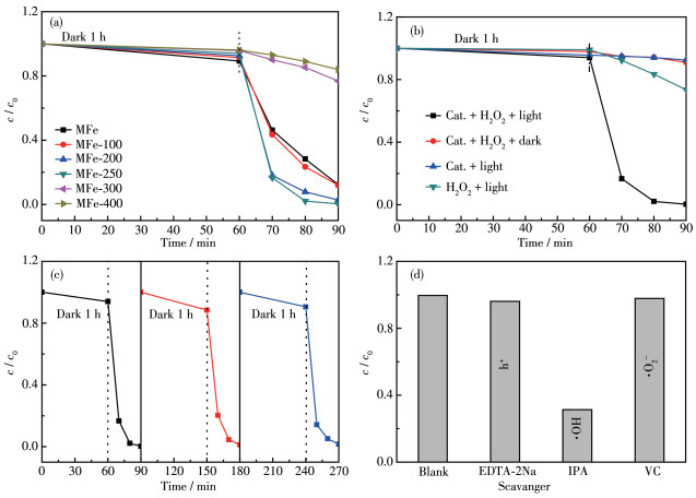
Control experiments were conducted to verify the photocatalytic degradation properties of MFe-250. Fig. 3b shows the concentration (c/c0) of MB in different photocatalytic degradation systems. In the absence of catalyst and H2O2, no degradation of MB under irradia- tion was observed[42]. The addition of H 2O2 to the MB solutions led to 27% degradation within 90 min of visi- ble-light irradiation. This resulted from the light- induced formation of ·OH upon the irradiation of H2O2. When only MFe-250 and visible light alone was em- ployed, 8% MB was degraded. This suggested an H2O2- dependence of the photoactivity of the catalyst. In the dark, a mixture of MFe-250 and H2O2 only led to a deg- radation rate of 9% MB within 90 min. Furthermore, 99% MB was degraded by using MFe-250 and H2O2 under visible light irradiation. Additionally, the chang- es in the UV-visible spectra of MB solution containing MFe-250 and H2O2 under visible-light irradiation (Fig. S6) corroborate the high catalytic performance of MFe- 250 in the presence of H2O2.
Catalyst stability is pivotal to the choice of practi- cal applications in photocatalysis. The stability of MFe- 250 was assessed by recycling it during several experi- ments. After each reaction, the recovered catalyst was washed with H2O and EtOH to remove adsorbed impu- rities, after which it was dried at 60 ℃. The same amount of sample was then added to the next experi- ment, which was conducted under the same conditions as the previous one. As shown in Fig. 3c, nearly no loss of activity was observed in MFe-250, even over three cycles. The stability of MFe-250 was confirmed by PXRD analysis, where the same results were obtained for both, the as-prepared and the recovered catalysts (Fig. S7). Hence, MFe-250 catalyst is both recyclable and stable under photocatalytic reaction conditions.
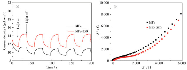
The origin of the excellent photocatalytic activity of MFe-250 was obtained from the photocurrent and EIS results. The photocurrent intensity of MFe-250 was almost twice that of MFe (Fig. 4a). Meanwhile, EIS, which reflects the charge separation capability of a pho- tocatalyst, revealed a higher light-induced charge sepa- ration rate in MFe-250 than in MFe. The Nyquist arc radius of MFe-250 was smaller than that of MFe (Fig. 4b), indicating a corresponding smaller charge/ electron transfer resistance in MFe-250 compared to that in MFe[2]. Therefore, the high charge separation efficiency of MFe-250 can be attributed to its low elec- tron transfer resistance. Thus, the thermal treatment improved the charge separation efficiency of MFe and ultimately enhanced its photocatalytic performance.
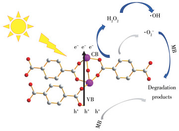
Hydroxyl radicals (·OH) play an important role in the photocatalytic degradation of MB[42]. Isopropanol (IPA) was used as a scavenger to detect ·OH generated at the surface of MFe-250[43]. To gain a deeper under- standing of the reaction mechanism, other scavengers such as EDTA-2Na[44-45] and VC[46] were used to detect the formation of photoexcited holes (h+) and superoxide radicals (·O2-) during the MB degradation experiments. Fig. 3d reveals a decrease in the MB photodegradation rate of MFe-250 from 99% to 96% following the addi- tion of EDTA-2Na. When VC was added, the photodeg- radation rate was 98%, whereas the addition of IPA sig- nificantly decreased the photodegradation rate to 31%, indicating that IPA greatly influences the degradation reaction. Hence, ·OH plays a crucial role in the photo- catalytic degradation of MB, whereas h+ and ·O2-are less important. Based on these results, a plausible mechanism for the photocatalytic degradation of MB was proposed (Fig. 5).
MFe-based quasi-MOF materials were synthe- sized by the solvothermal method to investigate the effect of the calcination temperature on their photocata- lytic activity towards MB degradation. Among the test- ed samples, MFe-250 showed the best efficiency in the photocatalytic degradation of MB. Furthermore, it exhibited high chemical stability. To study the reaction mechanisms involved in the process, a series of control experiments were conducted. MFe was synthesized and calcined at different temperatures in air. The results showed that most of MFe porous structure remained intact in MFe-250, which exhibited the best photocata- lytic degradation efficiency on MB among all tested ma- terials. According to the photocurrent and EIS results, the electronic transmission capability of MFe-250 exceeded that of MFe. The improvement in the photo- catalytic performance stems from the enhanced expo- sure of active sites and preservation of the framework. The porous structure favours the absorption of MB mol- ecules, whereas the exposed active sites promote photo- catalytic activity. In addition, scavenging experiments revealed that hydroxyl radicals are the main active spe- cies, whereas photoexcited holes and superoxide radi- cals are far less important. This study will likely inspire future designs and preparation methods of envi- ronmentally friendly photocatalysts.
Supporting information is available at http://www.wjhxxb.cn
Xu J X, Gao J Y, Liu Y, Li Q Y, Wang L. Fabrication of In2O3/Co3O4-Palygorskite Composites by the Pyrolysis of In/Co-MOFs for Efficient Degradation of Methylene Blue and Tetracycline[J]. Mater. Res. Bull., 2017, 91: 1-8. doi: 10.1016/j.materresbull.2017.03.018
Liu N, Huang W Y, Tang M Q, Yin C C, Gao B, Li Z M, Tang L, Lei J Q, Cui L F, Zhang X D. In-Situ Fabrication of Needle-Shaped MIL-53(Fe) with 1T-MoS2 and Study on Its Enhanced Photocatalytic Mechanism of Ibuprofen[J]. Chem. Eng. J., 2019, 359: 254-264. doi: 10.1016/j.cej.2018.11.143
Yi F Y, Zhang R, Wang H, Chen L F, Han L, Jiang H L, Xu Q. Metal-Organic Frameworks and Their Composites: Synthesis and Electrochemical Applications[J]. Small Methods, 2017, 1: 1700187. doi: 10.1002/smtd.201700187
Fang X, Zong B Y, Mao S. Metal-Organic Framework-Based Sensors for Environmental Contaminant Sensing[J]. Nano-Micro Lett., 2018, 10: 64. doi: 10.1007/s40820-018-0218-0
Chen L Y, Xu Q. Metal-Organic Framework Composites for Catalysis[J]. Matter, 2019, 1: 57-89. doi: 10.1016/j.matt.2019.05.018
Li H, Wang K C, Sun Y J, Lollar C T, Li J T, Zhou H C. Recent Advances in Gas Storage and Separation Using Metal-Organic Frameworks[J]. Mater. Today, 2018, 21: 108-121. doi: 10.1016/j.mattod.2017.07.006
Lu L L, Wu B Y, Shi W, Cheng P. Metal-Organic Framework-Derived Heterojunctions as Nanocatalysts for Photocatalytic Hydrogen Production[J]. Inorg. Chem. Front., 2019, 6: 3456-3467. doi: 10.1039/C9QI00964G
Liang Z B, Zhao R, Qiu T J, Zou R Q, Xu Q. Metal-Organic Framework-Derived Materials for Electrochemical Energy Applications[J]. EnergyChem, 2019, 1: 100001. doi: 10.1016/j.enchem.2019.100001
Li D D, Xu H Q, Jiao L, Jiang H L. Metal-Organic Frameworks for Catalysis: State of the Art, Challenges, and Opportunities[J]. EnergyChem, 2019, 1: 100005. doi: 10.1016/j.enchem.2019.100005
Meyer K, Ranocchiari M, Bokhoven J A V. Metal Organic Frameworks for Photo-Catalytic Water Splitting[J]. Energy Environ. Sci., 2015, 8: 1923-1937. doi: 10.1039/C5EE00161G
Zhu J J, Li P Z, Guo W H, Zhao Y L, Zou R Q. Titanium-Based Metal-Organic Frameworks for Photocatalytic Applications[J]. Coord. Chem. Rev., 2018, 359: 80-101. doi: 10.1016/j.ccr.2017.12.013
Shi Y, Yang A F, Cao C S, Zhao B. Applications of MOFs: Recent Advances in Photocatalytic Hydrogen Production from Water[J]. Coord. Chem. Rev., 2019, 390: 50-75. doi: 10.1016/j.ccr.2019.03.012
Nandasiri M I, Jambovane S R, McGrail B P, Schaef H T, Nune S K. Adsorption, Separation, and Catalytic Properties of Densified Metal-Organic Frameworks[J]. Coord. Chem. Rev., 2016, 311: 38-52. doi: 10.1016/j.ccr.2015.12.004
刘志强, 黄永清, 孙为银. 金属有机框架化合物对溶剂分子和有机小分子荧光识别与传感研究进展[J]. 无机化学学报, 2017,33,(11): 1959-1969. doi: 10.11862/CJIC.2017.244LIU Z Q, HUANG Y Q, SUN W Y. Progress in Fluorescent Recognition and Sensing of Solvent and Small Organic Molecules Based on Metal-Organic Frameworks[J]. Chinese J. Inorg. Chem., 2017, 33(11): 1959-1969. doi: 10.11862/CJIC.2017.244
李玉玲, 赵越, 孙为银. 混合配体构筑的两个锌(Ⅱ)和镉(Ⅱ)的金属有机框架化合物的合成、晶体结构及吸附和荧光性质[J]. 无机化学学报, 2020,36,(6): 1176-1184. LI Y L, ZHAO Y, SUN W Y. Two Zn (Ⅱ) and Cd (Ⅱ) Metal-Organic Frameworks with Mixed Ligands: Synthesis, Structure, Sorption and Luminescent Properties[J]. Chinese J. Inorg. Chem., 2020, 36(6): 1176-1184.
Fan K, Jin Z L, Yuan H, Hu H Y, Bi Y P. Construction of CuO-Modified Zeolitic Imidazolate Framework-9 for Photocatalytic Hydrogen Evolution[J]. Chin. J. Catal., 2017, 38: 2056-2066. doi: 10.1016/S1872-2067(17)62969-3
Hou Q Q, Wu Y, Zhou S, Wei Y Y, Caro J, Wang H H. Ultra-Tuning of the Aperture Size in Stiffened ZIF-8_Cm Frameworks with Mixed-Linker Strategy for Enhanced CO2/CH4 Separation[J]. Angew. Chem. Int. Ed., 2019, 58: 327-331. doi: 10.1002/anie.201811638
Liu S J, Liu J D, Hou X D, Xu T T, Tong J, Zhang J X, Ye B J, Liu B. Porous Liquid: A Stable ZIF-8 Colloid in Ionic Liquid with Permanent Porosity[J]. Langmuir, 2018, 34: 3654-3660. doi: 10.1021/acs.langmuir.7b04212
Wu Y P, Tian J W, Liu S, Li B, Zhao J, Ma L F, Li D S, Lan Y Q, Bu X H. Bi-Microporous Metal-Organic Frameworks with Cubane[M4(OH)4] (M=Ni, Co) Clusters and Pore-Space Partition for Electrocatalytic Methanol Oxidation Reaction[J]. Angew. Chem. Int. Ed., 2019, 58: 12185-12189. doi: 10.1002/anie.201907136
Liu S, Wang X, Yu H G, Wu Y P, Li B, Lan Y Q, Wu T, Zhang J, Li D S. Two New Pseudo-Isomeric Nickel (Ⅱ) Metal-Organic Frameworks with Efficient Electrocatalytic Activity Toward Methanol Oxidation[J]. Rare Met., 2021, 40: 489-498. doi: 10.1007/s12598-020-01596-x
Huang D D, Wu X Q, Tian J W, Wang X K, Zhou Z H, Li D S. Assembling of a Novel 3D Ag(Ⅰ)-MOFs with Mixed Ligands Tactics: Syntheses, Crystal Structure and Catalytic Degradation of Nitrophenol[J]. Chin. Chem. Lett., 2018, 29: 845-848. doi: 10.1016/j.cclet.2017.09.043
Xamena F X, Corma A, Garcia H. Applications for Metal-Organic Frameworks (MOFs) as Quantum Dot Semiconductors[J]. J. Phys. Chem. C, 2007, 111: 80-85. doi: 10.1021/jp063600e
Choi K M, Kim D, Rungtaweevoranit B, Trickett C A, Barmanbek J T D, Alshammari A S, Yang P D, Yaghi O M. Plasmon-Enhanced Photocatalytic CO2 Conversion within Metal-Organic Frameworks under Visible Light[J]. J. Am. Chem. Soc., 2017, 139: 356-362. doi: 10.1021/jacs.6b11027
Lan G X, Li Z, Veroneau S S, Zhu Y Y, Xu Z W, Wang C, Lin W B. Photosensitizing Metal-Organic Layers for Efficient Sunlight-Driven Carbon Dioxide Reduction[J]. J. Am. Chem. Soc., 2018, 140: 12369-12373. doi: 10.1021/jacs.8b08357
Li R, Wu S K, Wan X Y, Xu H X, Xiong Y J. Cu/TiO2 Octahedral-Shell Photocatalysts Derived from Metal-Organic Framework@Semiconductor Hybrid Structures[J]. Inorg. Chem. Front., 2016, 3: 104-110. doi: 10.1039/C5QI00205B
Su Y, Ao D, Liu H, Wang Y. MOF-Derived Yolk-Shell CdS Microcubes with Enhanced Visible-Light Photocatalytic Activity and Stability for hydrogen Evolution[J]. J. Mater. Chem. A, 2017, 5: 8680-8689. doi: 10.1039/C7TA00855D
Liu S J, Zhang C, Sun Y D, Chen Q, He L F, Zhang K, Zhang J, Liu B, Chen L F. Design of Metal-Organic Framework-Based Photocatalysts for Hydrogen Generation[J]. Coord. Chem. Rev., 2020, 413: 213266. doi: 10.1016/j.ccr.2020.213266
Zhang W, Wang Y, Ling L J, Wang X P, Chang H J, Li R, Duan W B, Liu B. Utilizing Crystals Defects to Boost Metal-Organic Frameworks Hydrogen Generation Abilities[J]. Microporous Mesoporous Mater., 2020, 294: 109943. doi: 10.1016/j.micromeso.2019.109943
Liang Q, Jin J, Liu C H, Xu S, Yao C, Li Z Y. Fabrication of the Ternary Heterojunction Cd05Zn0.5S@UIO-66@g-C3N4 for Enhanced Visible-Light Photocatalytic Hydrogen Evolution and Degradation of Organic Pollutants[J]. Inorg. Chem. Front., 2018, 5: 335-343. doi: 10.1039/C7QI00638A
Gong Y, Zhao X, Zhang H, Yang B, Xiao K, Guo T, Zhang J J, Shao H X, Wang Y B, Yu G. MOF-Derived Nitrogen Doped Carbon Modified g-C3N4 Heterostructure Composite with Enhanced Photocatalytic Activity for Bisphenol A Degradation with Peroxymonosulfate under Visible Light Irradiation[J]. Appl. Catal. B, 2018, 233: 35-45. doi: 10.1016/j.apcatb.2018.03.077
Yuan C, Cheng P F, Li J, Gao X L, Gao X S, Wang X, Jin M L, Nötzel R, Zhou G F, Zhang Z, Liu J M. ZIF-67 with Argon Annealing tReatment for Visible Light Responsive Degradation of Organic Dyes in a Wide pH Range[J]. Microporous Mesoporous Mater., 2019, 285: 13-20. doi: 10.1016/j.micromeso.2019.04.062
Liu S J, Zou Q C, Ma Y, Sun W, Li Y, Zhang J, Zhang C, He L F, Sun Y D, Chen Q, Liu B, Zhang H X, Zhang K. A Novel Amorphous CoSx/NH2-MIL-125 Composite for Photocatalytic Degradation of Rhodamine B under Visible Light[J]. J. Mater. Sci., 2020, 55: 16171-16183. doi: 10.1007/s10853-020-05210-4
Du Y, Chen R Z, Yao J F, Wang H T. Facile Fabrication of Porous ZnO by Thermal Treatment of Zeolitic Imidazolate Framework-8 and Its Photocatalytic Activity[J]. J. Alloys Compd., 2013, 551: 125-130. doi: 10.1016/j.jallcom.2012.10.045
Zhang Y, Zhou J B, Chen X, Feng Q Q, Cai W Q. MOF-Derived C-Doped ZnO Composites for Enhanced Photocatalytic Performance under Visible Light[J]. J. Alloys Compd., 2019, 777: 109-118. doi: 10.1016/j.jallcom.2018.10.383
Ma X, Wang L, Zhang Q, Jiang H L. Switching on the Photocatalysis of Metal-Organic Frameworks by Engineering Structural Defects[J]. Angew. Chem. Int. Ed., 2019, 58: 12175-12179. doi: 10.1002/anie.201907074
Ai L H, Zhang C H, Li L L, Jiang J. Iron Terephthalate Metal-Organic Framework: Revealing the Effective Activation of Hydrogen Peroxide for the Degradation of Organic Dye under Visible Light Irradiation[J]. Appl. Catal. B, 2014, 148-149: 191-200. doi: 10.1016/j.apcatb.2013.10.056
Tsumori N, Chen L Y, Wang Q J, Zhu Q L, Kitta M, Xu Q. Quasi-MOF: Exposing Inorganic Nodes to Guest Metal Nanoparticles for Drastically Enhanced Catalytic Activity[J]. Chem, 2018, 4: 845-856. doi: 10.1016/j.chempr.2018.03.009
Lin R B, Li S M, Wang J Y, Xu J P, Xu C H, Wang J, Li C X, Li Z Q. Facile Generation of Carbon Quantum Dots in MIL-53(Fe) Particles as Localized Electron Acceptors for Enhancing Their Photocatalytic Cr(Ⅵ) Reduction[J]. Inorg. Chem. Front., 2018, 5: 3170-3177.
Hu L X, Deng G H, Lu W C, Pang S W, Hu X. Deposition of CdS Nanoparticles on MIL-53(Fe) Metal-Organic Framework with Enhanced Photocatalytic Degradation of RhB under Visible Light Irradiation[J]. Appl. Surf. Sci., 2017, 410: 401-413. doi: 10.1016/j.apsusc.2017.03.140
Guo T, Wang K, Zhang G K, Wu X Y. A Novel α-Fe2O3@g-C3N4 Catalyst: Synthesis Derived from Fe-Based MOF and Its Superior Photo-Fenton Performance[J]. Appl. Surf. Sci., 2019, 469: 331-339. doi: 10.1016/j.apsusc.2018.10.183
Xu W K, Xue W J, Huang H L, Wang J S, Zhong C L, Mei D H. Morphology Controlled Synthesis of α-Fe2O3-x with Benzimidazole-Modified Fe-MOFs for Enhanced Photo-Fenton-like Catalysis[J]. Appl. Catal. B, 2021, 291: 120129. doi: 10.1016/j.apcatb.2021.120129
Li X Y, Pi Y H, Xia Q B, Li Z, Xiao J. TiO2 Encapsulated in Salicylaldehyde-NH2-MIL-101(Cr) for Enhanced Visible Light-Driven Photodegradation of MB[J]. Appl. Catal. B, 2016, 191: 192-201. doi: 10.1016/j.apcatb.2016.03.034
Yuan X Z, Wang H, Wu Y, Zeng G M, Chen X H, Leng L J, Wu Z B, Li H. One-Pot Self-Assembly and Photoreduction Synthesis of Silver Nanoparticle-Decorated Reduced Graphene Oxide/MIL-125(Ti) Photocatalyst with Improved Visible Light Photocatalytic Activity[J]. Appl. Organomet. Chem., 2016, 30: 289-296. doi: 10.1002/aoc.3430
Wang H, Yuan X Z, Wu Y, Zeng G M, Chen X H, Leng L J, Li H. Synthesis and Applications of Novel Graphitic Carbon Nitride/Metal-Organic Frameworks Mesoporous Photocatalyst for Dyes Removal[J]. Appl. Catal. B, 2015, 174-175: 445-454. doi: 10.1016/j.apcatb.2015.03.037
Wang H, Yuan X Z, Wu Y, Zeng G M, Chen X H, Leng L J, Wu Z B, Jiang L B, Li H. Facile Synthesis of Amino-Functionalized Titanium Metal-Organic Frameworks and Their Superior Visible-Light Photocatalytic Activity for Cr(Ⅵ) Reduction[J]. J. Hazard. Mater., 2015, 286: 187-194. doi: 10.1016/j.jhazmat.2014.11.039
Wang Q, Wang W, Zhong L L, Liu D M, Cao X Z, Cui F Y. Oxygen Vacancy-Rich 2D/2D BiOCl-g-C3N4 Ultrathin Heterostructure Nanosheets for Enhanced Visible-Light-Driven Photocatalytic Activity in Environmental Remediation[J]. Appl. Catal. B, 2018, 220: 290-302. doi: 10.1016/j.apcatb.2017.08.049

Figure 3 (a) Photocatalytic degradation of MB under visible-light irradiation over different samples; (b) Degradation rate of MFe-250 on MB under different conditions; (c) MB removal in the repeated tests over as-prepared MFe-250;(d) Photocatalytic degradation of MB over MFe-250 under visible light irradiation in the presence of different scavengers

 扫一扫看文章

扫一扫关注我们
 下载:
下载:

 下载:
下载:


