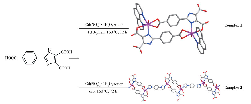
Scheme 1.
Synthesis routes of complexes 1 and 2
1,10-phen=1,10-phenanthroline, dib=1,4-bis(1-imidazolyl)benzene
Synthesis, Structure Regulation and Characterization of Cadmium(Ⅱ) Complexes Based on Imidazole Carboxylic Acid Ligands
Ding-Qi XIONG , Peng-Kui FU , Yu-Yan LI , Xiao-Yu ZHANG , Qing-Lin YANG , Mei-Mei JIA , Yan-Yan ZHU , Xiu-Yan DONG

In recent decades, complex has attracted lots of attention, due to its multi-functional crystalline materials and interesting structure, which are based on the coordination bonding interaction between metal ions/ clusters and bridging organic linkers. They stand out among polymer materials owing to their brilliant properties, for instance, large surface area[1], adjustable structure[2-3], and high porosity[4-5]. Considerable efforts have been made in synthesizing and researching new complexes, not only thanks to their intriguing structures and distinctive topologies, but also due to their promising applications as functional materials in many fields as luminescence[6-8], catalysis[9-11], chemical sensors[12-14], magnetism[15-18], gas storage and separation[19-21], and biology[22].
The fluorescent properties of d10 metal complexes have attracted the interest of many researchers. Especially, the d orbital of Cd(Ⅱ) ion was filled with electrons, which can effectively reduce the energy loss caused by d - d transitions, when connected to a π conjugated organic framework. It may exhibit good fluorescence properties[23-24].
In this work, we used 2-(4-carboxy-phenyl)-imidazole-4, 5-dicarboxylic acid (H3L) to connect with Cd(Ⅱ) and added auxiliary ligands to adjust the structure to construct complexes, The sp2 hybridization of N on the ring can increase the π electronic density. The introduction of a functional carboxyphenyl group at 2 position of the imidazole ring (H3L) can produce more coordination modes[25-26]. Both the O on the carboxyl group and the N on the imidazole can coordinate with Cd(Ⅱ), and N and O can form chelating coordination effect. So, H3L shows more advantages than other N- or Odonor ligands. Here, under the same conditions, we synthesized two new Cd(Ⅱ) complexes (Scheme 1) through introduce auxiliary ligand, and studied their properties.
1,10-phen=1,10-phenanthroline, dib=1,4-bis(1-imidazolyl)benzene
H3L was prepared according to the reported procedure[27-28]. All chemical reagents and solvents were purchased from a commercial source, which was used without undergoing the purification process. The FT - IR spectra were recorded in a range of 4 000-400 cm-1 on a Bruker VERTEX 70 spectrometer using KBr pellets. Elemental analyses (C, H, and N) were carried out on a VxRio EL elemental analyzer. Powder X-ray diffraction (PXRD) patterns were collected in a 2θ range of 5°-45° on a Philips PW 1710-based diffractometer with Cu Kα radiation (λ=0.154 184 nm) at room temperature, operated at 40 kV and 100 mA. Thermogravimetric analysis (TGA) was performed on a PerkinElmer TG-7 analyzer heated from room temperature to 800 ℃ under nitrogen at a heating rate of 10 ℃ ·min-1. Fluorescent analyses of the complexes were performed on an F-7100 Fluorescence spectrometer. UV - Vis DRS (UV - Vis Diffuse Reflectance Spectroscopy) spectra were recorded on a U-3900H spectrophotometer.
A mixture of Cd(NO3)2·4H2O (30.8 mg, 0.10 mmol), H3L (13.8 mg, 0.05 mmol), 1, 10-phen (9.9 mg, 0.05 mmol), and H2O (8 mL) was placed in a 25 mL Teflon - lined autoclave and heated to 160 ℃ for 3 d. When the mixture was cooled to room temperature, light yellow block - shaped crystals of 1 were obtained with 65% yield (based on H3L). Analysis Calcd. for C48H32Cd2N8O14(%): C 49.29, H 2.75, N, 9.58; Found (%): C 49.33, H 2.76, N 9.51. IR (KBr, cm-1): 3 428(s), 2 989(m), 2 826(w), 2 355(w), 2 066(w), 1 620(s), 1 486 (m), 1 395(s), 1 365(s), 1 176(m), 1 005(m), 790(m), 728(w), 621(m).
The synthesis of 2 used the same condition as 1 except that 1, 10-phen was replaced by dib. Clear colorless block crystals of 2 were obtained in 63% yield (based on H3L). Analysis Calcd. for C18H18CdN4O10(%): C 38.41, H 3.22, N 9.96; Found(%): C 38.43, H 3.23, N 9.99. IR (KBr, cm-1): 3 420(s), 2 985(m), 2 831(w), 2 716(w), 2 362(w), 2 062(w), 1 620(s), 1 490(m), 1 422 (m), 1 398(m), 1 365(s), 1 175(m), 1 049(w), 1 005(m), 882(w), 793(m).
The diffraction data were collected at 296(2) K for 1, with a Bruker APEX-Ⅱ CCD area detector diffractometer using φ and ω rotation scans and Mo Kα radiation (λ=0.071 073 nm). The crystallographic data of 2 were collected with Cu Kα radiation (λ=0.154 184 nm) on SuperNova, Dual, Cu at zero, Eos at 296(2) K. Then absorption corrections were carried out. The structures were solved by the direct method and refined by the full - matrix least - squares on F2 using the SHELX and Olex2 program[29-31]. The data of 1 were processed by squeeze to remove disordered solvent water molecules. Non-hydrogen atoms were refined with anisotropic, and the hydrogen atoms were included in the final refinement by using geometrical restrains and refined isotropically using the riding model. Crystal data and structure refinements for 1 and 2 and selected bond distances and angles are given in Table 1 and 2, respectively.
 下载:
导出CSV
下载:
导出CSV
| Parameter | Complex | |
| 1 | 2 | |
| Chemical formula | C48H32Cd2N8O14 | C18H19CdN4O10 |
| Formula weight | 1 169.61 | 563.77 |
| Crystal system | Triclinic | Triclinic |
| Space group | P1 | P1 |
| a / nm | 0.864 41(5) | 0.881 5(4) |
| b / nm | 1.063 69(5) | 1.134 57(4) |
| c / nm | 1.449 67(8) | 1.139 38(5) |
| α/(°) | 105.947(2) | 104.252(3) |
| β/(°) | 91.012(2) | 100.969(4) |
| γ/(°) | 100.591(2) | 95.618(3) |
| V / nm3 | 1.256 51(12) | 1.072 20(8) |
| Z | 1 | 2 |
| Dc / (g·cm-3) | 1.546 | 1.746 |
| μ / mm-1 | 0.919 | 8.741 |
| θ range / (°) | 4.062-54.104 | 8.212-133.172 |
| Crystal size / mm | 0.17×0.14×0.11 | 0.21×0.17×0.15 |
| Rint | 0.023 0 | 0.027 9 |
| F(000) | 584.0 | 566.0 |
| GOF | 1.058 | 1.048 |
| R1a, wR2b [I > 2σ(I)] | 0.029 3, 0.070 1 | 0.037 0, 0.098 8 |
| R1, wR2 (all data) | 0.033 7, 0.072 5 | 0.038 5, 0.101 0 |
| aR1=∑||Fo|-|Fc||/∑|Fo|; bwR2=[∑w(Fo2-Fc2)2/∑w(Fo2)2]1/2. | ||
下载:
导出CSV
| 1 | |||||
| Cd1—O5 | 0.231 60(18) | Cd1—N4 | 0.235 7(2) | Cd1—O6 | 0.256 81(18) |
| Cd1—O7 | 0.231 66(19) | Cd1—N3 | 0.235 9(2) | ||
| Cd1—N2#1 | 0.233 64(19) | Cd1—O2#1 | 0.247 43(17) | ||
| O5—Cd1—O7 | 85.58(7) | O7—Cd1—N3 | 85.35(9) | N3—Cd1—O2#1 | 81.49(7) |
| O5—Cd1—N2#1 | 115.68(7) | N2#1—Cd1—N3 | 150.92(8) | O5—Cd1—O6 | 53.37(6) |
| O7—Cd1—N2#1 | 85.13(7) | N4—Cd1—N3 | 70.69(10) | O7—Cd1—O6 | 128.90(7) |
| O5—Cd1—N4 | 107.76(7) | O5—Cd1—O2#1 | 164.70(6) | N2#1—Cd1—O6 | 87.26(6) |
| O7—Cd1—N4 | 152.34(8) | O7—Cd1—O2#1 | 80.63(7) | N4—Cd1—O6 | 76.70(7) |
| N2#1—Cd1—N4 | 109.07(8) | N2#1—Cd1—O2#1 | 69.87(6) | N3—Cd1—O6 | 119.73(8) |
| O5—Cd1—N3 | 90.86(8) | N4—Cd1—O2#1 | 82.29(7) | O2#1—Cd1—O6 | 141.79(6) |
| 2 | |||||
| Cd1—O0AA | 0.237 1(3) | Cd1—O6#1 | 0.229 8(3) | Cd1—N3 | 0.224 7(3) |
| Cd1—O1 | 0.246 3(3) | Cd1—O7 | 0.246 2(4) | ||
| Cd1—O5#1 | 0.261 6(4) | Cd1—N1 | 0.233 3(3) | ||
| O0AA—Cd1—O1 | 79.17(11) | O6#1—Cd1—O7 | 80.07(13) | N1—Cd1—O7 | 88.32(12) |
| O0AA—Cd1—O5#1 | 75.50(12) | O6#1—Cd1—N1 | 95.75(11) | N3—Cd1—O0AA | 94.83(12) |
| O0AA—Cd1—O7 | 162.35(13) | O7—Cd1—O1 | 83.51(13) | N3—Cd1—O1 | 82.30(11) |
| O1—Cd1—O5#1 | 149.07(11) | O7—Cd1—O5#1 | 122.02(13) | N3—Cd1—O5#1 | 82.44(14) |
| O6#1—Cd1—O0AA | 115.80(11) | N1—Cd1—O0AA | 82.62(10) | N3—Cd1—O6#1 | 109.97(12) |
| O6#1—Cd1—O1 | 158.63(12) | N1—Cd1—O1 | 70.07(10) | N3—Cd1—O7 | 86.16(14) |
| O6#1—Cd1—O5#1 | 52.17(12) | N1—Cd1—O5#1 | 122.96(13) | N3—Cd1—N1 | 152.27(11) |
| Symmetry codes: #1: 1-x, 1-y, 1-z for 1; #1: 2-x, 2-y, 2-z; #2: 1-x, 1-y, -z for 2. | |||||
CCDC: 2044127, 1; 2044128, 2.
Single X - ray crystallography shows that 1 based on H3L, 1, 10-phen, and Cd(Ⅱ) crystallizes in the triclinic system and P1 space group. The asymmetric unit of 1 contains two crystallographically independent Cd(Ⅱ) cation, two (HL)2- ligands, two 1, 10 - phen ligand, and two coordinated water molecules. As depicted in Fig. 1a, the Cd1 center is coordinated by three nitrogen atoms of one 1, 10 - phen ligand (N3 and N4) and one (HL)2- ligand (N2#1), four oxygen atoms of two different (HL)2- ligands (O2#1, O5, and O6), and one coordinated H2O (O7), adopting a distorted pentagonal bipyramid geometry. Two main ligands and two metal ions form a ring structure and then coordinate with the two chelating auxiliary ligands, and the remaining N and O of the ligand are not further coordinated with Cd which leads to a 0D structure (Fig. 1b). Intermolecular hydrogen bonding interactions (O—H…O, Table 3) between a coordinated water molecule and the carboxylate of (HL)2- ligands and a C—H… π interaction (Table 4) between the benzene ring and the imidazole ring of (HL)2- ligands are showed in Fig. 1c. The interaction of O—H…O and C—H…π (Fig. 1c, 1d), the π…π stacking interactions(Table 5) between benzene rings with the aid of 1, 10-phen (Fig. 1e) form supramolecular framework structure of complex 1 (Fig. 1f).
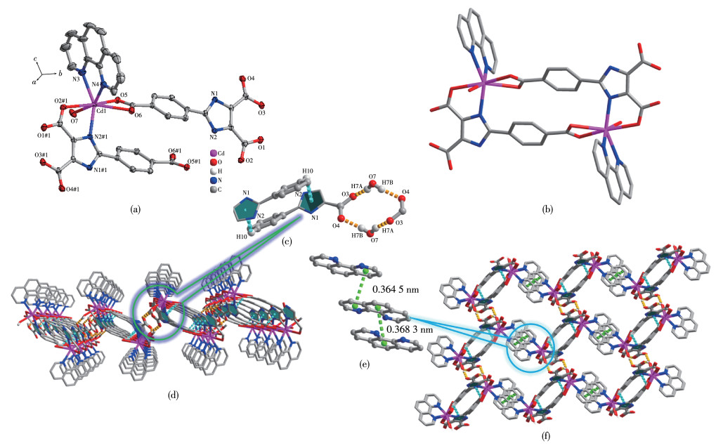
Cd: purple, N: blue, O: red, C: gray; Hydrogen atoms are omitted for clarity; Symmetry code: #1: 1-x, 1-y, 1-z
下载:
导出CSV
| D—H…A | d(D—H) / nm | d(H…A) / nm | d(D…A) / nm | ∠DHA / (°) |
| 1 | ||||
| O3—H3…O1 | 0.082 | 0.162 | 0.243 7(3) | 172 |
| O7—H7A…O3#2 | 0.088 | 0.184 | 0.271 1(3) | 167 |
| O7—H7B…O4#1 | 0.085 | 0.185 | 0.269 6(3) | 173 |
| C22—H2…O7 | 0.093 | 0.259 | 0.319 5(5) | 123 |
| 2 | ||||
| O0AA—H0AA…O3#2 | 0.087 | 0.202 | 0.284 6(4) | 159 |
| N2—H2…O9#4 | 0.086 | 0.200 | 0.286 1(5) | 173 |
| O2—H2…O3 | 0.090(9) | 0.155(9) | 0.245 1(5) | 173(15) |
| O0AA—H0AB…O1#1 | 0.087 | 0.201 | 0.286 5(5) | 167 |
| O0AA—H0AB…O2#1 | 0.087 | 0.251 | 0.317 5(5) | 134 |
| O7—H7A…O9#3 | 0.087 | 0.235 | 0.314 0(6) | 151 |
| O7—H7B…O8#3 | 0.087 | 0.197 | 0.278 8(8) | 156 |
| O9—H9B…O6#5 | 0.085 | 0.191 | 0.275 7(5) | 173 |
| C13—H13…O1 | 0.093 | 0.254 | 0.309 1(5) | 118 |
| C13—H13…O0AA#1 | 0.093 | 0.258 | 0.350 3(6) | 171 |
| Symmetry codes: #1: 1+x, -1+y, z; #2: -x, 1-y, 1-z for 1; #1: 1-x, 1-y, 1-z; #2: 1+x, y, z; #3: x, -1+y, z; #4: 1-x, 2-y, 2-z; #5: 2-x, 2-y, 2-z for 2. | ||||
下载:
导出CSV
| X—H…π | d(H…Cg) / nm | γ/(°) | ∠X—H…Cg / (°) | d(X…Cg) / nm | σ/(°) |
| C10—H10…Cg(1)#1 | 0.299 | 13.10 | 117 | 0.351 1(3) | 35 |
| *γ: angle between Cg-H vector and ring; σ: angle of X—H bond with π-plane (i.e., Perpendicular: 90°, Parallel: 0°); Symmetry codes: #1: -x, 1-y, 1-z; Rings: Cg(1): C2, C3, N2, C4, N1, C5. | |||||
下载:
导出CSV
| Cg…Cg | α/(°) | DC / nm | β/(°) | DZ / nm | S / nm |
| Cg(3)…Cg(3)#1 | 0.00(19) | 0.364 5(1) | 21.8 | 0.338 51(16) | 0.135 3 |
| Cg(5)…Cg(5)#2 | 0.00(2) | 0.368 3(2) | 19.4 | 0.347 3(2) | 0.122 6 |
| Cg(2)…Cg(3)#2 | 1.90(2) | 0.437 8(2) | 35.9 | 0.346 07(19) | 0.256 7 |
| Cg(4)…Cg(4)#3 | 0.00(11) | 0.459 42(15) | 47.3 | 0.311 32(10) | 0.337 8 |
| *α: dihedral angle between mean planes of the rings; DC: distance between ring centroids; β: angle between DC vector and normal to plane (i); DZ: perpendicular distance of the centroids of ring (i) on plane of ring (j); S: distance between ring centroid (i) and perpendicular projection of ring centroid (j) on ring (i); Symmetry codes: #1:2-x, 1-y, 2-z; #2:1-x, 1-y, 2-z; #3:1-x, 1-y, 1-z; Rings: Cg(2): N3, C22, C21, C20, C19 C23; Cg(3): N4, C13, C14, C15, C16, C24; Cg(4): C6, C7, C8, C9, C10, C11; Cg(5): C16, C17, C18, C19, C23, C24. | |||||
The single - crystal X - ray diffraction analysis reveals that 2 crystallizes in the triclinic crystal system with the space group of P1. The fundamental building unit of 2 consists of one crystallographically independent Cd(Ⅱ) ion, one (HL)2- ligands, half of dib ligand, and two coordinated water molecules. Cd1 adopts a distorted pentagonal bipyramid geometry, coordinating to three carboxylate oxygen atoms (O1, O5#2, and O6#2) from two different (HL)2- ligands, two water oxygen atoms (O7 and O0AA), and two nitrogen atoms from one dib ligand (N3) and one (HL)2- ligand (N1), respectively (Fig. 2a). Under the regulation of the auxiliary ligands, 2 formed a different structure from 1, but 2 has the same ring as 1, also composed of two main ligands and two metal ions. The difference between 1 and 2 is that the two coordination sites of 1, 10-phen in 1 are replaced by dib and water molecular in 2, and the dib acts as a bridging molecule to connect two rings to form a 1D chain structure (Fig. 2b). Intermolecular hydrogenbonding interactions (O—H…O and C—H…O, Talbe 3) between the coordinated water molecule and the carboxylate of (HL)2- ligand and (N—H…O, Talbe 3) between lattice water and (HL)2- are shown in Fig. 2c, and the intermolecular weak interaction forming supramolecular structure are shown in Fig. 2d.
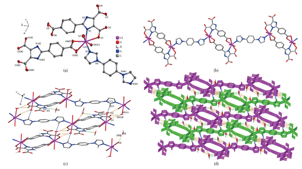
Cd: purple, N: blue, O: red, C: gray; Hydrogen atoms are omitted for clarity; Symmetry codes: #1: 1-x, 1-y, -z; #2: 2-x, 2-y, 2-z
The PXRD was carried out to test the phase purity of 1 and 2 (Fig. 3), and the experimental results are well consistent with the simulated data, which indicates the high purity of two complexes. The difference in peak intensity may be due to the different orientations of the sample.
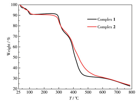
To test the thermal stability of 1 and 2, their thermal behaviors were investigated under a nitrogen atmosphere by TGA. As shown in Fig. 4, the TGA curve of 1 showed a slow weight loss near 100 ℃ which is consistent with the loss of lattice water (removed by SQUEEZE) molecules and one coordinated water molecules, and upon further heating, the structure was stable up to 275 ℃. The continuous heating up led to a decomposition. The TGA curve of 2 was similar to 1 and exhibited thermal stability up to 275 ℃, and then the structure started collapsing. The remaining weight was 23.66% (Calcd. 22.81%) at 800 ℃.
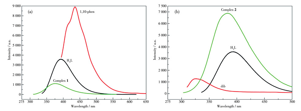
The solid - state photoluminescent properties of 1 and 2 were investigated at room temperature with the microcrystalline samples, and the luminescence spectra of free H3L, 1, 10-phen, and dib were also measured at the same condition. As shown in Fig. 5, the maximum emission of 1, 2 and ligand H3L, 1, 10-phen and dib are at 377 nm (λex=289 nm), 383 nm (λex=280 nm), 394 nm (λex=318 nm), 435 nm (λex=373 nm), 326 nm (λex=293 nm), respectively. The emission of 1 showed a blue shift relative to free 1, 10 - phen ligand (58 nm) and 2 showed a red shift relative to dib ligand (47 nm), both of them show blue shifts relative to H3L (17 nm for 1 and 11 nm for 2). Due to that the Cd(Ⅱ) ions with d10 configurations are difficult to oxidize or reduce, the emission of 1 and 2 couldn't be attributed to metal-toligand charge transfer (MLCT) or ligand-to-metal charge transfer (LMCT) [15]. Their photoluminescence could be assigned to intra-ligand fluorescence emission (π* → π) or ligand localized emission (π* →n), which are consistent with the reported Cd(Ⅱ) complexes[32-33]. The shift of the emission peaks may be ascribed to the coordination action of the ligands to Cd(Ⅱ) ions, which increases the energy between the ground state and excited state[34-35]. Although 1 and 2 have the same main ligand and center metal ions, 1 showed fluorescence quenching, while 2 showed fluorescence enhancement. The protons on the uncoordinated carboxyl group of the ligands in 1 are not removed, and the carboxyl group is used as a fluorescence quenching group, which may cause the fluorescence of 1 to be quenched[8]. The enhanced fluorescence of 2 may be due to the coordination between the ligands and the metal ion, which increases the rigidity of the ligands and reduces the non -radiative energy loss[36].
To analyze intermolecular interaction and surface electron distribution, we used crystalExplorer software to calculate the Hirshfeld surface explanation of the immediate environment of a molecile in the crystal. The 3D Hirshfeld surface and 2D fingerprint plots of 1 and 2 are shown in Fig. 6, and the 3D Hirshfeld surface was mapped with dnorm, shape index, and curvedness with standard high resolution. As shown in Fig. 6, the red regions on dnorm of 1 and 2 suggest that high electron densities may be due to the strong interaction, generally representing the formation of hydrogen bond or coordination bond; the blue regions have low electron densities and no obvious interaction; the white regions have moderate electron densities, which correspond to slightly weaker interaction, and commonly regarded as π…π stacked regions[37-38]. The shape index is an obvious indication of a subtle change of Hirshfeld's surface. The curvedness is the measurement of"how much shape"; the flat regions represent a low value of curvedness, while the sharp regions represent a high value of curvedness, indicating an interaction between adjacent molecules[39].
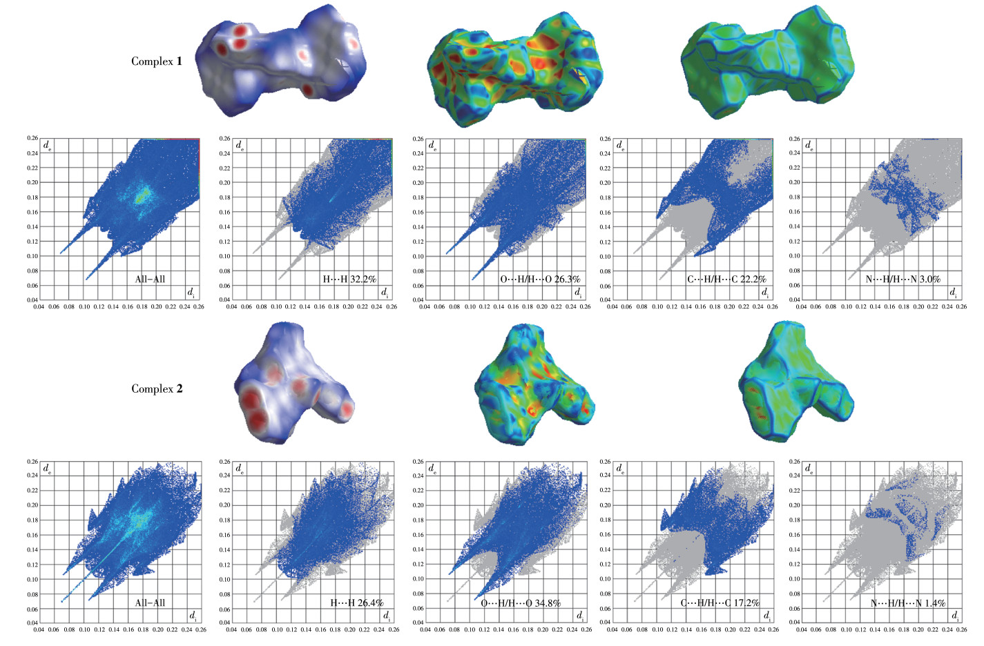
The 2D fingerprint plot can easily recognize the intermolecular interaction on the molecular surface. The C…H, O…H, N…H, and H…H interactions of 1 and 2 are shown in Fig. 6. For 1, the H…H, C…H/H… C, O…H/H…O and N…H/H…N interactions accounted for 32.2%, 22.2%, 26.3%, and 3.0% of Hirshfeld surface, respectively. For 2, the H…H, C…H/H…C, O…H/H…O and N…H/H…N interactions accounted for 26.4%, 17.2%, 34.8%, and 1.4% of Hirshfeld surface respectively. It is the interaction in 1 and 2 that makes them more stable.
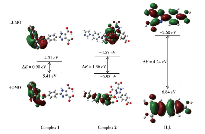
To prove the theoretical stability of 1 and 2, we applied Guassian09 software to optimize the structure and calculate the energies of 1, 2, and H3L, based on the method of density functional theory (DFT) with the level of B3LYP. Non-metal atoms were described by 631G and metal atoms were treated by LANL2DZ basis sets, which didn't consider the solvent effect[40-41]. Since the structures of 1 and 2 have been determined in the single crystal X-ray diffraction, it is possible to directly import data (CIF) and directly perform energy calculations without structural optimization.
The frontier molecular orbit energy levels, HOMO and LUMO, are two important indicators of molecular stability, where HOMO represents the ability to donate an electron, and LUMO represents the ability to accept an electron. The energy gaps (ΔE) between LUMO and HOMO represent the chemical stability of the molecule. The HOMO and LUMO levels of 1, 2, and H3L are shown in Fig. 7. The LUMO, HOMO, and ΔE of 1, 2, and H3L were -4.51, -5.41, and 0.90 eV; -4.57, -5.93, and 1.36 eV; -2.60, -6.84, and -4.24 eV, respectively. The energies of frontier orbit HOMO-LUMO are all negative, demonstrating that 1, 2, and H3L have chemical stability[42-43]. All the above indicates that 1, 2, and H3L are stable on the ground state, and the calculation method of DFT with B3LYP basis set is reasonable.
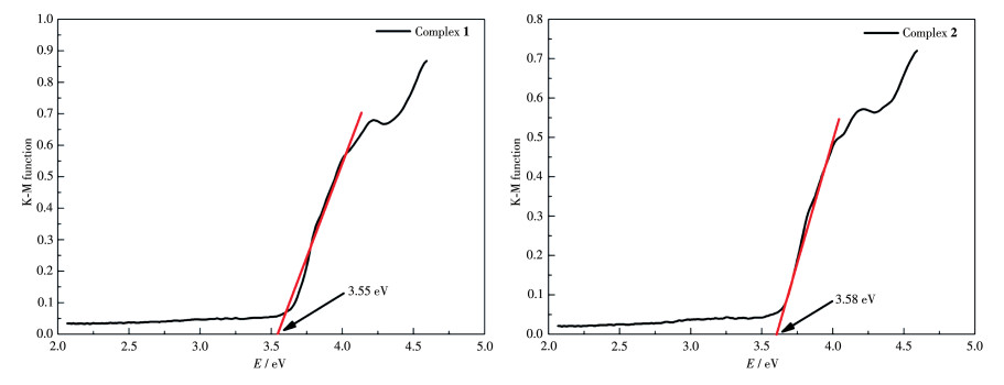
To test the candidate properties of 1 and 2 as semiconductors, the UV-Vis DRS spectra to determine the band gap (Eg) were tested (photoresponse wavelength region), based on Kubelka - Munk (K - M) function, using absorbance as the ordinate and energy as the abscissa to draw a graph and fit the linear section to obtain a linear equation. Its intercept on the x-axis is the band gap energy[44-45]. As shown in Fig. 8, Eg of 1 and 2 had similar values: 3.55 and 3.58 eV, respectively. The wide band gaps of 1 and 2 indicate that they are potential wide gap semiconductors materials[46].
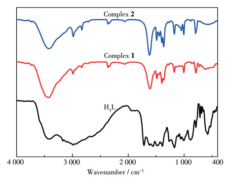
The FT-IR spectra of H3L, 1, and 2 are shown in Fig. 9. 1 and 2 had similar infrared absorption peaks to H3L. The absorption peaks at 3 420-3 430 cm-1 could be ascribed to the stretching vibration of N—H bond from H3L, and the absorption peaks between 1 400 and 1 630 cm-1 are attributed to the breathing vibrations of the aromatic ring and imidazole ring[47]. There was a strong absorption peak at 1 726 cm-1 for H3L but 1 and 2 had no absorption peak in the same region. This absorption peak could be attributed to the stretching vibration of C=O bond, and the absence of this absorption peak for 1 and 2 may be due to the coordination of the oxygen of carbonyl group with the metal ion.
In summary, two new Cd(Ⅱ) complexes were successfully synthesized by hydrothermal method. The structure analysis result shows that auxiliary ligand has an important effect on the crystal structure: bridge ligands can easier to form 1D structure, but chelating ligands tend to generate 0D structure. Through PXRD, TGA, luminescent, Hirshfeld surface analyses, and DFT calculation, we verified the phase purity of the complexes, tested their thermal stability and photoluminescence, calculated the intermolecular interaction, surface electron distribution, and the chemical stability of them. Besides, 1 and 2 showed wide optical band gaps, which indicates that they have potential application on wide band gap semiconductors materials.
Li Y L, Jin T, Ma G, Li Y C, Fan L Z, Li X H. Metal-Organic Framework Assisted and In-Situ Synthesis of Hollow CdS Nanostructures with High-Efficient Photocatalytic Hydrogen Evolution[J]. Dalton Trans., 2019, 48(17): 5649-5655. doi: 10.1039/C9DT00603F
Lu W G, Wei Z W, Gu Z Y, Liu T F, Park J, Park J, Tian J, Zhang M W, Zhang Q, Gentle T, Bosch M, Zhou H C. Tuning the Structure and Function of Metal-Organic Frameworks via Linker Design[J]. Chem. Soc. Rev., 2014, 43(16): 5561-5593. doi: 10.1039/C4CS00003J
Li N, Feng R, Zhu J, Chang Z, Bu X H. Conformation Versatility of Ligands in Coordination Polymers: From Structural Diversity to Properties and Applications[J]. Coord. Chem. Rev., 2018, 375: 558-586. doi: 10.1016/j.ccr.2018.05.016
Hönicke I M, Senkovska I, Bon V, Baburin I A, Bönisch N, Raschke S, Evans J D, Kaskel S. Balancing Mechanical Stability and Ultrahigh Porosity in Crystalline Framework Materials[J]. Angew. Chem. Int. Ed., 2018, 57(42): 13780-13783. doi: 10.1002/anie.201808240
Yuan J Q, Li J T, Che S T, Li G H, Liu X Y, Sun X D, Zou L F, Zhang L R, Liu Y L. Two Unique Copper Cluster-Based Metal-Organic Frameworks with High Performance for CO2 Adsorption and Separation[J]. Inorg. Chem. Front., 2019, 6(2): 556-561. doi: 10.1039/C8QI01315B
Cui Y J, Zhang J, He H J, Qian G D. Photonic Functional Metal-Organic Frameworks[J]. Chem. Soc. Rev., 2018, 47(15): 5740-5785. doi: 10.1039/C7CS00879A
An X X, Zhao Q, Mu H R, Dong W K. A New Half-Salamo-Based Homo-Trinuclear Nickel (Ⅱ) Complex: Crystal Structure, Hirshfeld Surface Analysis, and Fluorescence Properties[J]. Crystals, 2019, 9(2): 101. doi: 10.3390/cryst9020101
Dong X Y, Si C D, Fan Y, Hu D C, Yao X Q, Yang Y X, Liu J C. Effect of N-Donor Ligands and Metal Ions on the Coordination Polymers Based on a Semirigid Carboxylic Acid Ligand: Structures Analysis, Magnetic Properties, and Photoluminescence[J]. Cryst. Growth Des., 2016, 16(4): 2062-2073. doi: 10.1021/acs.cgd.5b01734
Chand S, Pal S C, Mondal M, Hota S, Pal A, Sahoo R, Das M C. 3D Co (Ⅱ)-MOFs with Varying Porosity and Open Metal Sites toward Multipurpose Heterogeneous Catalysis under Mild Conditions[J]. Cryst. Growth Des., 2019, 19(9): 5343-5353. doi: 10.1021/acs.cgd.9b00823
Liu G F, Qiao X X, Cai Y L, Xu J Y, Yan Y, Karadeniz B, Lü J, Cao R. Aluminum Metal-Organic Framework-Silver Nanoparticle Composites for Catalytic Reduction of Nitrophenols[J]. ACS Appl. Nano Mater., 2020, 3(11): 11426-11433. doi: 10.1021/acsanm.0c02516
Zhang H, Gao X W, Wang L, Zhao X S, Li Q Y, Wang X J. Microwave-Assisted Synthesis of Urea-Containing Zirconium Metal-Organic Framework for Heterogeneous Catalysis of Henry Reactions[J]. CrystEngComm, 2019, 21(9): 1358-1362. doi: 10.1039/C8CE02153H
Zhang M Y, Dai R D, Li B J, Hang T X, Xie J X, Lü J, Zhu X D. A Fluorescent Metal-Organic Framework Constructed from Semi-rigid Ligand for the Sensitive Sensing of 2, 4, 6-Trinitrophenol[J]. Cryst. Growth Des., 2020, 20(3): 1373-1377. doi: 10.1021/acs.cgd.9b01379
Hou X M, Yan C C, Xu X L, Liang A Q, Song Z W, Tang S F. Two-Dimensional Layered Lanthanide Diphosphonates: Synthesis, Structures and Sensing Properties toward Fe3+ and Cr2O72-[J]. Dalton Trans., 2020, 49(12): 3809-3815. doi: 10.1039/C9DT03531A
Dong W K, Sunday F A, Zhang Y, Sun Y X, Dong X Y. A Reversible "Turn-On" Fluorescent Sensor for Selective Detection of Zn2+[J]. Sens. Actuators B, 2017, 238: 723-734. doi: 10.1016/j.snb.2016.07.047
Zhang X Y, Yang Q L, Yun M, Si C D, An N, Jia M M, Liu J C, Dong X Y. Seven New Metal-Organic Frameworks Assembled from Semi-rigid Polycarboxylate and Auxiliary N-Donor Ligands: Syntheses, Structures and Properties[J]. Acta Crystallogr. Sect. B, 2020, 76: 1001-1017. doi: 10.1107/S2052520620012834
Fuller R O, Koutsantonis G A, Ogden M I. Magnetic Properties of Calixarene-Supported Metal Coordination Clusters[J]. Coord. Chem. Rev., 2020, 402: 213066. doi: 10.1016/j.ccr.2019.213066
Dhers S, Wilson R K, Rouzieres M, Clérac R, Brooker S. A One-Dimensional Coordination Polymer Assembled from a Macrocyclic Mn (Ⅲ)Single-Molecule Magnet and Terephthalate[J]. Cryst. Growth Des., 2020, 20(3): 1538-1542. doi: 10.1021/acs.cgd.9b01269
Navarro Y, Guedes G P, Cano J, Ocón P, Iglesias M J, Lloret F, López-Ortiz F. Synthesis, Structural Characterization and Electro-chemical and Magnetic Studies of M(hfac)2(M=CuⅡ, CoⅡ) and Nd (hfac)3 Complexes of 4-Amino-TEMPO[J]. Dalton Trans., 2020, 49(19): 6280-6294. doi: 10.1039/D0DT00541J
Wang T, Lin E, Peng Y L, Chen Y, Cheng P, Zhang Z J. Rational Design and Synthesis of Ultramicroporous Metal-Organic Frameworks for Gas Separation[J]. Coord. Chem. Rev., 2020, 423: 213485. doi: 10.1016/j.ccr.2020.213485
Wang Y, Jia X X, Yang H J, Wang Y X, Chen X T, Hong A N, Li J P, Bu X H, Feng P Y. A Strategy for Constructing Pore-Space-Partitioned MOFs with High Uptake Capacity for C2 Hydrocarbons and CO2[J]. Angew. Chem. Int. Ed., 2020, 59(43): 19027-19030. doi: 10.1002/anie.202008696
Li X Y, Li Y Z, Ma L N, Hou L, He C Z, Wang Y Y, Zhu Z H. Efficient Gas and Alcohols Uptake and Separation Driven by Two Types of Channels in a Porous MOF: Experimental and Theoretical Investigation[J]. J. Mater. Chem. A, 2020, 8(10): 5227-5233. doi: 10.1039/C9TA13322D
Pinto R V, Wang S J, Tavares S R, Pires J, Antunes F, Vimont A, Clet G, Daturi M, Maurin G, Serre C, Pinto M L. Tuning Cellular Biological Functions Through the Controlled Release of NO from a Porous Ti-MOF[J]. Angew. Chem. Int. Ed., 2020, 59(13): 5135-5143. doi: 10.1002/anie.201913135
Yuan G, Zhang C, Xu D J, Shao K Z, Li X M, Hao X R, Su Z M. Four d10 Metal Coordination Polymers Based on 2-(4-Carboxyphenyl)-1H-imidazole-4, 5-dicarboxylic Acid and Auxiliary N-Containing Ligands: Syntheses, Structures, Photoluminescence and Sensing Properties[J]. Polyhedron, 2020, 180: 114430. doi: 10.1016/j.poly.2020.114430
Zhang X T, Chen H T, Li B, Liu G Z, Liu X Z. Assembly of a Series of Coordination Polymers Built from Rigid Tetracarboxylate Ligand and Flexible Bis(imidazole) Linker: Syntheses, Structural Diversities, Luminescence Sensing, and Photocatalytic Properties[J]. Dalton Trans., 2018, 47(4): 1202-1213. doi: 10.1039/C7DT03761A
Sun Z L, Yu S H, Zhao L L, Wang J F, Li Z F, Li G. A Highly Stable Two-Dimensional Copper(Ⅱ) Organic Framework for Proton Conduction and Ammonia Impedance Sensing[J]. Chem. Eur. J., 2018, 24(42): 10829-10839. doi: 10.1002/chem.201801844
Mao N N, Zhang B Q, Yu F, Chen X, Zhuang G L, Wang Z X, Ouyang Z W, Zhang T L, Li B. Embedding 1D or 2D Cobalt-Carboxylate Substrates in 3D Coordination Polymers Exhibiting Slow Magnetic Relaxation Behaviors: Crystal Structures, High-Field EPR, and Magnetic Studies[J]. Dalton Trans., 2017, 46(14): 4786-4795. doi: 10.1039/C7DT00168A
Lebedev A V, Lebedeva A B, Sheludyakov V D, Kovaleva E A, Ustinnova O L, Shatunov V V. Synthesis and N-Alkylation of 2-Alkyl and 2-Arylimidazole-4, 5-dicarboxylic Acid Esters[J]. Russ. J. Gen. Chem., 2007, 77(5): 949-953. doi: 10.1134/S1070363207050234
Sharghi H, Aberi M, Doroodmand M M. One-Pot Synthesis of 2-Arylbenzimidazole, 2-Arylbenzothiazole and 2-Arylbenzoxazole Derivatives Using Vanadium(Ⅳ)-Salen Complex as Homogeneous Catalyst and Vanadium(Ⅳ)-Salen Complex Nanoparticles Immobilized onto Silica as a Heterogeneous Nanocatalyst[J]. J. Iran. Chem. Soc., 2012, 9: 189-204. doi: 10.1007/s13738-011-0045-4
Sheldrick G M. Crystal Structure Refinement with SHELXL[J]. Acta Crystallogr. Sect. C, 2015, C71: 3-8.
Dolomanov O V, Bourhis L J, Gildea R J, Howard J A K, Puschmann H. OLEX2:A Complete Structure Solution, Refinement and Analysis Program[J]. J. Appl. Cryst., 2009, 42: 339-341. doi: 10.1107/S0021889808042726
Bourhis L J, Dolomanov O V, Gildea R J, Howard J A K, Puschmann H. The Anatomy of a Comprehensive Constrained, Restrained Refinement Program for the Modern Computing Environment-Olex2 Dissected[J]. Acta Crystallogr. Sect. A, 2015, A71: 59-75.
Barbieri A, Accorsi G, Armaroli N. Luminescent Complexes beyond the Platinum Group: the d10 Avenue[J]. Chem. Commun., 2008, (19): 2185-2193. doi: 10.1039/b716650h
Zhang X T, Fan L M, Sun Z, Zhang W, Li D C, Dou J M, Han L. Syntheses, Structures, and Properties of a Series of Multidimensional Metal-Organic Polymers Based on 3, 3', 5, 5'-Biphenyltetracarboxylic Acid and N-Donor Ancillary Ligands[J]. Cryst. Growth Des., 2013, 13(2): 792-803. doi: 10.1021/cg301502u
Mao S S, Han X T, Li C, Huang G Z, Shen K S, Shi X K, Wu H L. Synthesis, Crystal Structure, Fluorescence and Electrochemical Properties of Two Ag(Ⅰ) Complexes Based on 2-(4'-Pyridyl)-benzoxazole/SPPh3 Ligands[J]. J. Coord. Chem., 2018, 71(20): 3330-3341. doi: 10.1080/00958972.2018.1514116
Wu Y L, Yang G P, Zhang Y D, Shi N N, Han J, Wang Y Y. New Luminescent Cd (Ⅱ)-MOF as Highly Selective Chemical Probe for Fe3+ in Aqueous Solution with Mixed Metal Ions[J]. RSC Adv., 2015, 5(110): 90772-90777. doi: 10.1039/C5RA18807E
Zhang L Y, Zhang J P, Lin Y Y, Chen X M. Syntheses, Structures, and Photoluminescence of Three Coordination Polymers of Cadmium Dicarboxylates[J]. Cryst. Growth Des., 2006, 6(7): 1684-1689. doi: 10.1021/cg060194f
Yan T, Zhou J, Zhu R R, Zhao Y R, Xue Z, Jia L, Wang Q, Du L, Zhao Q H. Two-Dimensional Excitonic Metal-Organic Framework: Design, Synthesis, Regulation, and Properties[J]. Inorg. Chem., 2019, 58(5): 3145-3155. doi: 10.1021/acs.inorgchem.8b03210
Pietrzak A, Modranka J, Wojciechowski J, Janecki T, Wolf W M. Topology of Ladder Supramolecular Assemblies in Azaheterocyclic Phosphonates. A Structural and Computational Approach[J]. Cryst. Growth Des., 2018, 18(1): 200-209. doi: 10.1021/acs.cgd.7b01087
Wang D W, Wang T, Du L, Zhou J, Yan T, Zhao Q H. Four Supramolecular Transition Metal (Ⅱ) Complexes Based on Triazole-Benzoic Acid Derivatives: Crystal Structure, Hirshfeld Surface Analysis, and Spectroscopic and Thermal Properties[J]. Struct. Chem., 2018, 29: 1013-1023. doi: 10.1007/s11224-018-1084-6
Chai L Q, Hu Q, Zhang K Y, Zhou L, Huang J J. Synthesis, Structural Characterization, Spectroscopic, and DFT Studies of Two Pentacoordinated Zinc (Ⅱ) Complexes Containing Quinazoline and 1, 10-Phenanthroline as Mixed Ligands[J]. J. Lumin., 2018, 203: 234-246. doi: 10.1016/j.jlumin.2018.06.058
Sen P, Mpeta L S, Mack J, Nyokong T. New Difluoroboron Complexes Based on N, O -Chelated Schiff Base Ligands: Synthesis, Characterization, DFT Calculations and Photophysical and Electrochemical Properties[J]. J. Lumin., 2020, 224: 117262. doi: 10.1016/j.jlumin.2020.117262
Zhang Z S, Zhang K Y, Chen L C, Li Y X, Chai L Q. Crystal Structure, Spectral Property, Antimicrobial Activity and DFT Calculation of N-(Coumarin-3-yl)-N'-(2-amino-5-phenyl-1, 3, 4-thiadiazol-2-yl) Urea[J]. J. Mol. Struct., 2017, 1145: 32-42. doi: 10.1016/j.molstruc.2017.05.078
Cai Y Y, Xu L Y, Chai L Q, Li Y X. Synthesis, Crystal Structure, TD/DFT Calculations and Hirshfeld Surface Analysis of 1-(4-((Benzo) dioxol-5-ylmethyleneamino) phenyl) Ethanone Oxime[J]. J. Mol. Struct., 2020, 1204: 127552. doi: 10.1016/j.molstruc.2019.127552
Guo J, Yang J, Liu Y Y, Ma J F. Two Novel 3D Metal-Organic Frameworks Based on Two Tetrahedral Ligands: Syntheses, Structures, Photoluminescence and Photocatalytic Properties[J]. CrystEngComm, 2012, 14(20): 6609-6617. doi: 10.1039/c2ce25588j
Du P, Yang Y, Yang J, Liu B K, Ma J F. Syntheses, Structures, Photoluminescence, Photocatalysis, and Photoelectronic Effects of 3D Mixed High-Connected Metal-Organic Frameworks Based on Octanuclear and Dodecanuclear Secondary Building Units[J]. Dalton Trans., 2013, 42: 1567-1580. doi: 10.1039/C2DT31964K
丁琦晖, 刘瑶瑶, 李鲁超, 黄永清, 赵越. 三个Zn(Ⅱ)/Co(Ⅱ)配位聚合物的合成、结构与光带能隙[J]. 无机化学学报, 2020,36,(11): 2014-2022. doi: 10.11862/CJIC.2020.245DING Q H, LIU Y Y, LI L C, HUANG Y Q, ZHAO Y. Syntheses, Structures and Optical Band Gaps of Three Zn(Ⅱ)/Co(Ⅱ) Coordination Polymers[J]. Chinese J. Inorg. Chem., 2020, 36(11): 2014-2022. doi: 10.11862/CJIC.2020.245
王彦斌, 于盟, 张雨, 苏琼, 董文魁. 基于非对称salamo型N2O4配体的四核镍(Ⅱ)和锌(Ⅱ)配合物的合成、晶体结构、Hirshfeld表面分析与荧光性质[J]. 无机化学学报, 2020,36,(10): 1967-1976. doi: 10.11862/CJIC.2020.215WANG Y B, YU M, ZHANG Y, SU Q, DONG W K. Syntheses, Crystal Structures, Hirshfeld Surfaces Analyses and Fluorescence Properties of Two Tetranuclear Nickel (Ⅱ) and Zinc (Ⅱ) Complexes Based on an Unsymmetrical Salamo-like N2O4-Donor Ligand[J]. Chinese J. Inorg. Chem., 2020, 36(10): 1967-1976. doi: 10.11862/CJIC.2020.215

Scheme 1 Synthesis routes of complexes 1 and 2
1,10-phen=1,10-phenanthroline, dib=1,4-bis(1-imidazolyl)benzene

Figure 1 (a) Coordination environment around Cd(Ⅱ) ions in 1 drawn with 30% probability displacement ellipsoids; (b) Unit cell of 1; (c) Hydrogen bonding in crystal of 1; (d) Supramolecular structure of 1 formed by hydrogen bonding; (e) π…π stacking in crystal of 1; (f) Supramolecular framework structure of 1 formed by hydrogen bonding and π…π stacking
Cd: purple, N: blue, O: red, C: gray; Hydrogen atoms are omitted for clarity; Symmetry code: #1: 1-x, 1-y, 1-z

Figure 2 (a) Coordination environment around Cd(Ⅱ) ions in 2 drawn with 30% probability displacement ellipsoids; (b) 1D chain structure of 2; (c) Hydrogen bonding in crystal of 2; (d) Supramolecular framework structure of 2 formed by hydrogen bonding
Cd: purple, N: blue, O: red, C: gray; Hydrogen atoms are omitted for clarity; Symmetry codes: #1: 1-x, 1-y, -z; #2: 2-x, 2-y, 2-z

Figure 6 Hirshfeld surfaces mapped with dnorm, shape index, curvedness, and 2D fingerprint plots of 1 and 2
Table 1. Crystallographic data and structure refinement parameters for 1 and 2
| Parameter | Complex | |
| 1 | 2 | |
| Chemical formula | C48H32Cd2N8O14 | C18H19CdN4O10 |
| Formula weight | 1 169.61 | 563.77 |
| Crystal system | Triclinic | Triclinic |
| Space group | P1 | P1 |
| a / nm | 0.864 41(5) | 0.881 5(4) |
| b / nm | 1.063 69(5) | 1.134 57(4) |
| c / nm | 1.449 67(8) | 1.139 38(5) |
| α/(°) | 105.947(2) | 104.252(3) |
| β/(°) | 91.012(2) | 100.969(4) |
| γ/(°) | 100.591(2) | 95.618(3) |
| V / nm3 | 1.256 51(12) | 1.072 20(8) |
| Z | 1 | 2 |
| Dc / (g·cm-3) | 1.546 | 1.746 |
| μ / mm-1 | 0.919 | 8.741 |
| θ range / (°) | 4.062-54.104 | 8.212-133.172 |
| Crystal size / mm | 0.17×0.14×0.11 | 0.21×0.17×0.15 |
| Rint | 0.023 0 | 0.027 9 |
| F(000) | 584.0 | 566.0 |
| GOF | 1.058 | 1.048 |
| R1a, wR2b [I > 2σ(I)] | 0.029 3, 0.070 1 | 0.037 0, 0.098 8 |
| R1, wR2 (all data) | 0.033 7, 0.072 5 | 0.038 5, 0.101 0 |
| aR1=∑||Fo|-|Fc||/∑|Fo|; bwR2=[∑w(Fo2-Fc2)2/∑w(Fo2)2]1/2. | ||
 下载: 导出CSV
Table 2. Selected bond lengths (nm) and angles (°) of 1 and 2
| 1 | |||||
| Cd1—O5 | 0.231 60(18) | Cd1—N4 | 0.235 7(2) | Cd1—O6 | 0.256 81(18) |
| Cd1—O7 | 0.231 66(19) | Cd1—N3 | 0.235 9(2) | ||
| Cd1—N2#1 | 0.233 64(19) | Cd1—O2#1 | 0.247 43(17) | ||
| O5—Cd1—O7 | 85.58(7) | O7—Cd1—N3 | 85.35(9) | N3—Cd1—O2#1 | 81.49(7) |
| O5—Cd1—N2#1 | 115.68(7) | N2#1—Cd1—N3 | 150.92(8) | O5—Cd1—O6 | 53.37(6) |
| O7—Cd1—N2#1 | 85.13(7) | N4—Cd1—N3 | 70.69(10) | O7—Cd1—O6 | 128.90(7) |
| O5—Cd1—N4 | 107.76(7) | O5—Cd1—O2#1 | 164.70(6) | N2#1—Cd1—O6 | 87.26(6) |
| O7—Cd1—N4 | 152.34(8) | O7—Cd1—O2#1 | 80.63(7) | N4—Cd1—O6 | 76.70(7) |
| N2#1—Cd1—N4 | 109.07(8) | N2#1—Cd1—O2#1 | 69.87(6) | N3—Cd1—O6 | 119.73(8) |
| O5—Cd1—N3 | 90.86(8) | N4—Cd1—O2#1 | 82.29(7) | O2#1—Cd1—O6 | 141.79(6) |
| 2 | |||||
| Cd1—O0AA | 0.237 1(3) | Cd1—O6#1 | 0.229 8(3) | Cd1—N3 | 0.224 7(3) |
| Cd1—O1 | 0.246 3(3) | Cd1—O7 | 0.246 2(4) | ||
| Cd1—O5#1 | 0.261 6(4) | Cd1—N1 | 0.233 3(3) | ||
| O0AA—Cd1—O1 | 79.17(11) | O6#1—Cd1—O7 | 80.07(13) | N1—Cd1—O7 | 88.32(12) |
| O0AA—Cd1—O5#1 | 75.50(12) | O6#1—Cd1—N1 | 95.75(11) | N3—Cd1—O0AA | 94.83(12) |
| O0AA—Cd1—O7 | 162.35(13) | O7—Cd1—O1 | 83.51(13) | N3—Cd1—O1 | 82.30(11) |
| O1—Cd1—O5#1 | 149.07(11) | O7—Cd1—O5#1 | 122.02(13) | N3—Cd1—O5#1 | 82.44(14) |
| O6#1—Cd1—O0AA | 115.80(11) | N1—Cd1—O0AA | 82.62(10) | N3—Cd1—O6#1 | 109.97(12) |
| O6#1—Cd1—O1 | 158.63(12) | N1—Cd1—O1 | 70.07(10) | N3—Cd1—O7 | 86.16(14) |
| O6#1—Cd1—O5#1 | 52.17(12) | N1—Cd1—O5#1 | 122.96(13) | N3—Cd1—N1 | 152.27(11) |
| Symmetry codes: #1: 1-x, 1-y, 1-z for 1; #1: 2-x, 2-y, 2-z; #2: 1-x, 1-y, -z for 2. | |||||
下载: 导出CSV
Table 3. Hydrogen bond parameters of 1 and 2
| D—H…A | d(D—H) / nm | d(H…A) / nm | d(D…A) / nm | ∠DHA / (°) |
| 1 | ||||
| O3—H3…O1 | 0.082 | 0.162 | 0.243 7(3) | 172 |
| O7—H7A…O3#2 | 0.088 | 0.184 | 0.271 1(3) | 167 |
| O7—H7B…O4#1 | 0.085 | 0.185 | 0.269 6(3) | 173 |
| C22—H2…O7 | 0.093 | 0.259 | 0.319 5(5) | 123 |
| 2 | ||||
| O0AA—H0AA…O3#2 | 0.087 | 0.202 | 0.284 6(4) | 159 |
| N2—H2…O9#4 | 0.086 | 0.200 | 0.286 1(5) | 173 |
| O2—H2…O3 | 0.090(9) | 0.155(9) | 0.245 1(5) | 173(15) |
| O0AA—H0AB…O1#1 | 0.087 | 0.201 | 0.286 5(5) | 167 |
| O0AA—H0AB…O2#1 | 0.087 | 0.251 | 0.317 5(5) | 134 |
| O7—H7A…O9#3 | 0.087 | 0.235 | 0.314 0(6) | 151 |
| O7—H7B…O8#3 | 0.087 | 0.197 | 0.278 8(8) | 156 |
| O9—H9B…O6#5 | 0.085 | 0.191 | 0.275 7(5) | 173 |
| C13—H13…O1 | 0.093 | 0.254 | 0.309 1(5) | 118 |
| C13—H13…O0AA#1 | 0.093 | 0.258 | 0.350 3(6) | 171 |
| Symmetry codes: #1: 1+x, -1+y, z; #2: -x, 1-y, 1-z for 1; #1: 1-x, 1-y, 1-z; #2: 1+x, y, z; #3: x, -1+y, z; #4: 1-x, 2-y, 2-z; #5: 2-x, 2-y, 2-z for 2. | ||||
下载: 导出CSV
Table 4. Structural parameters of C—H…π interaction in crystal of 1*
| X—H…π | d(H…Cg) / nm | γ/(°) | ∠X—H…Cg / (°) | d(X…Cg) / nm | σ/(°) |
| C10—H10…Cg(1)#1 | 0.299 | 13.10 | 117 | 0.351 1(3) | 35 |
| *γ: angle between Cg-H vector and ring; σ: angle of X—H bond with π-plane (i.e., Perpendicular: 90°, Parallel: 0°); Symmetry codes: #1: -x, 1-y, 1-z; Rings: Cg(1): C2, C3, N2, C4, N1, C5. | |||||
下载: 导出CSV
Table 5. Structural parameters of π…π interactions in crystal of 1*
| Cg…Cg | α/(°) | DC / nm | β/(°) | DZ / nm | S / nm |
| Cg(3)…Cg(3)#1 | 0.00(19) | 0.364 5(1) | 21.8 | 0.338 51(16) | 0.135 3 |
| Cg(5)…Cg(5)#2 | 0.00(2) | 0.368 3(2) | 19.4 | 0.347 3(2) | 0.122 6 |
| Cg(2)…Cg(3)#2 | 1.90(2) | 0.437 8(2) | 35.9 | 0.346 07(19) | 0.256 7 |
| Cg(4)…Cg(4)#3 | 0.00(11) | 0.459 42(15) | 47.3 | 0.311 32(10) | 0.337 8 |
| *α: dihedral angle between mean planes of the rings; DC: distance between ring centroids; β: angle between DC vector and normal to plane (i); DZ: perpendicular distance of the centroids of ring (i) on plane of ring (j); S: distance between ring centroid (i) and perpendicular projection of ring centroid (j) on ring (i); Symmetry codes: #1:2-x, 1-y, 2-z; #2:1-x, 1-y, 2-z; #3:1-x, 1-y, 1-z; Rings: Cg(2): N3, C22, C21, C20, C19 C23; Cg(3): N4, C13, C14, C15, C16, C24; Cg(4): C6, C7, C8, C9, C10, C11; Cg(5): C16, C17, C18, C19, C23, C24. | |||||
下载: 导出CSV

 扫一扫看文章

扫一扫关注我们