Scheme1.
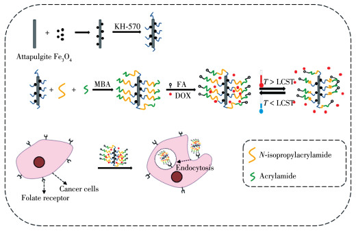
Schematic diagram of synthesis and targeting process of FA-Fe3O4/ATP-P(NIPAM-AAM)
Preparation and in Vitro Experiment of Attapulgite-based Microgels with Magnetic/Temperature Dual Sensitivities
Tian-Le LI , Hui ZHONG , Xiao-Rong LI , Jing ZHOU , Yi-Xin LIU , Wei-Cheng HU , Zhi-Peng CHENG , Cheng YAO

From the reports published on the International Agency for Research on Cancer (IARC), approximately 10 million new tumor cases arise and no fewer than 6 million patients die from the disease per year[1]. A main challenge in clinical treatment of tumor is how to accurately and high-efficiently deliver a nanocarrier which carries therapeutic agent to a tumor site to achieve ideal therapeutic effect. A drug delivery system is a means which enables the introduction of therapeutic drugs in the body and improves its effectiveness and safety by controlling the releasing rate, the releasing site and adjusting the releasing time. Drug delivery system is an interface between the patient and the drug[2]. In a bid to overcome in vivo adverse pharmacokinetic characteristics and non-specific distribution of most conventional chemotherapeutic agents, many advances in nanocarriers have been used in cancer targeted therapies[3-4].
Studies have been conducted on stimuliresponsive microgels and applied in many fields[5-8]. For their resemblance of physicochemical properties to those of the living tissues, these materials show good biocompatibilities, high water content, low interfacial tension, etc. N-isopropylacrylamide (NIPAM) is the most commonly and successfully researched materials among temperature-sensitive polymers. The lower critical solution temperature (LCST) of thermal-responsive nanogels based on NIPAM is about 32.0 ℃[9-10]. This temperature is close to that of the physiological human body. Furthermore, as the surrounding temperature increases, the structure of the polymer microgel undergoes a phase conversion from a swollen structure to collapse aggregation, which provides an opportunity for loading or releasing small molecules, potentially having expectable applications in tumor treatment[9-10]. Due to their reversible transition between dispersion and flocculation with the function of temperature, temperature variation can adjust the loading and releasing rate by PNIPAM-based nanocarriers. In addition, the LCST can adjust by adding comonomer into PNIPAM. Hydrophilic comonomer tends to increase the LCST and hydrophobic comonomer lowers the LCST, which provides an opportunity to embed or release small molecule, thus enabling drug delivery for tumor therapy[11].
In the last few years, organic and inorganic nanocomposites have become a hot spot in material science research. Attapulgite (ATP) is a hydrous magnesiumrich aluminosilicate clay mineral with chain layer structure, unique physical and chemical properties, including relatively permeability, good chemical stability and so on[12-13]. Magnetic iron oxide nanoparticles have attracted great attention in biomedical field owing to their good biocompatibility, low toxicity and high stability in physiological environment. Magnetic nanoparticles have unique characteristics and have been widely used in drug targeting and delivery for diagnosis and treatment[14]. It's easy to separate Fe3O4 nanoparticles with strong paramagnetism by an external magnetic field and this property is widely used in magnetic sepa- ration and presented excellent recyclability[15-17].
Folic acid (FA) is a kind of necessary vitamin necessary for a single-carbon transfer reaction. Since FA is important for the nucleotide biosynthesis sequences, it's consumed in large quantities through cell proliferation. Two membrane- associated proteins were used in normal cells. The former is the main approach in charge of metabolizing physiological folic acid present in almost whole cells. Most of them are found in degenerated epithelial cells and activated macrophages. They preferentially bound and internalized oxidized folate through receptor - mediated endocytosis. Though low concentrations of the reduced folate carrier may be sufficient to meet the requirements of most normal cells, folate receptor (FR) is often overexpressed in tumor cells, which may allow malignant cells to successfully contend for the vitamin when the supply is limited[15, 18].
Doxorubicin hydrochloride (DOX) has a wide range of biochemical effects in the body. It inhibits the synthesis of nucleic acids by embedding itself in DNA. This mechanism may eventually lead to the death of cancer cells and thus has anti- cancer effects. It has a significant influence on the chemotherapy of acute leukemia, lymphoma and other solid tumors. DOX is a satisfactory anticancer drug.
Herein, we synthesized FA-Fe3O4/ATP-P(NIPAM- AAM) based on emulsion polymerization. Due to the large surface area of ATP, the drug loading efficient is improved. The LCST of FA-Fe3O4/ATP-P(NIPAM- AAM) is well tuned by adjusting the composition of the two monomers before polymerization to adapt to human body temperature. Combining FA to the microgels through chemical bonds can increase the targeting ability. DOX is a model anticancer drug to carry out the in vitro test to verify the performance of the FA-Fe3O4/ATP-P(NIPAM-AAM) in identification and destruction cancer cells (Scheme 1).
N, N' - methylenebisacrylamide (MBA, 98%), N - isopropyl acrylamide (NIPAM, 99%), ammonium persulfate (APS, 98%) and sodium hyposulfite (SPS, 98%) were purchased from Acros Organics Co., Ltd. DMSO, toluene, ferric acetylacetonate (Fe(C5H7O2)3), triethylene glycol, N-hydroxysuccinimide (NHS, 98%), γ - methacryloxypropyl trimethoxysilane (KH-570), N-(3- dimethylaminopropyl)-N'-ethylcarbodllmidehydrochioricia (EDC, 98%), dialysis bag (MW: 8~14 kD) and folic acid (98%) were purchased from Aladdin Chemistry Co., Ltd. (Shanghai, China). Attapulgite was purchased from Hongjin Attapulgite Instruments (Xuyi, China). Sodium dodecyl sulfate (SDS) was purchased from Alpha Chemical Industry Co., Ltd. (3-4, 5-dimethylthiazol-2-yl)-2, 5-diphenyltetrazolium bromide (MTT) was obtained from Sigma - Aldrich (St. Louis, MO, USA). The fetal bovine serum (FBS) was from Corning (Medford, MA, USA). Dulbecco's modified eagle's medium (DMEM basic), penicillin - streptomycin solution and trypsin-EDTA (0.25%) were purchased from InvitrogenGibco (Carlsbad, CA, USA).
The morphologies and the particle size of the obtained samples were characterized by transmission electron microscopy (TEM, JEOL JEM-200CX, 120 kV) and scanning electron microscopy (SEM, JEOL JSM-6700F, 10 kV). The ultraviolet- visible (UV-Vis) spectroscopy was acquired with AVATAR- 360 FT-IR spectrophotometer from Nicolet Corporation. The magnetic properties of Fe3O4/ATP were characterized by vibrating sample magnetometer (VSM, AGFM, Iran) at 25 ℃. The crystal structure of the material was analyzed by an X - ray diffractometer with Cu Kα (λ =0.154 06 nm, X'pert-PRO). The tube voltage was 40 kV and the tube current was 60 mA. The scanning range was 5°~ 90°, and the scanning rate was 5 (°)·min-1.The MTT as-say was probed using a spectrophotometric plate reader (Elx808, Biotek, USA) at a wavelength of 570 nm in vitro uptake images and obtained by LSM700 laser con- focal microscope (nikon A1). The DLS was character- ized by Malvern particle size analyzer (Zetasizer Nano ZS) and the model of freeze dryer was FD-1A-50 from Beijing medical laboratory instrument Co. LTD.
First, 0.2 g of dried ATP and 1.000 g of Fe (C5H7O2)3 were mixed evenly in 30 mL of triethylene glycol under the ultrasonic condition. The mixture was heated to 270 ℃ for 2 h[19-20]. After cooling to room temperature, the prepared precipitate was washed with EtOH/deionized water for several times and dried in the vacuum oven over night at 50 ℃.
Silane coupling reagent KH-570 was used as surface modifier. The vinyl group of KH - 570 was introduced onto the surface of Fe3O4/ATP to copolymerize with NIPAM[21]. 1.0 mL of deionized water and 1.0 g of Fe3O4/ATP were dispersed in 100 mL of methylbenzene. Then, 3.0 mL of KH-570 was added into the mixture. In a 250 mL three-necked flask, the mixture was ultrasonificated for 40 min before stirred at 45~50 ℃ for 4 h. Finally, KH-570 modified Fe3O4/ATP was separated with a magnetic bar and sequentially washed with toluene, EtOH and deionized water. The KH-570- Fe3O4/ATP was dried at 65 ℃ in a vacuum oven.
The KH-570-Fe3O4/ATP was dispersed into deionized water to obtain a Fe3O4/ATP dispersion with a concentration of 0.04 g·mL-1. 0.4 g of NIPAM, the formulated amount of AAM (mass ratio of NIPAM/ AAM was 1:14), 5 mg of MBA and 2 mg of SDS were added in 2 mL of Fe3O4/ATP dispersion and then further diluted with 30 mL of deionized water[22-23]. After the deoxygeneration of above solution was carried out with nitrogen for 30 min, the temperature was raised to 70 ℃. 1 mL of APS (3 mg·mL-1) and 1 mL of SPS (2.5 mg·mL-1) were added into the solution with a syringe. The nitrogen atmosphere was maintained in the whole process. After being stirred for 6 h with a magnetic stirrer, the solution was cooled down to the room tempera- ture and then dialyzed with the deionized water for 7 d to remove unreacted monomers and impurities to obtain Fe3O4/ATP-P(NIPAM-AAM). The product was frozen by a refrigerator and then stored in a freeze dryer before use.
First, 0.034 mmol·L-1 of N-hydroxysuccinimide (NHS), 1.3 mg of 1 - ethyl- 3 - (3 - dimethylaminopropyl) carbodiimide (EDC) and 1.3 mg of FA were dissolved in DMSO. 20 mg of Fe3O4/ATP - P(NIPAM - AAM) was dispersed in 20 mL of PBS (pH 7.4) and adjust the pH value to 4.5~4.7. The above FA solution was added to the Fe3O4/ATP-P(NIPAM-AAM) solution and stirred at room temperature for 16 h in the dark[24-25]. After adjusting the pH value to 9.0 to end the reaction, reagents were dialyzed in PBS solution (pH 7.4) and deionized water (molecular weight cutoff of 14 kD) for 6 d. The resultant FA-Fe3O4/ATP-P(NIPAM-AAM) was dried for the following uses.
20 mg of Fe3O4/ATP-P(NIPAM-AAM) was dispersed in 20 mL of deionized water containing 5 mg of DOX. The mixture was stored in refrigerator at 4 ℃ to make the nanogel fully inflated[26-27]. The drug loading proceeded for 48 h.
The dialysis of above solution was conducted in deionized water for 4 h, whilst water was altered per half hour. The concentration of drug in outer water was measured by UV-visible spectrophotometer, thereby the drug loading rate was calculated. DOX absorbance was measured per half hour from 26 to 50 ℃ and the temperature gradient was 2 ℃[26-27].
The DOX loading and releasing amount was determined by UV-visible spectrophotometer at the wavelength of 225 nm.
Human liver cancer cells (HepG2) were purchased from American Type Culture Collection (ATCC). HepG2 cells were cultured in DMEM medium containing 10% (V/V) FBS and 1% (V/V) antibiotics at 37 ℃ in a HERA cell 150 incubator (Thermo Fisher Scientific Inc., USA).
The MTT assay was used to determine the cytotoxicity of FA - Fe3O4/ATP - P(NIPAM - AAM) of HepG2 according to the previous literature[28]. Briefly, cells were seeded in 96-well plate at a density of 2×10⁵ cells per well. After incubation overnight, cells were treated with different concentrations of samples. After 48 h of incubation, the medium of MTT solution was added. The cell survival rate was calculated according to the following formula:
|
$ {\rm{Cell}}{\kern 1pt} {\kern 1pt} {\kern 1pt} {\rm{viability = }}\frac{{{\rm{O}}{{\rm{D}}_{{\rm{sample group}}}} - {\rm{O}}{{\rm{D}}_{{\rm{blank group}}}}}}{{{\rm{O}}{{\rm{D}}_{{\rm{negative control}}}} - {\rm{O}}{{\rm{D}}_{{\rm{blank group}}}}}} \times 100\% $ |
Where, ODsample group is the absorbance of sample group; ODnegative group is the absorbance of control group; ODnegative group is the absorbance of blank group.
HepG2 cells were seeded in 96 - well plates at a density of 3×10⁵ cells per well and cultured incubator for an additional 16~20 h. The cells were treated with 200 μg·mL-1 of FA-Fe3O4/ATP-P(NIPAM-AAM) for indicated time points. Then the medium was removed. The cells were washed in following way: trice with PBS, fixed with 4% paraformaldehyde for 15 min, trice with PBS. Finally, cells were observed under a LSM700 laser confocal microscope after stained with an anti - fade mounting medium with DAPI.
Fig. 1A shows the XRD patterns of Fe3O4, Fe3O4/ ATP, and ATP. Six characteristic peaks (30.1°, 35.44°, 43.08°, 53.58°, 57.16°, and 62.78°) corresponding to the Fe3O4 nanoparticles (NPs) were also obtained for the Fe3O4/ATP, revealing that the Fe3O4 NPs were bonded to the ATP. What's more, the peak at 26.52° is well consistent to the primary diffraction plane of the ATP. The results indicated that the Fe 3O4/ATP were successfully synthesized[21].
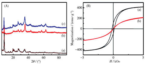
Fig. 1B exhibits the magnetic hysteresis loops of Fe3O4 and Fe3O4/ATP obtained with the VSM for magnetic fields from -5 to 5 kOe. The maximum values of magnetization saturation of the Fe3O4 and Fe3O4/ATP were 9.73 and 6.16 emu·g-1. Compared with Fe3O4, the saturation magnetization of Fe3O4/ATP was reduced.The difference in saturation magnetization was attributed to the non-magnetic ATP clay particles[21].
The TEM images depicted in Fig. 2a and 2b show the morphology and structure of Fe3O4/ATP and FA - Fe3O4/ATP-P(NIPAM-AAM). It is found that there is a gray layer on the edge of Fe3O4/ATP. This result indi-cates that the coating of temperature responsive poly- mer on the Fe3O4/ATP is successful.
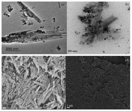
The surface morphology of the Fe3O4/ATP and FA- Fe3O4/ATP-P(NIPAM-AAM) was probed by SEM. Fig. 2c and 2d are the SEM images of the Fe3O4/ATP and FA-Fe3O4/ATP-P(NIPAM-AAM) respectively. As a result, the irregular surface of Fe3O4/ATP implies that a layer of P(NIPAM-AAM) gel was coated on the surface of FA-Fe3O4/ATP particles.
Fig. 3A presents the IR spectra of ATP, Fe3O4, Fe3O4/ATP, KH-570-Fe3O4/ATP, P(NIPAM-AAM) and Fe3O4/ATP-P(NIPAM-AAM), respectively. The curve of ATP shows an apparent peak at 1 035 cm-1, corresponding to the stretching vibration peak of Si—O of attapulgite clay. The stretching vibration peak observed in the curve of Fe 3O4 was at 580 cm-1 according to the Fe—O of Fe3O4. The curve c in Fig. 3A presents the characteristic absorption peaks of Fe3O4 and ATP. Compared with Fe3O4/ATP, the stretching vibration peaks of —CH3 and —CH2 appeared in the curve of KH- 570 -Fe3O4/ATP, which are at about 2 876 and 2 930 cm-1, indicating the successful modification of KH- 570. For P(NIPAM-AAM), the peak at 1 385 and 1 456 cm-1 are C—H stretching vibration peaks on isopropyl, 1 547 cm-1 is the bending vibration peak of —NH, and 2 876, 2 930 and 2 973 cm-1 are the stretching vibration peaks of —CH3 and —CH2, 1 644 cm-1 is the stretching vibration peak of the C=O double bond. 1 035 cm-1 is the bending vibration peak of Si—O. Compared with P(NIPAM-AAM), there are characteristic absorption peaks of Si-O and Fe-O for Fe3O4/ATP-P (NIPAM-AAM), indicating the successful grafting of Fe3O4/ATP-P(NIPAM-AAM)[19].
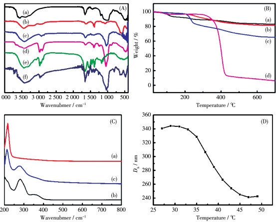
Fig. 3B shows the TG curves of ATP, Fe3O4/ATP, KH-570-Fe3O4/ATP and Fe3O4/ATP-P(NIPAM-AAM). From the picture, we can see that compared to KH-570/ Fe3O4/ATP, the weight loss of Fe3O4/ATP-P(NIPAM- AAM) increased, indicating that P(NIPAM-AAM) has been coated onto KH-570-Fe3O4/ATP successfully.
Fig. 3C shows the UV - Vis spectra of FA, Fe3O4/ ATP-P(NIPAM-AAM) and FA-Fe3O4/ATP-P(NIPAM- AAM). The absorption peak of FA was 225 nm and the absorption peak of Fe3O4/ATP-P(NIPAM-AAM) was 280 nm. From the above results, the FA-Fe3O4/ATP- P (NIPAM-AAM) was synthesized successfully.
As shown in Fig. 3D, the thermo-sensitive profile of FA-Fe3O4/ATP-P(NIPAM-AAM) was characterized in the temperature range of 27.0~49.0 ℃ by DLS monitoring hydrodynamic diameter (Dh). It is clear that compared with the LCST (32.0 ℃) of PNIPAM microgel,the LCST of FA-Fe3O4/ATP-P(NIPAM-AAM) was around 38.5 ℃ due to the hydrophilic group of acrylamide. The temperature is close to that of human body and it's expected to be applied to the study of targeted sustained release cancer drugs.
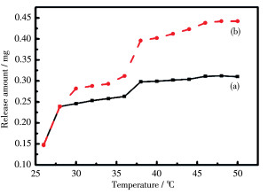
Fig. 4 exhibits the DOX releasing amount spectra of P(NIPAM - AAM) and Fe3O4/ATP - P(NIPAM - AAM) from 26.0 to 28.0 ℃. In Fig 4a, the increase of releasing amount of DOX was obvious. It may be ascribed to the free DOX which was unloaded on P(NIPAM - AAM) microgel. The shape of P(NIPAM-AAM) and Fe3O4/ ATP-P(NIPAM-AAM) curves was similar between 26.0~30.0 ℃, namely two- stage releases. There was a sharp hopping on curve of Fe3O4/ATP-P(NIPAM-AAM) at around 38.5 ℃ due to the swell - shrink of nanogel corresponding to the analysis of DLS. Compared with P(NIPAM-AAM), the loading efficiency of Fe3O4/ATP- P(NIPAM-AAM) rose from 62% to 79% and the releasing efficiency increased from 36% to 56%. The advantage of ATP is apparent to raise the adsorption capacity and the releasing amount.
To evaluate the safety of FA-Fe3O4/ATP-P(NIPAM - AAM), HepG2 cells were treated with 0, 6.25, 12.5, 25, 50, and 100 μg·mL-1 DOX, 0, 31.65, 63.29, 126.58, 253.16, and 506.32 μg·mL-1 FA-Fe3O4/ATP- P(NIPAM-AAM) and 0, 25.4, 50.79, 101.58, 203.16, and 406.32 μg·mL-1 DOX loaded FA-Fe3O4/ATP- P(NIPAM-AAM), respectively. MTT was used to detect cell viability. As shown in Fig. 5a, after treatment with different concentrations of FA-Fe3O4/ATP-P(NIPAM-AAM) for 48 h, no significant cytotoxicity was observed on HepG2 cells. The cell viability was above 1.0, indicating that FA-Fe3O4/ATP-P(NIPAM-AAM) promoted the growth, rather than inhibited, the cell viability. The result indicates that the synthesized FA-Fe3O4/ATP- P(NIPAM-AAM) polymer is safe and can be utilized as a media for drug delivery in biomedical field.
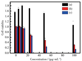
From Fig. 5b and 5c, a dose-dependent inhibitory effect of DOX and DOX loaded FA-Fe 3O4/ATP-P(NIPAM - AAM) was observed on HepG2 cells. The cytotoxicity increased as the concentration increased. However, the cytotoxicity of free DOX was weaker than that of DOX loaded FA-Fe3O4/ATP-P(NIPAM-AAM). This is mainly attributed to the targeting ability of FA. The IC50 (the concentration at which cell proliferation is half inhibited) values of free DOX and DOX loaded FA-Fe3O4/ATP-P(NIPAM-AAM) were 46 and 19 μg· mL-1 respectively. The inhibitory effect on cancer cells of DOX loaded FA-Fe3O4/ATP-P(NIPAM-AAM) was stronger than that of free DOX.
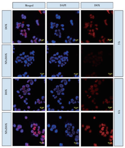
For further research on the in vitro targeting of liver cancer cells by FA-Fe3O4/ATP-P(NIPAM-AAM), DOX was chosen as model drugs to identify the cellular uptake of microgels. A qualitative test of cellular uptake was performed using confocal laser scanning microscope (CLSM) of HepG2 cells after 3 or 6 h of exposure. It should be noted that the blue fluorescent signal was the nucleus of DAPI negative staining and the surrounding red fluorescent signal was the intake of DOX. As shown in Fig. 6, DOX loaded FA-Fe3O4/ATP-P(NIPAM-AAM) was swallowed intracellularly by HepG2 cells after incubating HepG2 cells with DOX loaded FA-Fe3O4/ATP-P(NIPAM-AAM) for 3 h. A red fluorescent signal was observed arising from the cytoplasmic matrix. CLSM test's results showed that when the time is prolonged to 6 h, the red fluorescent signal in the cells of the free DOX group was almost unchanged when compared with the HepG2 cells incubated for 3 h. In contrast, the red fluorescent signal of FA- Fe3O4/ATP - P(NIPAM - AAM) - DOX was significantly enhanced relative to the group incubated for 6 h. It can verify its targeting ability.
In summary, magnetic and temperature - sensitive nanoparticle microgels FA-Fe3O4/ATP-P(NIPAM-AAM) with targeting were synthesized by emulsion polymerization method. The phase transition temperature rose from 32.0 to 38.5 ℃, which is slightly higher than that of human body and suitable for cell experiment in vitro. In vitro cell test results show that the FA-Fe3O4/ATP-P(NIPAM-AAM) exhibits good biocompatibility. At the same time, the microgels with targeting ability can effectively prolong the time of drug release and action. In vitro drug release experiments show that ATP can increase drug loading and drug release. This material is expected to accurately release anticancer drugs to tumor cells so as to reduce side effects, which is expanding the application of targeted tumor therapy.
Iyer A. K, Singh A, Ganta S, Amiji M M. Adv. Drug Delivery Rev., 2013, 65(13/14):1784-1802
Jain K K. Methods Mol. Biol., 2008, 437:1-50
Peng J, Zhang W L, Ai S L, Zhang Y H, Liu J Y, Liu J, He P X, Li Y. Nanotechnology, 2019, 30(11):115701 doi: 10.1088/1361-6528/aaf8e4
Li R, Nie W, Yang W. Mater. Res. Express, 2019, 6(7):075048 doi: 10.1088/2053-1591/ab179e
Hopkins S, Carter S. R, Haycock J W, Fullwood N J, MacNeil S, Rimmer S. Soft Matter, 2009, 5(19):3701-3712
Matsune H, Ono T, Yoshida R, Yamamoto T, Kishida M. Chem. Lett., 2019, 48:1058-1060 doi: 10.1246/cl.190353
Sakai K, Sawa M, Nomura K, Endo T, Tsuchiya K, Sakamoto K. Chem. Lett., 2016, 45:655-657 doi: 10.1246/cl.160231
Motiei M, Dreifuss T, Sadan T, Omer N, Blumenfeld-Katzir T, Frago-georgi E. Chem. Lett., 2019, 48:291-294 doi: 10.1246/cl.180780
Chen S J, Zhang Q Y, Gu J W, Ma M L, Zhang L, Zhou J, Zhou Y Y. Colloid. Polym. Sci., 2015, 290(12):1207-1213
Xu G. Wu W T, Wang Y, Pang W, Lu F. Nanotechnology, 2006, 17(10):2458-2465
Zhang K J, Zhou D, Wang Z G, Zhang Y H, He P X. Nanotechnology, 2019, 30(35):355604 doi: 10.1088/1361-6528/ab209d
Li X M, Zhong H, Li X R, Jia F F, Cheng Z P, Zhang L L, Yin J Z, An L T, Guo L P. Mater. Sci. Eng. C, 2014, 45:170-175 doi: 10.1016/j.msec.2014.08.056
Han Y, Chen H F, Chen D J. J. Mater. Sci., 2010, 45(9):2372-2380 doi: 10.1007/s10853-009-4203-3
Wang D, Duan H, Lu J H, Lv C. J. Mater. Chem. A, 2017, 5(10):5088-5097 doi: 10.1039/C6TA09772C
Lu Y J, Low P S. Adv. Drug Deliver. Rev., 2012, 54(5):342-352
Sahoo S L, Liu C H, Wu W C. RSC Adv., 2017, 7:22468-22478 doi: 10.1039/C7RA02084H
Dutta B, Nema A, Shetake N G, Gupta J, Hassan P A. Mater. Sci. Eng. C, 2020, 112:110915 doi: 10.1016/j.msec.2020.110915
Yang P, Zeng H, Liu J. J. Mater. Chem. B, 2013, 1(39):5298-5308 doi: 10.1039/c3tb20975j
Fu M, Zhang Z P. Mater. Lett., 2018, 226:43-46 doi: 10.1016/j.matlet.2018.04.128
Kim Y H, Sim B, Choi H J. Colloids Surf. A, 2016, 507:103-109 doi: 10.1016/j.colsurfa.2016.07.095
Zhao Y J, Chen Y, Li M S, Zhou S Y, Xue A L, Xing W H. J. Hazard Mater., 2009, 171(1/2/3):640-646
Wang C N, Wang Y Y, Jin Y L, Xu T, Yuan L, Fang J H. J. Nanosci. Nanotechnol., 2015, 15(9):6784-6789 doi: 10.1166/jnn.2015.11111
Han H D, Shin B C, Choi H S. Eur. J. Pharm. Biopharm., 2006, 62(1):110-116
Jo A, Lim D, Kim H. Sci. Rep., 2017, 7:41090 doi: 10.1038/srep41090
Ma C B, Shi Y, Pena D A, Peng L L, Yu G H. Angew. Chem., 2015, 127(25):7484-7488 doi: 10.1002/ange.201501705
Chen Y, Chen Y B, Nan J Y, Wang C, Chu F. J. Appl. Polym. Sci., 2012, 124(6):4678-4685
Li A H, Ma H J, Feng S Y, Liu J. RSC Adv., 2016, 6(39):33138-33147 doi: 10.1039/C5RA27839B
Zhang J, Jiang Y, Li Y, Li W, Hu W. Carbohydr. Polym., 2019, 230:115576

Scheme1 Schematic diagram of synthesis and targeting process of FA-Fe3O4/ATP-P(NIPAM-AAM)

Figure 1 (A) XRD patterns of (a) ATP, (b) Fe3O4 and (c) Fe3O4/ATP; (B) VSM patterns of (a) Fe3O4 and (b) Fe3O4/ATP

Figure 2 TEM images of (a) Fe3O4/ATP and (b) Fe3O4/ATP-P(NIPAM-AAM); SEM images of (c) Fe3O4/ATP and (d) Fe3O4/ATP-P(NIPAM-AAM)

Figure 3 (A) IR spectra of (a) ATP, (b) Fe3O4, (c) Fe3O4/ATP, (d) KH-570-Fe3O4/ATP, (e) P(NIPAM-AAM) amd (f) Fe3O4/ATP-P(NIPAM-AAM); (B) TG curves of (a) ATP, (b)Fe3O4/ATP, (c) KH-570-Fe3O4/ATP and (d) Fe3O4/ATP-P(NIPAM-AAM); (C) UV-Vis spectra of (a) FA, (b) Fe3O4/ATP-P(NIPAM-AAM) and (c) FA-Fe3O4/ATP-P(NIPAM-AAM); (D) DLS curve of FA-Fe3O4/ATP-P(NIPAM-AAM)

Figure 5 MTT test of (a) FA-Fe3O4/ATP-P(NIPAM-AAM), (b) free DOX and (c) DOX loaded FA-Fe3O4/ATP- P(NIPAM-AAM)

 扫一扫看文章

扫一扫关注我们
 下载:
下载:
 下载:
下载:

