Figure 1.
Structures of ligands HOBPT and HABPT
Crystal Structure, Electrochemistry and Spectral Properties of Two 2-Substituted-4, 6-di(2-pyridyl)-1, 3, 5-triazine Bridged Dinuclear Ruthenium Complexes
Man-Li CAO , Yu-Qi HE , Wen-Ting LIU , Wei YIN , Su-Yang YAO

The Ru(Ⅱ) polypyridyl complexes have been deep-ly researched because of their excellent redox and pho-tophysical properties and good activity against cancer cell lines, promising applications as solar energy con-version, molecular electronics, therapeutic agents and so on[1-5]. Among them, multinuclear Ru(Ⅱ) polypyridyl complexes, especially dinuclear ones bridged by poly-pyridyl-like bridging ligands, have drawn our interests, because they can afford electronic coupling between the linked partners to allow, in turn, intercomponent energy and/or electron-transfer processes. The ability of the bridging ligand to transfer the electronic charge, and energy gap of the metal components, as well as the coordination environments, have great influence on the photophysical and electrochemical properties of the complexes, which means that they have potential appli-cations for bioinorganic chemistry and theoretical aspects of electron-transfer mechanisms[6]. To obtain these complexes, many bridging ligands having two or more bidentate sets of coordination sites were designed and synthesized. Complexes based on rigid polypyridyl-like bridging ligands: 2, 4, 6-tris(2-pyridyl)triazine (tpt)[6-7], 2, 3-bis(2-pyridyl)pyrazine (dpp)[8-9], 2, 2'-bipy-rimidine (bpm)[10], 2, 3-bis(2-pyridyl)quinoxaline (dpq)[11], 2, 3-bis(2-pyridyl)-benzoquinoxaline (dpb)[11], 3, 6-bis(2-pyridyl)-1, 2, 4, 5-tetrazine (bptz)[12], 2, 2'-bis(1, 2, 4-triazi-no[5, 6-f]phenanthren-3-yl)-4, 4'-bipyridine (btpb)[13], 1, 4-phenylene-bis(1 -pyridin-2-ylimidazo[1, 5-a]pyridine[14], and so on[15-18], have been reported.
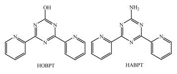
In this paper, we report two new bridging ligands, 4, 6-bi(2-pyridyl)-1, 3, 5-triazin-2-ol (HOBPT)[19-23] and 2-amino -4, 6-bi(2- pyridyl)-1, 3, 5-triazine (HABPT)[24-25], whose structures are shown in Fig. 1. The ligands were assembled into dinuclear Ru(Ⅱ) polypyridyl complexes [Ru2(OBPT) (bpy)4] (PF6)3·3H2O·0.5CH3CH2OH (1) and [Ru2(HABPT)(bpy) 4](PF6)4·0.5H2O (2) (bpy=2, 2'-bipyri-dine), and the single crystal structures, spectral and electrochemical properties of the complexes were deter-mined. In the complexes, there are different degrees of metal - metal communication between two ruthenium centers through the bridge. And moreover, although they are similar in structure, they are different in ste-reoisomeric forms: 1 shows in the rac-(ΔΔ) form, but 2 is in the meso-(ΔΛ) form.
The reagents and solvents employed were commer-cially available and were used as received without fur-ther purification. The C, H and N microanalyses were carried out with a Vario EL elemental analyzer. The FT-IR spectra were recorded from KBr pellets in a range of 400~4 000 cm-1 on a Bruker TENSOR 27 spectrome-ter. Electron spray ionization mass spectra (ESI-MS) were obtained on a LCQ DECA XP quadrupole ion trap mass spectrometer with methanol as the carrier solvent. The optical ultraviolet-visible (UV-Vis) absorption spectra were obtained using a UV-3150 UV-Vis spec-trophotometer of SHIMADZU corporation at room tem-perature, and the solvents used for spectral experi-ments were distilled from CaH2 and kept over molecu-lar sieves (type 4A). The luminescence spectra were ob-tained using a FLSP 920 combined fluorescence life-time and steady state spectrometer of Edinburgh Instru-ments LTD.
The cyclic voltammetry (CV) and differential pulse voltammetry (DPV) measurements were made with a CHI-660 electrochemical workstation. The experiments were performed with a standard three-electrode arrangement: working electrode, glassy carbon electrode; quasi reference electrode, Ag-AgCl electrode; counter electrode, Pt wire electrode. Back-ground correction was accomplished by subtracting the current curves of the blank electrolyte (containing the supporting electrolyte with same concentration) from the experimental curve. The experiments were per-formed with 1 mmol·L-1 solutions of the complexes in dried acetonitrile at a scan rate of 100 mV·s-1 with 0.1 mol·L-1 Bu4NPF6as supporting electrolyte.
The ligands HOBPT[26], HABPT[27] and the com-plex cis - [Ru(bpy)2Cl2]·2H2O[28] were prepared accord-ing to the literature methods.
Under an atmosphere of dry nitrogen, the mixture of HOBPT (0.125 g, 0.50 mmol), cis-[Ru(bpy)2Cl2]·2H2O(0.52 g, 1.00 mmol) in ethylene glycol (15 mL) and water (5 mL) was refluxed at 110 ℃ for 12 h.The crude product was purified by column chromatography on silica using MeCN/H2O/sat. aq. KNO3(40:4:1, V/V) as the eluent. After counterion exchange with KPF6 and drying in vacuo, a red powder was obtained. Yield:0.455 g, 57.6%. The single-crystals of 1 were grown from acetonitrile and ethanol solution by slow evapora-tion of ether at room temperature. ESI-MS (m/z):1 367[Ru2(OBPT)(bpy)4+2PF6]+; 664.2 [Ru(OBPT)(bpy)2]+; 611.7 [Ru2(OBPT)(bpy)4+PF6]2+; 359.3 [Ru2(OBPT)(bpy)4]3+.Anal.Calcd. for C54H49F18N13O4.5P3Ru2(%):C, 40.81; H, 3.11; N, 11.46. Found(%): C, 40.68; H, 3.22; N, 11.62. FT - IR (cm-1): 3 651(m), 3 429(s), 3 084(m), 1 676(s), 1 630(m), 1 604(m), 1 564(w), 1 495(vs), 1 468(vs), 1 445 (s), 1 427(m), 1 385(s), 1 313(w), 1 246(w), 1 163(w), 1 101(w), 1 028(w), 839(vs), 762(vs), 732(m), 703(vw), 681(vw), 658(vw), 559(vs), 422(vw).
Complex 2 was prepared with a similar procedure only with HABPT instead of HOBPT. After counterion exchange with KPF6 and drying in vacuo, a dark red powder was obtained. Yield: 53.4%. The single-crystals were grown from acetonitrile and ethanol solution by slow evaporation of ether at room temperature. ESI-MS (m/z):1 365.9 [Ru2(HABPT) (bpy)4+2PF6]+; 663.1 [Ru(HABPT) (bpy)2]+; 358.7[Ru2(HABPT)(bpy)4]3+.Anal. Calcd. for C53H43F24N14O0.5P4Ru2(%):C, 38.21; H, 2.60; N, 11.77.Found(%): C, 38.30; H, 2.72; N, 11.68.FT-IR (KBr, cm-1): 3 649(w), 3 490(s), 3 390(s), 3 122 (m), 3 084(m), 2 363(w), 1 624(s), 1 606(m), 1 546(s), 1 468(s), 1 448(s), 1 429(m), 1 412(m), 1 385(s), 1 315 (w), 1 302(vw), 1 244(m), 1 196(w), 1 165(w), 1 028(w), 841(vs), 768(s), 731(m), 692(vw), 648(vw), 633(m), 559 (vs), 424(vw).
Diffraction intensities for the complexes were col-lected at 293 K on a Bruker Smart Apex CCD diffrac-tometer with graphite-monochromated Mo Kα radiation (λ=0.071 073 nm). The structure was solved with the SHELXS[29]structure solution program using direct methods and refined with the SHELXL[30] refinement package using least squares minimization. All non-hydrogen atoms were refined anisotropically. The organic hydrogen atoms were generated geometrically (C-H 0.096 nm, N-H 0.086 nm). The hydrogen atoms of solvents were located from difference maps and refined isotropically. The solvents in 2 were squeezed by SQUEEZE program. Crystal data as well as details of data collection and refinement for the complexes are summarized in Table 1. Selected bond distances and bond angles are listed in Table 2.
 下载:
导出CSV
下载:
导出CSV
| Complex | 1 | 2 |
| Molecular formula | C54H49F18N13O4.5P3Ru2 | C53H43F24N14O0.5P4Ru2 |
| Formula weight | 1 589.11 | 1 666.03 |
| Crystal system | Monoclinic | Orthorhombic |
| Space group | C2/c | Pca21 |
| a / nm | 2.452 9(3) | 1.287 8(2) |
| b / nm | 1.411 1(1) | 2.427 0(4) |
| c / nm | 3.980 1(4) | 2.169 6(3) |
| β/(°) | 93.834(2) | |
| V/ nm3 | 13.746(2) | 6.781(2) |
| Z | 8 | 4 |
| Dc / (g·cm-3) | 1.536 | 1.632 |
| Absorption coefficient / mm-1 | 0.611 | 0.654 |
| F(000) | 6 360 | 3 308 |
| Crystal size / mm | 0.300×0.250×0.100 | 0.260×0.250×0.210 |
| θ range for data collection / (°) | 1.664~25.999 | 2.056~24.999 |
| Limiting indices | -30 ≤ h ≤ 30, -17 ≤ k ≤ 17, -49 ≤ l ≤ 49 | -15 ≤ h ≤ 8, -15 ≤ k ≤ 28, -24 ≤ l ≤ 10 |
| Reflection collected, unique | 66 533, 13 491 (Rint=0.050 8) | 14 107, 7 098 (Rint=0.074 3) |
| Reflection observed | 10 132 | 4 414 |
| Data, restraint, parameter | 13 491, 45, 815 | 7 098, 149, 732 |
| Goodness-of-fit on F2 | 1.002 | 1.024 |
| Final R indices [I > 2σ(I)]a, b | R1=0.076 4, wR2=0.231 2 | R1=0.072 7, wR2=0.158 1 |
| R indices (all data)a, b | R1=0.095 8, wR2=0.257 1 | R1=0.110 3, wR2=0.174 2 |
| a R1=∑||Fo|-|Fc||/∑|Fo|, b wR2=[∑w(Fo2-Fc2)2/∑w(Fo2)2]1/2. | ||
下载:
导出CSV
| 1 | |||||
| Ru1-N1 | 0.206 1(5) | Ru2-N6 | 0.204 3(6) | C47-N10 | 0.139 2(7) |
| Ru1-N2 | 0.206 8(5) | Ru2-N7 | 0.204 9(6) | C47-N12 | 0.140 2(8) |
| Ru1-N3 | 0.204 5(5) | Ru2-N8 | 0.205 8(5) | C47-O1 | 0.121 4(7) |
| Ru1-N4 | 0.207 3(5) | Ru2-N12 | 0.208 1(5) | C48-N11 | 0.133 2(8) |
| Ru1-N9 | 0.205 4(5) | Ru2-N13 | 0.207 4(6) | C48-N12 | 0.132 7(8) |
| Ru1-N10 | 0.209 5(5) | C46-N10 | 0.133 4(8) | ||
| Ru2-N5 | 0.206 1(5) | C46-N11 | 0.133 7(8) | ||
| N1-Ru1-N2 | 78.7(2) | N4-Ru1-N10 | 90.3(2) | N6-Ru2-N7 | 96.7(3) |
| N3-Ru1-N4 | 78.5(2) | N2-Ru1-N4 | 172.7(2) | N7-Ru2-N5 | 85.8(2) |
| N9-Ru1-N10 | 78.4(2) | N1-Ru1-N10 | 170.9(2) | N8-Ru2-N5 | 97.7(2) |
| N3-Ru1-N1 | 90.3(2) | N2-Ru1-N10 | 95.1(2) | N6-Ru2-N13 | 89.5(3) |
| N9-Ru1-N1 | 95.1(2) | N3-Ru1-N9 | 172.0(2) | N8-Ru2-N13 | 95.5(2) |
| N3-Ru1-N2 | 95.8(2) | N8-Ru2-N12 | 85.4(2) | N5-Ru2-N13 | 98.0(2) |
| N9-Ru1-N2 | 91.0(2) | N6-Ru2-N8 | 174.2(2) | N6-Ru2-N12 | 98.5(2) |
| N9-Ru1-N4 | 94.8(2) | N6-Ru2-N5 | 78.5(3) | N7-Ru2-N12 | 98.4(2) |
| N1-Ru1-N4 | 96.6(2) | N7-Ru2-N8 | 78.4(2) | N7-Ru2-N13 | 173.2(2) |
| N3-Ru1-N10 | 96.9(2) | N13-Ru2-N12 | 78.0(2) | N5-Ru2-N12 | 175.2(2) |
| 2 | |||||
| Ru1-N1 | 0.203 6(6) | Ru2-N6 | 0.2082(8) | C47-N14 | 0.129(2) |
| Ru1-N2 | 0.203 7(7) | Ru2-N7 | 0.2035(8) | C47-N10 | 0.139 0(9) |
| Ru1-N3 | 0.205 2(8) | Ru2-N8 | 0.2040(7) | C47-N12 | 0.139 0(8) |
| Ru1-N4 | 0.207 3(9) | Ru2-N12 | 0.2117(5) | C48-N11 | 0.139 0(9) |
| Ru1-N9 | 0.206 4(8) | Ru2-N13 | 0.2073(8) | C48-N12 | 0.139 0(8) |
| Ru1-N10 | 0.212 8(5) | C46-N11 | 0.1390(8) | ||
| Ru2-N5 | 0.207 6(8) | C46-N10 | 0.1390(8) | ||
| N1-Ru1-N3 | 85.9(4) | N9-Ru1-N10 | 77.6(3) | N7-Ru2-N8 | 78.8(4) |
| N1-Ru1-N4 | 99.7(4) | N2-Ru1-N3 | 95.8(5) | N7-Ru2-N12 | 95.5(4) |
| N1-Ru1-N9 | 93.3(4) | N2-Ru1-N4 | 173.9(4) | N7-Ru2-N13 | 89.6(4) |
| N1-Ru1-N10 | 170.4(3) | N2-Ru1-N9 | 89.6(4) | N8-Ru2-N5 | 87.3(4) |
| N1-Ru1-N2 | 79.3(4) | N2-Ru1-N10 | 97.3(4) | N8-Ru2-N6 | 97.3(4) |
| N3-Ru1-N4 | 78.0(5) | N5-Ru2-N6 | 78.4(4) | N8-Ru2-N12 | 170.3(4) |
| N3-Ru1-N9 | 174.2(5) | N5-Ru2-N12 | 101.3(3) | N8-Ru2-N13 | 94.3(4) |
| N3-Ru1-N10 | 103.5(3) | N6-Ru2-N12 | 89.1(3) | N13-Ru2-N5 | 174.0(4) |
| N4-Ru1-N10 | 84.6(4) | N7-Ru2-N5 | 96.4(4) | N13-Ru2-N6 | 95.6(4) |
| N9-Ru1-N4 | 96.5(4) | N7-Ru2-N6 | 173.7(4) | N13-Ru2-N12 | 77.7(3) |
CCDC: 2026074, 1; 2026075, 2.
The ligand HOBPT was prepared by the reaction of 2-cyanopyridine and urea in the presence of sodium hydride as a catalyst in DMSO solution according to the previous report[26]. The ligand HABPT was prepared by the reaction of 2-cyanopyridine and guanidine hydrochloride in an ethanol solution according to the previous report and recrystallized with methanol[27]. The ligands are soluble in common organic solvents, such as CH3OH, CH3CN, DMF.
In general, HOBPT and HABPT are potential mul-titopic ligands with different coordination modes, such as tridentate terpyridine-like, bidentate bipyridine-like and tetradentate bis-bipyridine-like[19-25]. Indeed, reac-tion of cis-[Ru(bpy)2Cl2]·2H2O with HOBPT or HABPT in a 2:1 ratio in ethylene glycol/water mixture solution afforded dinuclear complexes. Crystals of complex [Ru2(OBPT) (bpy)4] (PF6) 3·3H2O·0.5CH3CH 2OH (1) and complex [Ru2(HABPT) (bpy) 4] (PF6)4·0.5H2O (2), were grown from acetonitrile and ethanol solution by slow evaporation of ether at room temperature. The structure reveals that 1 and 2 are dinuclear structures, in which the ligands act as tetradentate bis-bipyridine-like bridging two Ru(Ⅱ) ions via the triazine ring in chelat-ing way.
Single-crystal X-ray diffraction analysis reveals that complex 1 crystallizes in the monoclinic C2/c space group. The asymmetric unit of complex 1 con-tains one dinuclear [Ru2(OBPT) (bpy) 4]3+ cation, along with three PF6- anions and some solvent molecules.
The structure of the cation[Ru2(OBPT) (bpy)4]3+ is shown in Fig. 2. Each metal ion is coordinated by two N atoms from a bidentate coordination set of HOBPT and four N atoms from two bpy ligands to form a distorted octahedral geometry. The distances of Ru-N are in a range of 0.204 3(6)~0.209 5(5) nm, which are comparable to those of reported dinuclear ruthenium complexes[6-18]. The"bite angles"of the terminal chelating ligands vary between 78.4(2)° and 78.7(3)°, which are typical for the ruthenium polypyridyl complexes. The metal centers are coordinated at each bidentate coordination site of HOBPT with N-Ru-N angles of 78.0(2)° and 78.4(2)°.
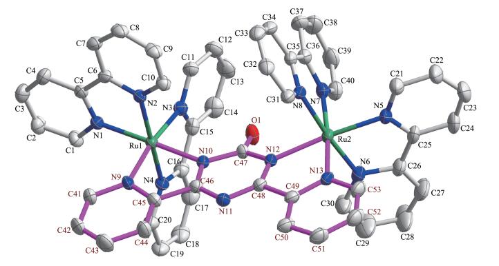
All hydrogen atoms on carbon atoms are omitted for clarity
The bridging ligand HOBPT acts as a bis-bidentate ligand to connect two Ru(Ⅱ) ions. The distance between two Ru(Ⅱ) ions is 0.619 8 nm. For the bridging ligand, the bond length of O1-C47 is 0.121 4(8) nm, indicating that it is a typical double bond. In the triazine ring, the C47-N10 and C47-N12 bond distances are 0.139 2(7) and 0.140 2(8) nm, respectively, whereas the remaining four C-N distances (in a range of 0.132 7(8)~0.133 7(7)nm) are similar to the mean C-N bond distances report-ed for other complexes of this ligand[19-23]. This indi-cates that the bridging ligand is deprotonated and shows negative charge. This is also supported by the fact of only three PF6- anions are present in an asym-metric unit.
The bridging ligand does not show good planarity. In the triazine ring, the five atoms N10, C46, N11, C48, and N12 make a good plane (average deviation is 0.000 70 nm) and behave like a delocalized pentadi - enide moiety. The deviation of C47 from this least - squares plane is 0.016 21 nm. The angles between two pyridyl rings and the triazine ring are 5.7° and 7.0°, respectively.
In the crystal structure, there are plenty of strong hydrogen bonds existing between the lattice water mol-ecules and cations, which are shown in Fig. 3 and listed in Table 3. The lattice water molecules form two five-membered rings via O-H…O hydrogen bonds, which link two cations to form hydrogen-bonded dimers via O1W-H1WA…O1. The hydrogen - bonded dimers and PF6-anions are then linked to three dimensional frame-work via abundant C-H…F hydrogen bonds.
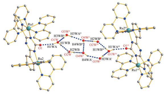
Most hydrogen atoms are omitted for clarity; Symmetry codes: i-x, -y-3, -z; iix+1/2, y-1/2, z; iii-x+1/2, -y-7/2, -z
下载:
导出CSV
| D-H…A | d(D-H) / nm | d(H…A) / nm | d(D…A) / nm | ∠DHA / (°) |
| O1W-H1WA…O1 | 0.085 | 0.211 | 0.293(2) | 161.9 |
| O1W-H1WB…O2W | 0.086 | 0.182 | 0.267(2) | 167.0 |
| O2W-H2WB…O4Wi | 0.087 | 0.172 | 0.255(3) | 156.8 |
| O3W-H3WA…O4W | 0.088 | 0.208 | 0.296(4) | 178.2 |
| O3W-H3WB…O1Wii | 0.086 | 0.200 | 0.284(3) | 168.1 |
| O4W-H4WA…O3W | 0.087 | 0.221 | 0.296(4) | 145.2 |
| O4W-H4WB…O4Wiii | 0.088 | 0.186 | 0.269(6) | 156.5 |
| Symmetry codes: i -x, -y-3, -z; ii x-1/2, y+1/2, z; iii -x-1/2, -y-5/2, -z. | ||||
Complex 2 crystallizes in the orthorhombic Pca21 space group. The asymmetric unit contains one [Ru2 (HABPT)(bpy)4]4+ cation, four PF6- anions and half of a lattice water molecule. The solvents were squeezed by SQUEEZE program and the components are sup-ported by elemental analysis data.
The structure of the cation in 2 is shown in Fig. 4. HABPT ligand also acts as a bis -bidentate ligand and coordinates to two Ru(Ⅱ) ions, with two bpy ligands complementing the remaining four coordination sites for each metal ion to form a distorted octahedral geome-try. The distances of Ru- N are in a range of 0.203 5(8)~ 0.212 8(5) nm and the bite angles are in a range of 77.6(3)°~79.3(4)°, which are comparable to those of di-nuclear ruthenium complexes[6-18]. The bond length of N14-C47 is 0.129(2) nm, which is longer than the C-O bond (0.121 4 nm) in 1. Moreover, we can easily find four PF6- anions and two hydrogen atoms on N14 on the diffraction maps. Therefore, the ligand HABPT is un-deprotonated and maintains electroneutral[24-25]. The distance between two Ru(Ⅱ) ions is 0.633 7 nm, which is slightly longer than that in 1. The bridging ligand HABPT is more distorted than OBPT- in 1. In the triazine ring, the five atoms N10, C46, N11, C48 and N12 make a plane (average deviation 0.001 06 nm), and the deviation of C47 from this least- squares plane is 0.006 7 nm. The angles between two pyridyl rings and the triazine ring are 6.4° and 9.2°, respectively.
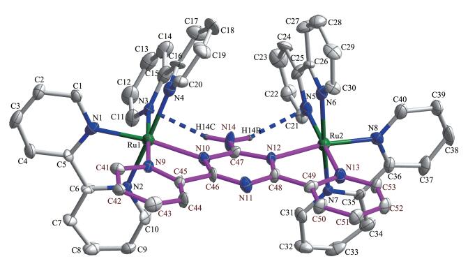
Hydrogen atoms except for protons on N14 are omitted for clarity
In 2, the intramolecular hydrogen bonds (N14… N5 0.307(2) nm, ∠N14-H14B…N5=143.3°; N14…N3 0.309(2) nm, ∠N14-H14A…N3=146.3°) existing in the cation. The cations and PF6- anions are then linked to three dimensional framework via abundant C-H…F hydrogen bonds.
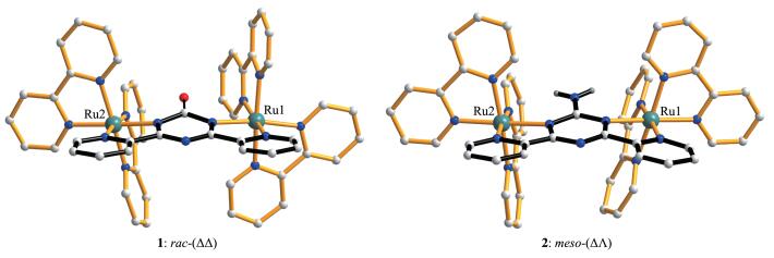
It is worth mentioning that the stereoisomeric forms of 1 and 2 are different. According to the refer-ence, the terminal bidentate polypyridyl ligands 'above'and'below'the plane of the bridging ligand bear a significantly different relationship in the rac and meso diastereoisomers[9]. For the complexes of this type, the terminal rings above the plane of the bridge are approximately parallel for the meso diastereoiso-mers and orthogonal for the rac diastereoisomers. Therefore, we can find that complex 1 is in rac - (ΔΔ) form while 2 is in meso-(ΔΛ) form, as shown in Fig. 5.
Electrochemical behaviors of the complexes in acetonitrile were studied through CV and DPV meth-ods using Ag/AgCl reference electrode and Bu4NPF 6 as the supporting electrolyte. The CV and DPV of the complexes are displayed in Fig. 6.
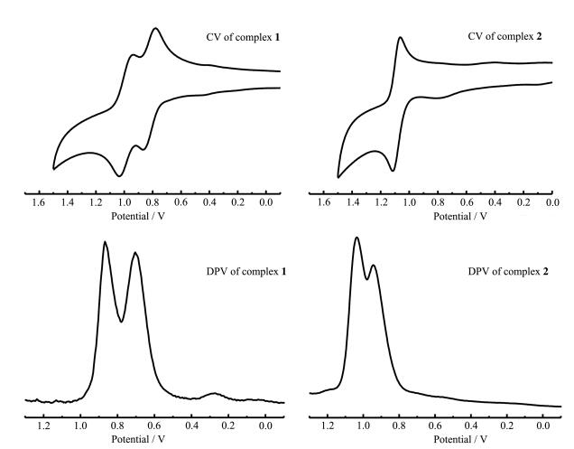
As we can see, two sequential redox couples at E1/2, 1=0.779 V andE1/2, 2=0.943 V were evident on the CV of dinuclear ruthenium complex 1. The two irre-versible oxidation processes correspond to sequential one electron oxidation from Ru(Ⅱ) to Ru(Ⅲ), which occur at different potentials for the two metal centers, an indi-cation of electronic coupling through the bridging ligands. DPV also confirms the splitting between two half - wave potentials of complex 1. The extent of elec-tronic interaction between the two metal centers in homobimetallic complexes can be estimated from the differences between the two oxidation potentials (ΔE1/2=|E1/2, 2-E1/2, 1|)[31]. The ΔE1/2 value obtained in this way is found to be 0.164 V. For complex 2, two redox couples could be found on the DPV curve, the splitting between two metal - based oxidation peaks (ΔE) was 0.092 V, which indicates that the metal -metal interactions are relatively weak. However, there was only one reversible redox couple observed at 1.064 V in the CV curve, this could be caused by the adsorption of the species onto the surface of the working electrode. The compropor-tionation constant Kc (Kc=10ΔE/0.059) [32] values (602 for complex 1 and 36 for complex 2) for the equilibrium RuⅡ-RuⅡ +RuⅢ-RuⅢ ⇌ 2RuⅡ-RuⅢ, also suggest the presence of electronic coupling between the metal centers.
Comparing the data, we can find that the poten-tials of 1 are slightly less positive than those of 2. This is probably because the bridging ligand of 1 is deprot-onated, and the electron density at the Ru(Ⅱ) center is larger, making its oxidation potential less-positive values.
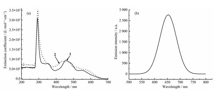
Absorption spectral profiles of the complexes in acetonitrile are presented in Fig. 7, and the related data are summarized in Table 4. In order to compare the relative intensity of the absorption bands, isovolume solutions (50 μmol·L-1) of the complexes were used.
 下载:
导出CSV
下载:
导出CSV
| Complex | λmax / nm (ε / (L·mol-1·cm-1)) | |||||||
| UV region | Visible region | |||||||
| 1 | 292 (3.09×10⁻³) | 353 (7.75×10⁻²) | 458 (9.52×10⁻²) | 484 (8.13×10⁻²) | 550 (4.30×10⁻²) | |||
| 2 | 294 (3.54×10⁻³) | 436 (9.06×10⁻²) | 466 (8.86×10⁻²) | 517 (5.98×10⁻²) | 568 (4.75×10⁻²) | |||
The highly intense absorption bands observed in the UV region arised mainly due to the π - π* transi-tions within the ligands[6, 31, 33]. The strong absorption band at 292 nm for 1 and 294 nm for 2 can be attribut-ed to the π - π* transition of bpy ligand. The 353 nm absorption band of 1, can be assigned to the π-π* tran-sition of OBPT- ligand. The moderately intense broad bands in the visible region can be attributed to M (dπ) → L(π*) MLCT transitions. In the absorption spectral profiles, it was observed that the MLCT bands in the complexes underwent gradual blue-shifts from 1 to 2. This is probably because that the bridging ligand of 1 is deprotonated. The electron density at the Ru (Ⅱ)center is larger, its oxidation potential is a less-positive value, and the MLCT band energy is smaller. This is consistent with the electrochemical behaviors.
The emission spectra of both complexes have been measured in degassed CH3CN solution at room temperature. Interestingly, complex 1 had a strong emission maximum at 653 nm with excitation wave-length at 450 nm (Fig. 7b), while complex 2 was not lu-minescent. This is probably because the proton of the triazine ring of ligand HABPT caused the quenching of complex 2, the intensity of emission has been rapidly reduced.
In summary, we have synthesized two new bridged dinuclear Ru(Ⅱ) complexes based on 2-substituted-4, 6 - di(2-pyridyl)-1, 3, 5-triazine and studied their single - crystal structures, electrochemical behaviors, UV-Vis absorption and luminescence properties at room tem-perature. The X-ray structures reveal that both com-plexes are dinuclear structures, and each Ru(Ⅱ) center are in distorted octahedral geometry with the bridging ligand in 1 being deprotonated but that of 2 being not. Moreover, two complexes are different in stereoisomer-ic forms: 1 shows in the rac - (ΔΔ) form, but 2 in the meso-(ΔΛ) form. Electrochemical studies (CV and DPV) display two metal centered oxidations which demonstrated the presence of metal-metal electronic in-teraction through bridging ligands, and the oxidation potentials of 1 are less positive than those of 2, which may be attributed to the deprotonation of the ligand. The MLCT bands in the complexes undergo gradual blue-shifts from 1 to 2, and moreover, the lumines-cence of 1 exhibits a strong emission but 2 is not lumi-nescent. It seems that the electron charge of bridging ligand will significantly affect the spectral and electro-chemical properties of the dinuclear ruthenium complexes.
D'Alessandro D M, Keene F R. Chem. Rev., 2006, 106(6):2270-2298 doi: 10.1021/cr050010o
Kaim W, Lahiri G K. Angew. Chem. Int. Ed., 2007, 46:1778-1796 doi: 10.1002/anie.200602737
D'Alessandro D M, Keene F R. Chem. Soc. Rev., 2006, 35:424-440 doi: 10.1002/chin.200637201
Gill M R, Thomas J A. Chem. Soc. Rev., 2012, 41:3179-3192 doi: 10.1039/c2cs15299a
Li X, Gorle A K, Sundaraneedi M K, et al. Coord. Chem. Rev., 2018, 375:134-147 doi: 10.1016/j.ccr.2017.11.011
Paul P, Tyagi B, Bilakhiya A K, et al. Inorg. Chem., 2000, 39:14-22 doi: 10.1021/ic990938x
Berger R M, Ellis D D. Inorg. Chim. Acta, 1996, 241:1-4 doi: 10.1016/0020-1693(95)04771-9
D'Alessandro D M, Topley A C, Davies M S, et al. Chem. Eur. J., 2006, 12:4873-4884 doi: 10.1002/chem.200501483
Keene F R. Chem. Soc. Rev., 1998, 27:185-194 doi: 10.1039/a827185z
Buck D P, Spillane C B, Collins J G, et al. Mol. BioSyst., 2008, 4:851-854 doi: 10.1039/b803216e
D'Alessandro D M, Junk P C, Keene F R. Supramol. Chem., 2005, 17(7):529-546 doi: 10.1080/10610270500310537
Lenis-Rojas O A, Roma-Rodrigues C, Fernandes A R, et al. Inorg. Chem., 2017, 56:7127-7144 doi: 10.1021/acs.inorgchem.7b00790
Jiang C W. Inorg. Chim. Acta, 2005, 358:1231-1236 doi: 10.1016/j.ica.2004.11.011
Guckian A L, Doering M, Ciesielski M, et al. Dalton Trans., 2004:3943-3949 doi: 10.1039/b409189b
O'Reilly F M, Kelly J M. New J. Chem., 1998, 22:215-217 doi: 10.1039/a800148k
Smith J A, Morgan J L, Turley A G, et al. Dalton Trans., 2006:3179-3187 doi: 10.1002/chem.200801790
Gill M R, Garcia-Lara J, Foster S J, et al. Nat. Chem., 2009, 1:662-667 doi: 10.1038/nchem.406
Cheng F X, Chen J S, Tang N, et al. Transition Met. Chem., 2012, 37:721-726 doi: 10.1007/s11243-012-9643-y
Cao M L, Wu J J, Mo H J, et al. J. Am. Chem. Soc., 2009, 131:3458-3459 doi: 10.1021/ja810107a
Cao M L, Hao H G, Zhang W X, et al. Inorg. Chem., 2008, 47:8126-8133 doi: 10.1021/ic800585d
Wu J J, Cao M L, Zhang J Y, et al. RSC Adv., 2012, 2:12718-12723 doi: 10.1039/c2ra22549b
Wu J J, Xue W, Cao M L, et al. CrystEngComm, 2011, 13:5495-5501 doi: 10.1039/c1ce05370a
Wu J J, Ye Y X, Qiu Y Y, et al. Inorg. Chem., 2013, 52:6450-6456 doi: 10.1021/ic400340y
Drew M G B, Hudson M J, Iveson P B, et al. J. Chem. Soc. Dalton Trans., 2000:2711-2720
Cao M L, Mo H J, Ye B H. Cryst. Growth Des., 2009, 9:546-554 doi: 10.1021/cg800852d
Wieprecht T, Dubs M J, Schlingloff G. Int. Patent, WO2005105303-A1. 2005-11-10.
Case F H, Koft E. J. Am. Chem. Soc., 1959, 81:905-906 doi: 10.1021/ja01513a037
Sprintschnik G, Sprintschnik H W, Kirsch P P, et al. J. Am. Chem. Soc., 1977, 99:4947-4954 doi: 10.1021/ja00457a010
Sheldrick G M. Acta Crystallogr. Sect. A, 2008, A64:112-122
Sheldrick G M. Acta Crystallogr. Sect. C, 2015, C71:3-8
Mardanya S, Karmakar S, Mondal D, et al. Inorg. Chem., 2016, 55(7):3475-3489 doi: 10.1021/acs.inorgchem.5b02912
Richardson D E, Taube H. Inorg. Chem., 1981, 20:1278-1285 doi: 10.1021/ic50218a062
Juris A, Balzani V. Coord. Chem. Rev., 1988, 84:85-277 doi: 10.1016/0010-8545(88)80032-8

Figure 2 ORTEP drawing of the cation in 1 with 20% thermal ellipsoids
All hydrogen atoms on carbon atoms are omitted for clarity

Figure 3 Hydrogen bonds between lattice water molecules and cations in 1
Most hydrogen atoms are omitted for clarity; Symmetry codes: i-x, -y-3, -z; iix+1/2, y-1/2, z; iii-x+1/2, -y-7/2, -z

Figure 4 ORTEP drawing of the cation in 2 with 20% thermal ellipsoids
Hydrogen atoms except for protons on N14 are omitted for clarity

Figure 6 CV and DPV curves of 1 and 2 in acetonitrile containing 0.1 mol·L-1 of Bu4NPF6 as the supporting electrolyte

Figure 7 (a) UV-Vis absorption spectra of complexes 1 (solid line) and 2 (dash line) and (b) emission spectra of complex 1 at room temperature
Table 1. Crystal data and structure refinement for complexes 1 and 2
| Complex | 1 | 2 |
| Molecular formula | C54H49F18N13O4.5P3Ru2 | C53H43F24N14O0.5P4Ru2 |
| Formula weight | 1 589.11 | 1 666.03 |
| Crystal system | Monoclinic | Orthorhombic |
| Space group | C2/c | Pca21 |
| a / nm | 2.452 9(3) | 1.287 8(2) |
| b / nm | 1.411 1(1) | 2.427 0(4) |
| c / nm | 3.980 1(4) | 2.169 6(3) |
| β/(°) | 93.834(2) | |
| V/ nm3 | 13.746(2) | 6.781(2) |
| Z | 8 | 4 |
| Dc / (g·cm-3) | 1.536 | 1.632 |
| Absorption coefficient / mm-1 | 0.611 | 0.654 |
| F(000) | 6 360 | 3 308 |
| Crystal size / mm | 0.300×0.250×0.100 | 0.260×0.250×0.210 |
| θ range for data collection / (°) | 1.664~25.999 | 2.056~24.999 |
| Limiting indices | -30 ≤ h ≤ 30, -17 ≤ k ≤ 17, -49 ≤ l ≤ 49 | -15 ≤ h ≤ 8, -15 ≤ k ≤ 28, -24 ≤ l ≤ 10 |
| Reflection collected, unique | 66 533, 13 491 (Rint=0.050 8) | 14 107, 7 098 (Rint=0.074 3) |
| Reflection observed | 10 132 | 4 414 |
| Data, restraint, parameter | 13 491, 45, 815 | 7 098, 149, 732 |
| Goodness-of-fit on F2 | 1.002 | 1.024 |
| Final R indices [I > 2σ(I)]a, b | R1=0.076 4, wR2=0.231 2 | R1=0.072 7, wR2=0.158 1 |
| R indices (all data)a, b | R1=0.095 8, wR2=0.257 1 | R1=0.110 3, wR2=0.174 2 |
| a R1=∑||Fo|-|Fc||/∑|Fo|, b wR2=[∑w(Fo2-Fc2)2/∑w(Fo2)2]1/2. | ||
 下载: 导出CSV
Table 2. Selected bond lengths (nm) and angles (°) of complexes 1 and 2
| 1 | |||||
| Ru1-N1 | 0.206 1(5) | Ru2-N6 | 0.204 3(6) | C47-N10 | 0.139 2(7) |
| Ru1-N2 | 0.206 8(5) | Ru2-N7 | 0.204 9(6) | C47-N12 | 0.140 2(8) |
| Ru1-N3 | 0.204 5(5) | Ru2-N8 | 0.205 8(5) | C47-O1 | 0.121 4(7) |
| Ru1-N4 | 0.207 3(5) | Ru2-N12 | 0.208 1(5) | C48-N11 | 0.133 2(8) |
| Ru1-N9 | 0.205 4(5) | Ru2-N13 | 0.207 4(6) | C48-N12 | 0.132 7(8) |
| Ru1-N10 | 0.209 5(5) | C46-N10 | 0.133 4(8) | ||
| Ru2-N5 | 0.206 1(5) | C46-N11 | 0.133 7(8) | ||
| N1-Ru1-N2 | 78.7(2) | N4-Ru1-N10 | 90.3(2) | N6-Ru2-N7 | 96.7(3) |
| N3-Ru1-N4 | 78.5(2) | N2-Ru1-N4 | 172.7(2) | N7-Ru2-N5 | 85.8(2) |
| N9-Ru1-N10 | 78.4(2) | N1-Ru1-N10 | 170.9(2) | N8-Ru2-N5 | 97.7(2) |
| N3-Ru1-N1 | 90.3(2) | N2-Ru1-N10 | 95.1(2) | N6-Ru2-N13 | 89.5(3) |
| N9-Ru1-N1 | 95.1(2) | N3-Ru1-N9 | 172.0(2) | N8-Ru2-N13 | 95.5(2) |
| N3-Ru1-N2 | 95.8(2) | N8-Ru2-N12 | 85.4(2) | N5-Ru2-N13 | 98.0(2) |
| N9-Ru1-N2 | 91.0(2) | N6-Ru2-N8 | 174.2(2) | N6-Ru2-N12 | 98.5(2) |
| N9-Ru1-N4 | 94.8(2) | N6-Ru2-N5 | 78.5(3) | N7-Ru2-N12 | 98.4(2) |
| N1-Ru1-N4 | 96.6(2) | N7-Ru2-N8 | 78.4(2) | N7-Ru2-N13 | 173.2(2) |
| N3-Ru1-N10 | 96.9(2) | N13-Ru2-N12 | 78.0(2) | N5-Ru2-N12 | 175.2(2) |
| 2 | |||||
| Ru1-N1 | 0.203 6(6) | Ru2-N6 | 0.2082(8) | C47-N14 | 0.129(2) |
| Ru1-N2 | 0.203 7(7) | Ru2-N7 | 0.2035(8) | C47-N10 | 0.139 0(9) |
| Ru1-N3 | 0.205 2(8) | Ru2-N8 | 0.2040(7) | C47-N12 | 0.139 0(8) |
| Ru1-N4 | 0.207 3(9) | Ru2-N12 | 0.2117(5) | C48-N11 | 0.139 0(9) |
| Ru1-N9 | 0.206 4(8) | Ru2-N13 | 0.2073(8) | C48-N12 | 0.139 0(8) |
| Ru1-N10 | 0.212 8(5) | C46-N11 | 0.1390(8) | ||
| Ru2-N5 | 0.207 6(8) | C46-N10 | 0.1390(8) | ||
| N1-Ru1-N3 | 85.9(4) | N9-Ru1-N10 | 77.6(3) | N7-Ru2-N8 | 78.8(4) |
| N1-Ru1-N4 | 99.7(4) | N2-Ru1-N3 | 95.8(5) | N7-Ru2-N12 | 95.5(4) |
| N1-Ru1-N9 | 93.3(4) | N2-Ru1-N4 | 173.9(4) | N7-Ru2-N13 | 89.6(4) |
| N1-Ru1-N10 | 170.4(3) | N2-Ru1-N9 | 89.6(4) | N8-Ru2-N5 | 87.3(4) |
| N1-Ru1-N2 | 79.3(4) | N2-Ru1-N10 | 97.3(4) | N8-Ru2-N6 | 97.3(4) |
| N3-Ru1-N4 | 78.0(5) | N5-Ru2-N6 | 78.4(4) | N8-Ru2-N12 | 170.3(4) |
| N3-Ru1-N9 | 174.2(5) | N5-Ru2-N12 | 101.3(3) | N8-Ru2-N13 | 94.3(4) |
| N3-Ru1-N10 | 103.5(3) | N6-Ru2-N12 | 89.1(3) | N13-Ru2-N5 | 174.0(4) |
| N4-Ru1-N10 | 84.6(4) | N7-Ru2-N5 | 96.4(4) | N13-Ru2-N6 | 95.6(4) |
| N9-Ru1-N4 | 96.5(4) | N7-Ru2-N6 | 173.7(4) | N13-Ru2-N12 | 77.7(3) |
下载: 导出CSV
Table 3. Hydrogen bonds between lattice water molecules and cations in 1
| D-H…A | d(D-H) / nm | d(H…A) / nm | d(D…A) / nm | ∠DHA / (°) |
| O1W-H1WA…O1 | 0.085 | 0.211 | 0.293(2) | 161.9 |
| O1W-H1WB…O2W | 0.086 | 0.182 | 0.267(2) | 167.0 |
| O2W-H2WB…O4Wi | 0.087 | 0.172 | 0.255(3) | 156.8 |
| O3W-H3WA…O4W | 0.088 | 0.208 | 0.296(4) | 178.2 |
| O3W-H3WB…O1Wii | 0.086 | 0.200 | 0.284(3) | 168.1 |
| O4W-H4WA…O3W | 0.087 | 0.221 | 0.296(4) | 145.2 |
| O4W-H4WB…O4Wiii | 0.088 | 0.186 | 0.269(6) | 156.5 |
| Symmetry codes: i -x, -y-3, -z; ii x-1/2, y+1/2, z; iii -x-1/2, -y-5/2, -z. | ||||
下载: 导出CSV
Table 4. Absorption spectra of complexes 1 and 2 in acetonitrile
| Complex | λmax / nm (ε / (L·mol-1·cm-1)) | |||||||
| UV region | Visible region | |||||||
| 1 | 292 (3.09×10⁻³) | 353 (7.75×10⁻²) | 458 (9.52×10⁻²) | 484 (8.13×10⁻²) | 550 (4.30×10⁻²) | |||
| 2 | 294 (3.54×10⁻³) | 436 (9.06×10⁻²) | 466 (8.86×10⁻²) | 517 (5.98×10⁻²) | 568 (4.75×10⁻²) | |||
下载: 导出CSV

 扫一扫看文章

扫一扫关注我们