Scheme 1.
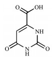
Structure of orotic acid
Synthesis, Structure and Hydrogen-Bonding Interaction of Three Metal-Orotic Acid Complexes
Pei-Pei CUI , Yue ZHAO , Peng WANG , Rui JIN , De-Jie JIAO , Xiu-Ling ZHANG , Wen-Ning YAN

Metal - organic complexes are an interesting and useful class of compound. During the past decades, the synthesis and characterization of metal-organic com-plexes is rapidly developing in chemical science[1-3]. The reason is that their interesting structures and topol-ogy are diverse on the one hand, on the other hand, their potential applications are tempting, such as gas storage and separation, biomedical applications, envi-ronmental pollution control and catalysis[4-7]. Metal-organic complexes as a rapidly developing class of organic-inorganic hybrid materials are constructed from metal ion/cluster nodes and functional organic ligands through coordination bonds. According to the composition, the certain features of ligands are crucial, such as flexibility, functionality and versatile binding modes[8-10]. In order to research the biomedical applica-tions of complexes, the biocompatibility must be con-sidered as important as the function. Thus, more and more natural biomolecules as ligands were considered to be used. Nucleobase is one of the first such ligands used to synthesize complexes in the form of crystals successfully. In 2009, zinc-adeninate framework bio-MOF-1 with crystal structure was reported by Rosi group[11]. After that more and more complexes as crystals were synthesized based on biomolecules, such as bio-MOF-100 and medi-MOF-1[12-15]. However, compared to other types of metal - organic complexes, the number of metal-biomolecule complexes is very lit-tle. Thus, it is necessary to research the method using biomolecules as ligands to synthesize crystalline metal-organic complexes.
In this work, we choose orotic acid (H3Or) as ligand. H3Or (vitamin B13) as a uracil carboxylic acid is one simple natural biomolecule ligand, and it plays a key role in the biosynthesis of pyrimidine bases of nucleic acids. Therefore, H3Or has biological activi-ty[16-20]. On the other side, comparing with ligands only containing carboxylate or pyridyl group, as we all know, the reports on pyridyl - carboxylate ligands are also less to now. The carboxylate group can have vari-able coordination modes and strong coordination abili-ties, while the pyridyl-containing ligands have been demonstrated to be powerful in construction of com-plexes with novel structures and topologies[21-23]. From the analysis of structure, orotic acid as ligand has the advantages of both carboxylic acid and pyridine (Scheme 1). However, because of the structure with low symmetry, it is difficult to synthesize ideal crystals using H3Or as ligand[24-27]. In this work, we designed and synthesized three new complexes, namely {[Cu (HOr)2] ·2NH2(CH3)2}n (R - 1), [Co2(HOr)2(bipy) (H2O)6] · 2H2O (R - 2) and {[Ni2(HOr)2(1, 3-dpp)2(H2O)2] [Ni(HOr) (1, 3-dpp)(H2O)]2·(1, 3-dpp)·2H2O}n (R- 3) (H3Or=orotic acid, bipy=4, 4' - bipyridine and 1, 3 - dpp=1, 3 - di(4 - pyridyl)propane). R-1 is mononuclear complex with 2D sheet structure, and R-2 is a simple monomolecular complex. R-3 is a complicated structure containing 2D layers and 1D chains, which looks like sandwich. In the complexes, intermolecular hydrogen bonding inter-actions exist, which result in 3D supramolecular frame-works, respectively. In addition, the thermal stability and infrared spectra property of the complexes were investigated.
All commercially available chemicals and sol-vents are of reagent grade and were used as received without further purification. Elemental analysis for C, H and N was performed on a Perkin-Elmer 240 C Ele-mental Analyzer. FT-IR spectra was recorded in a range of 400~4 000 cm-1 on a Bruker Vector 22 FT-IR spectrophotometer using KBr pellets. Thermogravimet-ric analysis (TGA) was performed on a simultaneous SDT 2960 thermal analyzer under nitrogen with a heat-ing rate of 10 ℃·min-1.
H3Or (15.6 mg, 0.1 mmol) and biphenyl - 4, 4' - dicarboxylic acid (24.2 mg, 0.1 mmol) were added in DMF (8 mL) and stirred. After two hours, CuSO4·5H 2O (25.0 mg, 0.1 mmol) was added. Then, the mixture was sealed in a 20 mL Teflon - lined steel bomb for half an hour and heated at 90 ℃ for three days. Blue block sin-gle crystals of R -1 were obtained in 35% yield. Anal. Calcd. for C14H20N6O8Cu(%): C, 36.25; H, 4.35; N, 18.12. Found(%): C, 36.61; H, 4.16; N, 18.51. IR (KBr pellet, cm-1): 3 120(s), 1 651(s), 1 480(m), 1 401(s), 1 342(m), 1 220(s), 1 029(m), 947(m), 847(w), 791(s), 685(w), 592(w) (Fig.S3a).
H3Or (1.56 g, 10 mmol) and KOH (1.68 g, 30 mmol) were stirred in 100 mL H2O for four hours, and this solution was called S-1. 1.0 mL S-1, 1.0 mL Co(OAc)2·4H 2O (0.1 mol·L-1) aqueous solution and 4, 4' - bipyridine (15.6 mg, 0.1 mmol) were added in 8.0 mL H2O and stirred for two hours. The resultant solu-tion was sealed in a 20 mL Teflon-lined stainless -steel container and heated at 90 ℃ for 3 days. Orange block single crystals of R-2 were obtained in 65% yield. Anal. Calcd. for C20H28N6O16Co2(%): C, 33.07; H, 3.89; N, 11.57. Found(%): C, 33.21; H, 3.56; N, 11.72. IR (KBr pellet, cm-1): 3 364(s), 1 623(s), 1 480(m), 1 384 (s), 1 321(m), 1 217(m), 1 015(m), 805(s), 633(m), 576 (w) (Fig.S3b).
1.0 mL S-1, 1.0 mL Ni(OAc)2·4H2O (0.1 mol·L-1) aqueous solution and 1, 3 - di(4 - pyridyl)propane (19.8 mg, 0.1 mmol) were added in 8.0 mL H 2O and stirred for two hours. The resultant solution was sealed in a 20 mL Teflon-lined stainless - steel container and heated at 150 ℃ for 3 days. Green block single crystals of R - 3 were obtained in 52% yield. Anal. Calcd. for C85H 90N18O22Ni4(%): C, 52.34; H, 4.65; N, 12.93. Found (%): C, 52.65; H, 4.56; N, 12.51. IR (KBr pellet, cm-1): 3 404(s), 1 620(s), 1 469(m), 1 378(s), 1 328(w), 1 220 (s), 1 071(m), 948(m), 853(w), 795(s), 674(w), 615(w) (Fig.S3c).
Diffraction data for R-1, R-2 and R-3 were col-lected on a Bruker Smart Apex Ⅱ CCD with graphite monochromated Mo Kα radiation source (λ=0.071 073 nm) in φ- ω scan mode. The diffraction data were inte-grated by using SAINT program[28], which was also used for the intensity corrections for the Lorentz and polar-ization effects. Semi - empirical absorption corrections were applied using SADABS program[29]. All the struc-tures were solved by direct methods using SHELXS - 97[30] and the non - hydrogen atoms were refined on F2 by full-matrix least-squares procedures with SHELXL-2018[31]. The final formulas were calculated from TGA and elemental analysis. Hydrogen atoms except those of water molecules were generated geometrically and refined isotropically using the riding model. The hydro-gen atoms of coordinated water molecules were found directly. The details of the crystal parameters, data col-lection and refinements for R-1, R-2 and R-3 are sum-marized in Table 1. Selected bond lengths and angles for R-1, R-2 and R-3 are listed in Table S1 (Support-ing information). The parameters of hydrogen bonds for R-1, R-2 and R-3 are listed in Table S2.
 下载:
导出CSV
下载:
导出CSV
| Complex | R-1 | R-2 | R-3 |
| Formula | C14H20N6O8Cu | C20H28N6O16Co2 | C85H90N18O22Ni4 |
| Formula weight | 463.90 | 726.34 | 1 950.58 |
| Crystal system | Monoclinic | Triclinic | Triclinic |
| Space group | P21/c | P1 | P1 |
| a / nm | 1.003 94(18) | 0.605 9(5) | 1.078 5(3) |
| b / nm | 0.977 15(18) | 1.069 0(5) | 1.486 9(5) |
| c / nm | 0.901 68(16) | 1.168 9(5) | 2.802 0(8) |
| α/(°) | 66.807(5) | 82.737(5) | |
| β/(°) | 92.401(2) | 83.841(5) | 86.264(5) |
| γ/(°) | 85.606(5) | 82.435(5) | |
| V / nm3 | 0.883 8(3) | 0.691 4(7) | 4.413(2) |
| Z | 2 | 1 | 2 |
| Dc / (g·cm-3) | 1.743 | 1.744 | 1.468 |
| μ / mm-1 | 1.297 | 1.288 | 0.924 |
| F(000) | 478 | 372 | 2 028 |
| Unique reflection | 1 775 | 2 714 | 10 426 |
| Observed reflection [I > 2σ(I)] | 2 002 | 3 032 | 16 512 |
| Parameter | 135 | 199 | 1168 |
| GOF | 1.004 | 1.075 | 1.036 |
| Final R indices [I > 2σ(I)]a, b | R1=0.037 1, wR2=0.122 3 | R1=0.039 9, wR2=0.133 1 | R1=0.069 8, wR2=0.204 2 |
| R indices (all data) | R1=0.044 1, wR2=0.142 1 | R1=0.044 7, wR2=0.140 6 | R1=0.108 7, wR2=0.227 0 |
| Largest difference peak and hole / (e·nm-3) | 760 and -524 | 786 and -664 | 2 059 and -638 |
| aR1=Σ||Fo|-|Fc||/Σ|Fo|; b wR2=[Σw(Fo2-Fc2)2]/Σw(Fo2)2]1/2. | |||
CCDC: 1983479, R-1; 1983481, R-2; 1983482, R-3.
The crystal structure of R-1 was determined by single-crystal X-ray diffraction analysis. R-1 is a mono-nuclear complex crystallizing in P21/c space group. The asymmetric unit of R-1 consists of one Cu(Ⅱ) ion, one HOr2- ligand, and free one dimethylamine ion (Fig. S1). Each Cu(Ⅱ) is coordinated by two chelated sym-metry related HOr2- ligands through carboxylate oxy-gen O1 and N1 in the equatorial plane resulting in [Cu(HOr)2] secondary building unit (SBU). In the trans axial positions, there are two other oxygen atoms (O2ii and O2iii) from other [Cu(HOr) 2] SBUs linked with Cu(Ⅱ), and the distance between Cu(Ⅱ) and O2ii or O2iii is 0.273 3 nm measured by Mercury program, which results in a 4+2 coordination environment around each Cu(Ⅱ) (Fig. 1a). The coordination environment is similar to the reported complex {[Cu(H2iso)2] ·H 2 O}n (H3iso= isoorotic acid) [15]. Such [Cu(HOr)2] SBUs adjacent to each other link to form a 2D sheet parallel to bc plane (Fig. 1b). Adjacent 2D sheets through H-bonding inter-action connect to form a 3D framework (Fig. 1c) and the holes of the framework are occupied by dimethylamine molecules (Fig. 1d).
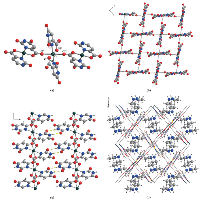
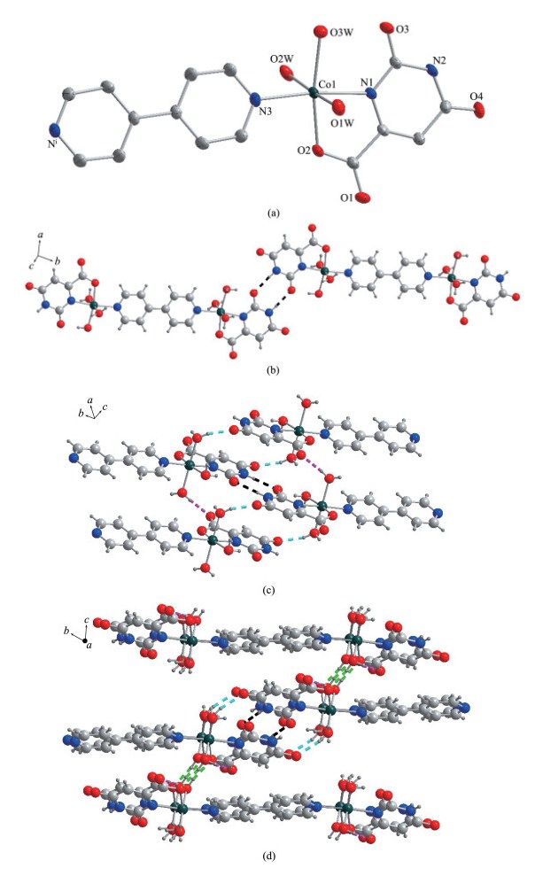
R-2 as a monomolecular complex crystallizes in the triclinic P1 space group by single crystal X-ray structural analysis. The asymmetric unit of R-2 con-sists of one Co(Ⅱ)ion, one HOr2- ligand, half 4, 4'-bipy ligand and three coordinated water molecules. The coordination number of Co(Ⅱ)ion is six. Co(Ⅱ)ion is the center with distorted octahedron coordination geome-try, in which the equatorial positions are occupied by N1, N3, O2 and O3W, and the axial positions are occu-pied by coordinated water molecules (Fig. 2a). Two [Co(HOr) (H2O)3] units link together by 4, 4'-bipy to form [Co2(HOr)2(4, 4'-bipy) (H2O)6] dimer. Between [Co2 (HOr)2(4, 4'-bipy) (H2O)6] dimers, there are hydro-gen bonding interactions. Such dimers connect together to form a 1D chain by N2- H2…O3 (Fig. 2b and Table S2). Though intermolecular hydrogen bonding interac-tion from two axial positions O1W and O2W, the chains link together to form irregular layer structure (Fig. 2c and Table S2). The 2D layers are further extended into a 3D structure though O2W-H2O2…O2 interaction (Fig. 2d and Table S2).
As far as we know, R-3 has the most complex structure based on H3Or as ligand in the reports until now[15-27]. Crystallographic analysis indicates that R-3 crystallizes in the triclinic space group P1 with compli-cated sandwich structure. In the structure of R-3, except free 1, 3-dpp and water molecules, there are six different coordination modes of Ni(Ⅱ).
Ni1 is coordinated with distorted octahedron coor-dination geometry, in which the equatorial positions are occupied by O1A, O6A, N3A and N6A, and the axi-al positions are occupied by N1A and N8A (Fig. 3a). Among them, O1A and N3A come from one HOr2-, while O6A and N6A come from another HOr2-. For Ni2, it is coordinated with O2A, O5A, O1W, O2W, N2A and N7A. Among them, O2A and O5A come from different HOr2-, while N2A and N7A come from differ-ent 1, 3 - dpp molecules. Thus, the carboxyl groups of HOr2- bridge adjacent Ni1 and Ni2 to form a chain along a axis (Fig. 3b). Along c axis, the adjacent chains connect the auxiliary ligand of 1, 3-dpp to form a layer (Fig. 3b).
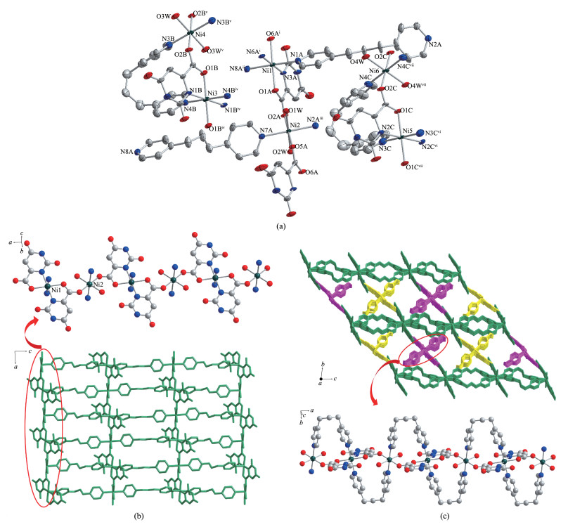
Except Ni1 and Ni2, the coordination numbers of Ni3, Ni4, Ni5 and Ni6 are also six. Besides, the coordi-nated mode of Ni3 is similar to Ni5, and Ni4 is similar to Ni6. For Ni3, it is coordinated by four N atoms and two O atoms with distorted octahedron coordination geometry. Besides, two O1B atoms and two N1B atoms occupy the equatorial positions and two N4B from dif-ferent 1, 3-dpp molecules occupy the axial positions for Ni3 (Fig. 3a). The carboxyl group of HOr2- ligand and N3B and N4B of 1, 3-dpp bridge adjacent Ni3 and Ni4. The remainder positions of Ni4 are coordinated with water molecules. Such adjacent Ni3 and Ni4 are linked by bridging the carboxyl groups and 1, 3-dpp molecules to form a chain along a axis, which is same to Ni5 and Ni6 (Fig. 3c). Between the adjacent layers based on Ni1 and Ni2, the chains based on Ni3~Ni6 fill. Thus, the sandwich structure is formed (Fig. 3c). Interestingly, in the sandwich structure, intermolecular hydrogen bonding interaction also exists between the layers and the chains (Fig.S2 and Table S2).
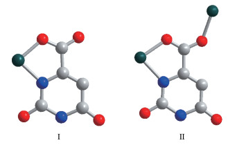
Comparing with other organic ligands, the reports on biomolecules are less. In this work, three metal-organic complexes were synthesized and characterized based on orotic acid (H3Or), a simple natural biomole-cule. In the complexes, although all H3Or molecules loss two protons and becomes HOr2-, there are two coor-dination modes of HOr2- ligand (Fig. 4). In the struc-tures of R-1 and R-2, all HOr2- ligands adopt chelate mode (mode Ⅰ), in which one oxygen atom of the car-boxylate group and one adjacent nitrogen atom coordi-nate one metal ion, and other oxygen and nitrogen atoms are not involved in coordination. This chelate mode is the main reason leading to the low dimensional structure of R-1 and R-2. Similarly, the complicated sandwich structure of R-3 is also inseparable from the coordination mode of HOr2- ligand. In R-3, all HOr2-ligands adopt another coordination mode (mode Ⅱ). In mode Ⅱ, one oxygen atom of the carboxylate group with chelate mode links one metal ion as mode Ⅰ, and the other oxygen atom of the carboxylate group with bridge mode links another metal ion. Thus, the struc-ture of R-3 is more complex.
In addition, the deprotonation of carboxylate group for H3Or was confirmed as described above by crystal structural analyses as well as by IR spectra. In IR spectra, no obvious bands between 1 680 and 1 760 cm-1 were found, which are originated from the carbox-ylic group (- COOH) (Fig.S3). Although biphenyl-4, 4'-dicarboxylic acid does not exist in the final structures of R-1, it should be pointed out that it may play impor-tant role in the formation of R-1. Because without addi-tion of biphenyl-4, 4' -dicarboxylic acid in the synthetic reactions, no crystals were obtained.
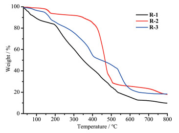
The thermal stability of complexes is important for their application research, so thermogravimetric analy-ses (TGA) of R-1, R-2 and R-3 were measured. The thermal stability was examined by TGA in the N2 atmo-sphere from 30 to 800 ℃ (Fig. 5). The result indicates that R-1 showed a weight loss of 19.2% before 200 ℃, which corresponds to the release of dimethylamine (Calcd. 19.8%). Above 200 ℃, R-1 began to decom-pose. For R-2, the first weight loss of 5.1% from 30 to 170 ℃ is in accordance with the loss of free water mole-cules (Calcd. 5.0%). The further weight loss was observed at about 350 ℃, owing to the decomposition of R-2. For R-3, before 180 ℃ the weight loss was 12.8% and it results from the loss of free water and 1, 3-dpp molecules (Calcd. 12.1%).
In summary, three metal-organic complexes based on orotic acid were synthesized and characterized. The orotic acid losses two protons and there are two coordi-nation modes in the complexes. R-1 is 2D sheet struc-ture, R-2 is a simple monomolecular complex and R-3 is a complex sandwich structure containing layers and chains. Three-dimensional frameworks of the complex-es can be formed by intermolecular hydrogen bonding interactions. Furthermore, the thermal stability and infrared spectra property of the complexes were researched.
Supporting information is available at http://www.wjhxxb.cn
Cui P P, Wang P, Zhao Y, et al. Cryst. Growth Des., 2019, 19:1454-1470 doi: 10.1021/acs.cgd.8b01628
Wang Z F, Zhang S, Chen Y, et al. Chem. Soc. Rev., 2020, 49:708-735 doi: 10.1039/C9CS00827F
Xiao X, Zou L L, Pang H, et al. Chem. Soc. Rev., 2020, 49:301-331 doi: 10.1039/C7CS00614D
Cui P P, Zhang X D, Wang P, et al. Inorg. Chem., 2017, 56:14157-14163 doi: 10.1021/acs.inorgchem.7b02235
Begum S, Hassan Z, Bräse S, et al. Acc. Chem. Res., 2019, 52:1598-1610 doi: 10.1021/acs.accounts.9b00039
Ma X J, Chai Y T, Li P, et al. Acc. Chem. Res., 2019, 52:1461-1470
Kang Y S, Lu Y, Chen K, et al. Coord. Chem. Rev., 2019, 378:262-280 doi: 10.1016/j.ccr.2018.02.009
Cui P P, Wu J L, Zhao X L, et al. Cryst Growth Des., 2011, 11:5182-5187 doi: 10.1021/cg201181s
Fan W D, Wang X, Zhang X R, et al. ACS Cent. Sci., 2019, 5:1261-1268 doi: 10.1021/acscentsci.9b00423
Cui P P, Cui L F, Zhao X H, et al. Chin. J. Struct. Chem., 2017, 36:1526-1531
An J, Geib S J, Rosi N L. J. Am. Chem. Soc., 2009, 131:8376-8377 doi: 10.1021/ja902972w
An J, Farha O K, Hupp J T, et al. Nat. Commun., 2012, 3:1-6
Li T, Kozlowski M T, Doud E A, et al. J. Am. Chem. Soc., 2013, 135:11688-11691 doi: 10.1021/ja403810k
Su H M, Sun F X, Jia J T, et al. Chem. Commun., 2015, 51:5774-5777 doi: 10.1039/C4CC10159F
Haldar R, Gurunatha K L, Sikdar N, et al. Inorg. Chem. Front., 2015, 2:278-289 doi: 10.1039/C4QI00129J
Erer H, Yeşilel O Z, Darcan C, et al. Polyhedron, 2011, 30:2406-2413 doi: 10.1016/j.poly.2011.06.026
Köse D A, Zumreoglu-Karan B, Sahin O, et al. J. Mol. Struct., 2006, 789:147-151 doi: 10.1016/j.molstruc.2005.12.030
Yeşilel O Z, Soylu M S, Ölmez H, et al. Polyhedron, 2006, 25:2985-2292 doi: 10.1016/j.poly.2006.05.010
Yeşilel O Z, Erer H, Buyukgungor O. CrystEngComm, 2011, 13:1339-1349 doi: 10.1039/C0CE00308E
Kundu S, Nagapradeep N, Mohapatra B, et al. CrystEngComm, 2016, 18:4807-4817 doi: 10.1039/C6CE00719H
Raptopoulou C P, Tangoulis V, Psycharis V. Inorg. Chem., 2000, 39:4452-4459 doi: 10.1021/ic9914084
Yesilel O Z, Erer H, Büyükgüngör O. Polyhedron, 2010, 29:1815-1821 doi: 10.1016/j.poly.2010.02.037
Li X, Bing Y, Zha M Q, et al. CrystEngComm, 2011, 13:6373-6376 doi: 10.1039/c1ce05727h
Xu H R, Zhang Q C, Ren Y P, et al. CrystEngComm, 2011, 13:6361-6364 doi: 10.1039/c1ce05623a
Stankiewicz J, Tomas M, Dobrinovitch I T, et al. Chem. Mater., 2014, 26:5282-5287 doi: 10.1021/cm502142a
Mukiza J, Hosten E C, Gerber T I A. Polyhedron, 2015, 98:251-258 doi: 10.1016/j.poly.2015.06.006
Yadav A, Lama P, Bienko A, et al. Polyhedron, 2018, 141:247-261 doi: 10.1016/j.poly.2017.11.047
SAINT, Program for Data Extraction and Reduction, Bruker AXS, Inc., Madison, WI, 2001.
Sheldrick G M. SADABS, University of Göttingen, Germany, 2003.
Sheldrick G M. SHELXS-97, Program for Crystal Structure Solution, University of Göttingen, Germany, 1997.
Sheldrick G M. SHELXL-2018, Program for Crystal Structure Refinement, University of Göttingen, Germany, 2018.

Figure 1 (a) Coordination environment around Cu(Ⅱ) center(Hydrogen atoms have been omitted for clarity; Symmetry codes: i 1-x, 1-y, 1-z; ii x, 1/2-y, 1/2+z; iii 1-x, 1/2+y, 1/2-z); (b) 2D sheet based on [Cu(HOr)2] SBUs parallel to bc plane; (c) H-bonding between 2D sheet along a and c axes showed by the yellow dashed line; (d) 3D structure of R-1

Figure 2 (a) Coordination environment of Co(Ⅱ)ion in R-2 with 50% thermal ellipsoid probability (Hydrogen atoms have been omitted for clarity; Symmetry code: i -x, - 1-y, 2 -z); (b) 1D chain formed by N2-H2…O3 intermolecular hydrogen bonding interaction shown by dashed black line; (c) Intermolecular hydrogen bonding interaction at two axial positions (purple: O2W-H1O2…O1, blue: O1W-H2O1…O4); (d) 3D structure of R-2 with hydrogen bonds indicated by dashed lines (black: N2-H2…O3, purple: O2W-H1O2…O1, green: O2W-H2O2…O2, blue: O1W-H2O1…O4)

Figure 3 (a) Coordination environment of Ni(Ⅱ) in R-3 with ellipsoids drawn at 50% probability level (The hydrogen atoms and free 1, 3-dpp and water molecules are omitted for clarity; Symmetry codes: i 1+x, y, z; ii 1-x, 1-y, 1-z; iii 1-x, 1-y, -z; iv 1-x, -y, 1-z; v 2-x, -y, 1 -z; vi -x, -y, -z; vii 1-x, -y, -z); (b) Chains based on Ni1, Ni2 and bridging HOr2- ligands linked together by 1, 3-dpp to form 2D layer in R-3; (c) 3D sandwich structure of R-3 containing the chains formed by Ni3-Ni6 and the layers formed by Ni1 and Ni2 (the layers formed by Ni1 and Ni2 are shown in green; the chains formed by Ni3 and Ni4 are shown in yellow; the chains formed by Ni5 and Ni6 are shown in purple)
Table 1. Crystal data and structure refinements for complexes R-1, R-2 and R-3
| Complex | R-1 | R-2 | R-3 |
| Formula | C14H20N6O8Cu | C20H28N6O16Co2 | C85H90N18O22Ni4 |
| Formula weight | 463.90 | 726.34 | 1 950.58 |
| Crystal system | Monoclinic | Triclinic | Triclinic |
| Space group | P21/c | P1 | P1 |
| a / nm | 1.003 94(18) | 0.605 9(5) | 1.078 5(3) |
| b / nm | 0.977 15(18) | 1.069 0(5) | 1.486 9(5) |
| c / nm | 0.901 68(16) | 1.168 9(5) | 2.802 0(8) |
| α/(°) | 66.807(5) | 82.737(5) | |
| β/(°) | 92.401(2) | 83.841(5) | 86.264(5) |
| γ/(°) | 85.606(5) | 82.435(5) | |
| V / nm3 | 0.883 8(3) | 0.691 4(7) | 4.413(2) |
| Z | 2 | 1 | 2 |
| Dc / (g·cm-3) | 1.743 | 1.744 | 1.468 |
| μ / mm-1 | 1.297 | 1.288 | 0.924 |
| F(000) | 478 | 372 | 2 028 |
| Unique reflection | 1 775 | 2 714 | 10 426 |
| Observed reflection [I > 2σ(I)] | 2 002 | 3 032 | 16 512 |
| Parameter | 135 | 199 | 1168 |
| GOF | 1.004 | 1.075 | 1.036 |
| Final R indices [I > 2σ(I)]a, b | R1=0.037 1, wR2=0.122 3 | R1=0.039 9, wR2=0.133 1 | R1=0.069 8, wR2=0.204 2 |
| R indices (all data) | R1=0.044 1, wR2=0.142 1 | R1=0.044 7, wR2=0.140 6 | R1=0.108 7, wR2=0.227 0 |
| Largest difference peak and hole / (e·nm-3) | 760 and -524 | 786 and -664 | 2 059 and -638 |
| aR1=Σ||Fo|-|Fc||/Σ|Fo|; b wR2=[Σw(Fo2-Fc2)2]/Σw(Fo2)2]1/2. | |||
 下载: 导出CSV

 扫一扫看文章

扫一扫关注我们