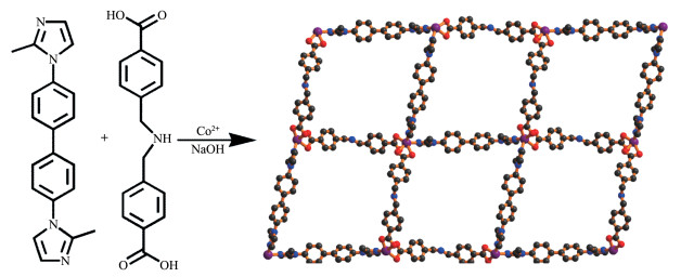
Scheme 1.
Synthesis of 1

As is known to all, Fe3+ is a class of ample triva-lent metal ion for all organisms and plays an significant role in environmental and biological systems due to its importance and function in various crucial processes, such as electron transfer in DNA and RNA formation and biological metabolisms[1-2]. Both iron shortage or excess will give rise to various serious function condi-tion disorders, such as agrypnia, skin diseases, de-creased immunity, and iron deficiency anemia (IDA). Huntington's, Alzheimer's and Parkinson's diseases also have been related to the abnormal distribution of the iron[3-4]. Though Fe3+ is very important for organ-isms, excess Fe3+ will cause environmental pollution[5]. So, how to effectively sense Fe3+ ion is an extremely im-portant issue for life system that needs to be given more attention[6-7]. Nowadays, Fe3+ ions are commonly analyzed using large - scale instruments such as atomic emission/absorption spectrometry, inductively coupled plasma-mass spectrometry (ICP -MS), X -ray dispersion, voltammetry, some of which are limited in their charac-terization. However, all of these ways have limitations, such as sophisticated instrumentation, complicated pre-treatment procedures, being time-consuming, and easi-ly interfered by other metal ions, which make them less efficient for fast, facile and exclusive determination of Fe3+ ion in the daily life. Thus, it is very necessary to develop novel techniques that can be easily applied to exclusively detect Fe3+ ion[8-9].
The coordination polymers (CPs) or metal- organic frameworks (MOFs) as a new type of crystalline materi-als has been aroused widespread concern in view of their extensive applications in catalysis[10-11], magne-tism[12-13], gas storage[14-15], fluorescent sensor[16-24], and so on. Among them, luminescent MOF - based chemosen-sors have afforded great interest due to their particular aspects such as monitoring in real-time, quick response, high selectivity, and high sensitivity, so numerous CPs or MOFs have been synthesized as sensors for the de-tection of ions, explosives, and small molecules in wa-ter system.
The mixed - ligand MOFs derived from 4, 4' - oxy- dibenzoic acid or 4, 4'-iminodibenzoic acid and N- donor linkers have attracted intensive interest due to their ability to incorporate the virtues of different functional groups and to easily obtain controlled architecture by changing one of the ligands. Meanwhile, the organic ar-omatic dicarboxylic acid ligands, 4, 4' - oxydibenzoic ac-id or 4, 4' - iminodibenzoic acid, have been extensively applied in the construction of MOFs due to their versa-tile coordination modes and high structural stability. Herein we present a new synthetic approach for a metal - organic coordination framework by employing the Co2+ and a newly ligand H3bcba(4, 4'-biscarboxyl-N, N-dibenzylamine) which has two carboxylate groups at the terminal position, and one aliphatic amine group between the benzoxy groups. These different coordina-tion groups would react with equatorial and axial posi-tions of paddle-wheel type dimer to extend an open framework. In addition, the N - containing auxiliary ligand bis(imidazole) has been proven to be good candi-dates for constructing multifunctional MOFs due to their length and flexibility. We have selected 1, 1' - bi-phenyl-4, 4'-diylbis(4-methyl-1H-imidazole) (bdmi) as the organic linker, which features three special charac-teristics: (i) as a relatively rigid ligand, bdmi has two coordination fashions, and the free rotation of the imid-azolyl ring and benzene ring can improve the flexibility of the polymeric frameworks; (ii) the long size makes it a potential candidate to generate CPs of entangled to-pology; (iii) the good fluorescent characteristic of bdmi may endow the resulting products some interesting lu-minescent properties.
Based on the above consideration, to investigate the effect of the coordination modes of H3bcba and bdmi ancillary ligand on the structural assembly and diversi-ty, we designed and obtained a new CP, {[Co(H3bcba) (bdmi)]·H2O}n (1) (Scheme 1), which has been charac-terized by single-crystal X-ray diffraction, IR spectros-copy, thermogravimetry, and elemental analysis. Its luminescence properties have been investigated.
All chemicals were commercially available and used as received without further purification. H3bcba was synthesized according to the reference[25]. Powder X -ray diffraction (PXRD) data were collected on a Bruk-er D8 ADVANCE X -ray diffractometer with Cu Kα radiation (λ =0.154 18 nm) at generator voltage of 40 kV, generator current of 40 mA with a scanning range of 5°~50°. The simulation of PXRD pattern was carried out by the single -crystal data and diffraction -crystal module of the Mercury 2.0 program available free of charge via http://www.iucr.org. The purity and homoge-neity of the bulk products were determined by compar-ing the simulated and experimental PXRD patterns. The elemental analyses (C, H, N) were performed on a Perkin -Elmer 240C apparatus. FT-IR spectra were re-corded in a range of 4 000~450 cm-1 on a PerkinElmer Frontier spectrometer. Thermogravimetric analyses (TG) were performed under nitrogen with a heating rate of 10 ℃·min-1 using a PerkinElmer Thermogravimetric Analyzer TGA4000. Photoluminescence spectra were measured on a Varian Cary Eclipse fluorescence spec-trophotometer with a xenon arc lamp as the light source. In the measurement of the emission spectrum and the excitation spectrum, the widths of the excita-tion slit and the emission slit were 5 and 10 nm, respec-tively. The crystal structure was determined by a Rigaku XtaLAB Mini (ROW) diffractometer.
A mixture of Co(NO3)2·6H2O (29.1 mg, 0.1 mmol), H3bcba (28.5 mg, 0.2 mmol), bdmi (31.5 mg, 0.1 mmol), NaOH (8.0 mg, 0.2 mmol), H2O (10 mL) and C2H5OH (3 mL) was placed in a Teflon-lined stainless steel vessel (25 mL), and then the vessel was sealed and heated at 145 ℃ for 3 days. After gradually cooling to room temperature at a rate of 10 ℃ ·h-1, red block - shaped crystals of 1 were collected from filtration, washed with distilled water and dried in the air (Yield: 45% based on Co). Elemental analysis Calcd. for C36H 33N5O5Co(%): C, 64.04; H, 4.89; N, 10.38. Found (%): C, 64.01; H, 4.92; N, 10.35. IR (cm-1): 3 409s, 3 120s, 1 608m, 1 520m, 1 353s, 1 290m, 1 135m, 995m, 767m, 659m.
The luminescence properties of 1 were investigat-ed in the solid state and various analytes at room tem-perature. For sensing of cations and anions, 2.0 mg of a grounded powder samples of 1 was immersed in 2.0 mL aqueous solution of M(NO3)x (Mx+=Ag+, Al3+, Cd2+, Co2+, Cr3+, Cu2+, K+, Li+, Mn2+, Mg2+, Ni2+, Pb2+, Zn2+, Fe2+ and Fe3+, 1.0 mmol·L-1) or KyX (Xy-=CO32-, Cl-, SO42-, HCO3-, F-, Ac-, I-, PO43-, C2O42-, SiO32-, S2O82-, IO3-, H2PO4-, SCN-, OH -, BF4-, CrO42- and Cr 2O72-, 1.0 mmol·L-1). Then the solid- liquid mixture was ultrasoni-cated for 30 min to form steady turbid suspension of 1@Mx+ or 1@Xy- for the fluorescence measurements. The fluorescent intensities of these 1@Mx+ or 1 @Xy-suspensions were immediately recorded at room tem-perature and compared.
The 2.0 mg grounded powder sample of 1 was dis-persed in 2.0 mL H2O solution of target analyte Fe(NO3)3 with different concentrations (0~0.26 mmol·L-1). Then the solid - liquid mixture was ultrasonicated for 30 min to form steady turbid suspension of 1@Fe3+ for the fluo-rescence measurements. The fluorescent intensities of these 1 @Fe3+ suspensions were immediately recorded at room temperature and compared.
The aqueous solutions with pH value of 0 and 12 were configured respectively. A powder sample of 1 (2.0 mg) was dispersed in these aqueous solution (2.0 mL), then the solid - liquid mixture was ultrasonicated for 30 min to form steady turbid suspension of 1, and the fluorescence emission spectrum of 1 was recorded.
Single - crystal data collections were performed on a Rigaku XtaLAB Mini (ROW) diffractometer with graphite-monochromatized Mo Kα radiation (λ =0.071 073 nm) at 296(2) K. Using Olex2[26], the structure was solved with the SHELXTL[27] structure solution program using Intrinsic Phasing and refined with the SHELXL[28] refinement package using Least Squares minimization. All non-hydrogen atoms were refined with anisotropic displacement parameters. C, N and O-bound H atoms were placed in calculated positions (dC-H= 0.093 nm for benzene and imidazole -CH, 0.097 nm for methylene - CH2-, 0.096 nm for methyl - CH3; dN-H= 0.086 nm for -NH; dO-H=0.085 nm for H2O) and were in-cluded in the refinement in the riding model approxi-mation, with Uiso(H) set to 1.2 Ueq(C or N) for benzene and imidazole -CH, methylene -CH2- and -NH, with Uiso (H) set to 1.5Ueq(C or O) for methyl -CH3 and H2O. The disordered atoms (N1, C8 and C20) were split in two parts and refined with an occupancy ratio of 0.63:0.37. DFIX and EADP instructions from ShelXL have been applied to constrain the disordered atoms associated N-C, C - C distances and their anisotropic displacement parameters. Further details of the structure determina-tions are summarized in Table S1 (Supporting informa-tion). Selected bond lengths and bond angles for 1 are listed in Table S2. Hydrogen bonds for 1 are listed in Table S3.
CCDC: 1973191.
The single-crystal X-ray diffraction structural analysis has revealed that 1 is a two-dimensional metal-organic framework, whose asymmetric unit is com-prised of one Co2+, one partly deprotonated divalent an-ion Hbcba2- ligand, one bimb ligand, and one free H2O. As shown in Fig. 1a, each crystallographic independent Co(Ⅱ) metal centers with a distorted octahedral (CoN2O4) geometry lies in the crystal structure (Fig. 1a and Table S2). Co1 lies in a distorted octahedral coordination sphere, the equatorial plane of which consists of three carboxylate oxygen atoms (O1, O3ii and O4ii) (Symmetry code: ii -1/2-x, 1/2+y, 1/2-z) from two symmetry-relat-ed Hbcba2- ligands and one bdmi nitrogen atom (N2), another carboxylate oxygen atom (O2) and symmetry - related bdmi nitrogen atom (N5i) (Symmetry code: i 5/2-x, -1/2+y, 1/2-z) in the axial coordination site. The distances of the Co - O bonds range from 0.203 5(4) to 0.247 5(4) nm, the lengths of the Co - N bond are 0.205 6(5) and 0.208 6(4) nm, both of which are in the normal range. It is worth mentioning that the Co1 - O2 distance is 0.247 5(4) nm, suggesting a non-negligible interaction with the uncoordinated carboxylate oxygen atom which can be described as a semi -chelating coor-dination mode, implying the Co1 ion in a distorted octa-hedron environment.
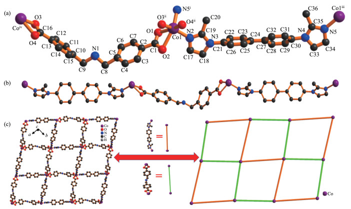
Symmetry codes: i5/2-x, -1/2+y, 1/2-z; ii-1/2-x, 1/2+y, 1/2-z; iii5/2-x, 1/2+y, 1/2-z; iv-1/2-x, -1/2+y, 1/2-z
The partly deprotonated Hbcba2- ligand connects with two Co(Ⅱ)ions, with two carboxylate groups acting as bidentate chelate fashion. The Hbcba2- ligands bend to coordinate Co(Ⅱ)ions to form a 1D framework with a Co…Co separation of 1.591(4) nm (Fig. 1b). In addition, it should be noted that these independent sets of bdmi spacers, which establish a physical bridge be-tween Co ions with a Co…Co separation of 1.731(4) nm. Both Hbcba2- and bdmi reside at an alternate hori-zontal and vertical of arrangement, resulting in the for-mation of an irregular parallelogram 2D structure with dimensions of 1.59 nm×1.73 nm along c - axis, which are occupied by the distorted solvent water molecules (Fig. 1c). In 1, the μ2 -bridging bdmi ligands also adopt the cis - conformation, different from each other by the dihedral angle between the imidazolyl and phenyl rings.
From a topological point of view[29], each Co(Ⅱ)links two Hbcba2- ligands and two bdmi ligands as a four - connected node. The Hbcba2- or bdmi ligands bridging two metal ions serve as linkers. Thus, the 2D structure of 1 can be classified as a four-connected (4, 4) grid layer topology for (Co)4(Hbcba2-)2(bdmi)2.
Intermolecular O-H…N, N-H…O, C-H…N and C-H…O hydrogen bonds are originated from the non - coordinated imino group -NH (N1, acceptor and donor), the solvate H2O (O5, donor and acceptor) and the car-boxylate group (O1/O2/O3 and O4, acceptors). These hydrogen bonds alternate by translation along the c axis and link the grid layers in reversely alternating parallel arrangements, supporting the supramolecular architecture.
Three C-H… π interactions, i. e. C4-H4…Cg8ix (6-membered ring (Cg8) C21-C22-C23-C24-C25-C26, Symmetry code: ix 1-x, 1-y, 1-z), C20-H20E…Cg5i (5-membered ring (Cg5) N4-C33-C34-N5-C35, Symmetry code: i 5/2- x, -1/2+y, 1/2-z), and C36-H36C…Cg6x (6 -membered ring (Cg6) C2-C3-C4-C5-C6-C7, Symmetry code: x 3/2-x, 1/2+y, 1/2-z) have distances of 0.294, 0.243 and 0.281 nm, respectively, which interlink the adjacent chains. These interactions also exist between flanking benzene rings of Hbcba2-, bdmi and imidazole. Thus, the molecules are extended into an interwoven 3D supramolecular architecture through O-H…N, N-H…O, C-H…N, C-H…O and C-H…π interactions.
As seen in the experimental section, we conduct-ed a series of spectral measurements to explore the flu-orescence response of 1 which interacted with 15 differ-ent cations and 18 different anions. Interestingly, the fluorescence emission of 1 was almost entirely quenched in the Fe(NO 3)3 solution, while a moderate strength reduction were produced in 1@Al3+, 1@Cr3+, 1@S2O82-, 1 @Cr2O 72-, 1@CrO42- and 1@BF- suspen-sions, but a slight intensity changes were observed in other ions(Fig. 2a, b). Due to the significant quenching phenomenon of 1 towards Fe3+, the quenching effect of Fe3+ was subsequently examined as a function of Fe(NO3)3 in a concentration range of 0~0.26 mmol·L-1. As shown in Fig. 2c, when the Fe3+ concentration was in-creased from 0 to 0.26 mmol·L-1, the fluorescence gradually decreased, and the quenching efficiency was nearly 93.8% when the concentration of Fe3+ ions reached 0.26 mmol·L-1.
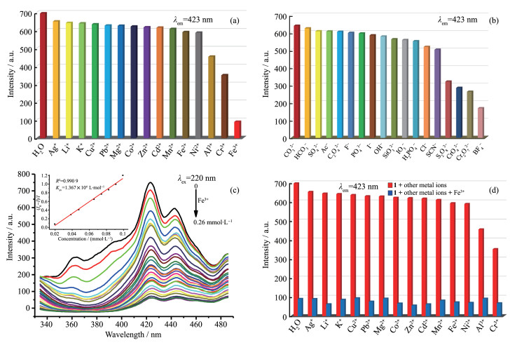
As is well known, the fluorescent quenching effi-ciency can be quantitatively accounted for the Stern - Volmer formula: I0/I=1+KsvcM (I0: initial fluorescent intensity of 1; I: fluorescence intensity after adding iron ion; cM: concentration of Fe3+ ions; Ksv: Stern - Vol-mer constant (L·mol-1)). According to the Stern-Volmer plots, there was a good linear relationship in the range of Fe3+ concentration from 0 to 0.26 mmol·L-1 (Ksv=13 670 L·mol-1, R2=0.990 9). According to the equa-tion 3σ/k (σ: standard error; k: slope), the detection limit of 1 detection Fe3+ was calculated to be 2.17 μmol·L-1. It implies that 1 can selectively sense Fe3+ ions[30-31].
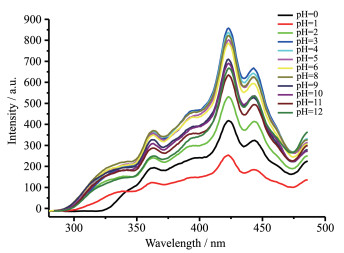
Considering multiple cations in industrial waste-water, anti-interference experiments were subsequently performed to inspect the influence of mixed solutions containing Fe3+ ions and various other cations on the lu-minescence. As shown in Fig. 2d, initially, the fluores-cence intensity of 1 showed a negligible change with the addition of other metal ions. However, the lumines-cence intensities rapidly decreased with the addition of Fe3+ ions to the mixed solution of 1 and other metal ions. The decrease of the fluorescence intensities indi-cate that 1 can detect Fe3+ ions even in the presence of other metal ions. In order to adapt to detection in more complicated environments, we further explored the flu-orescence intensity of 1 with different pH values (Fig. 3). As expected, 1 exhibited satisfactory fluores-cence intensity when the pH value ranged from 3 to 12, indicating that we could carry out fluorescence detec-tion by 1 in a wide range of acid-base medium. There-fore, these results show that 1 has better anti-interference ability and can be applied to detect Fe3+ ions in real en-vironment.
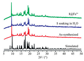
To verify the purity and stability of 1, we explored the PXRD experiments of 1 at room temperature. We added 1 to the Fe(NO3)3 (1mmol·L-1) solution, soaked it for 12 hours, multiple rinsed and collected 1, and final-ly dried it at room temperature for 48 hours before per-forming a PXRD test. At the same time, after being im-mersed in the aqueous solution for 48 hours, the sam-ples of 1 were collected and dried at room temperature for PXRD test. The PXRD measurement results showed that under the above two conditions, compared with the initial and simulated structures indexes, the structure of the sample after immersion remained basi-cally unchanged (Fig. 4). These results show that 1 has very high water - stability and no structural change has occurred after the detection of Fe3+, so it can be used to detect Fe3+ in aqueous solutions.
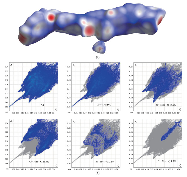
By using CrystalExplorer software[32] to calculate the Hirshfeld surface, dnorm and 2D fingerprints were further drawn to clarify the molecular interactions and surface environment. There is an obvious correspon-dence between the two maps[33-34]. As shown in Fig. 5a, the red parts represent intense interactions that similar to hydrogen bond and coordination bond effects. In order to quantitatively study the effect of intermolecu-lar interaction on the molecular surface, a two - dimen-sional fingerprint was analyzed[35]. For compound 1, 48.0% H…H interactions and 26.8% C…H/H…C in-teractions accounted for approximately half of the total. Many types of hydrogen bonds might be the main cause of O…H/H…O interactions, which value achieve 14.0%. The other hydrogen bonds of C-H…N possibly contained 3.5% N…H/H…N interactions. Besides, compound 1 had an C…C (π…π) interaction being on-ly 1.5%, which was very significant in the formation of structure (Fig. 5b). So, it is found that the Hirshfeld sur-face analysis results are well consistent with the crystal structure of 1.
In summary, a 4- connected (4, 4) topology 2D Co(Ⅱ)-based luminescent CP(1) has been successfully ob-tained by solvothermal way. 1 exhibits exceptional fluo-rescence and it can be used to detect Fe3+ ions in aque-ous solution with high selectivity and anti-interference. The quenching constant (KSV) value of 1 towards Fe3+ was as high as 13 670 L·mol-1, and the limit of detec-tion of Fe3+ could reach 2.17 μmol·L-1. These results indicate that 1 can be applied as a potential lumines-cent sensing material for quantitative detection of Fe3+ ion in biological and environmental areas.
Acknowledgements: The support of the Natural Science Foundation of Guangxi (Grant No. 2018GXNSFAA281174) are gratefully acknowledged. The authors also acknowledge the fi-nancial supports from the Natural Science Foundation of Qin-zhou University (Grant No.2016PY-GJ01) and the Opening Proj-ect of Guangxi Colleges and Universities Key Laboratory of Bei-bu Gulf Oil and Natural Gas Resource Effective Utilization (Grants No.2017KLOG11, 2017KLOG14).
Supporting information is available at http://www.wjhxxb.cn
Chen L H, McBranch D W, Wang H L, et al. Proc. Natl. Acad. Sci. USA, 1999, 96:12287-12292 doi: 10.1073/pnas.96.22.12287
Patel S K S, Kumar P, Kalia V C. Int. J. Hydrogen Energy, 2012, 37:10590-10603 doi: 10.1016/j.ijhydene.2012.04.045
Ward R J, Zucca F A, Duyn J H, et al. Lancet Neurol., 2014, 13:1045-1060 doi: 10.1016/S1474-4422(14)70117-6
渠星宇, 边永军, 白杨, 等.无机化学学报, 2019, 35(4):649-657QU Xing-Yu, BIAN Yong-Jun, BAI Yang, et al. Chinese J. Inorg. Chem., 2019, 35(4):649-657
Nwachukwu M A, Feng H, Alinnor J. Procedia Environ. Sci., 2011, 4:310-322 doi: 10.1016/j.proenv.2011.03.036
赵越, 翟玲玲, 孙为银.无机化学学报, 2014, 30(8):99-105ZHAO Yue, ZHAI Ling-Ling, SUN Wei-Yin. Chinese J. Inorg. Chem., 2014, 30(8):99-105
Wang B, Yang Q, Guo C, et al. ACS Appl. Mater. Interfaces, 2017, 9:10286-10295 doi: 10.1021/acsami.7b00918
Chen S G, Shi Z Z, Qin L, et al. Cryst. Growth. Des., 2016, 17:67-72
Wang T, Liu Q H, Gao Y, et al. Chin. Chem. Lett., 2016, 27:497-501 doi: 10.1016/j.cclet.2016.01.011
Liu J W, Chen L F, Cui H, et al. Chem. Soc. Rev., 2014, 43:6011-6061 doi: 10.1039/C4CS00094C
刘媛媛, 张慧敏, 王鑫蕊, 等.无机化学学报, 2018, 34(4):791-799LIU Yuan-Yuan, ZHANG Hui-Min, WANG Xin-Rui, et al. Chinese J. Inorg. Chem., 2018, 34(4):791-799
侯向阳, 王潇, 付峰, 等.无机化学学报, 2013, 29(10):2245-2250HOU Xiang-Yang, WANG Xiao, FU Feng, et al. Chinese J. Inorg. Chem., 2013, 29(10):2245-2250
Li J X, Du Z X, Zhu B L, et al. Inorg. Chem. Commun., 2011, 14:522-525 doi: 10.1016/j.inoche.2011.01.012
Li J X, Du Z X, Wang L Z, et al. Inorg. Chim. Acta, 2011, 376:479-485 doi: 10.1016/j.ica.2011.07.013
Liao H P, Wang H M, Ding H M, et al. J. Mater. Chem. A, 2016, 4:7416-7421 doi: 10.1039/C6TA00483K
Du Z X, Li J X. Inorg. Chim. Acta, 2015, 436:159-162 doi: 10.1016/j.ica.2015.07.036
黄玉婷, 欧阳兴梅, 等.无机化学学报, 2005, 21(10):1479-1482HUANG Yu-Ting, OUYANG Xing-Mei, Okamura T, et al. Chinese J. Inorg. Chem., 2005, 21(10):1479-1482
Han Y H, Zeng D R, Li J M, et al. J. Coord. Chem., 2018, 71:16-18
Li J X, Du Z X, Wang J G, et al. Inorg. Chem. Commun., 2012, 15:243-247 doi: 10.1016/j.inoche.2011.10.036
Li J X, Guo W B, Du Z X, et al. Inorg. Chim. Acta, 2011, 375:290-297 doi: 10.1016/j.ica.2011.05.018
Rajak R, Saraf M, Verma S K, et al. Inorg. Chem., 2019, 58:16065-16074 doi: 10.1021/acs.inorgchem.9b02611
Li J X, Du Z X. J. Coord. Chem., 2016, 69:2563-2572 doi: 10.1080/00958972.2016.1216106
Xu T Y, Wang H, Li J M, et al. Inorg. Chim. Acta, 2019, 493:72-80 doi: 10.1016/j.ica.2019.05.002
刘志强, 曹师虎, 张哲, 等.无机化学学报, 2019, 35(11):2145-2151LIU Zhi-Qiang, CAO Shi-Hu, ZHANG Zhe, et al. Chinese J. Inorg. Chem., 2019, 35(11):2145-2151
Horike S, Hasegawa S, Tanaka D, et al. Chem. Commun., 2008, 37:4436-4438
Dolomanov O V, Bourhis L J, Gildea R J, et al. J. Appl. Crys-tallogr., 2009, 42:339-341 doi: 10.1107/S0021889808042726
Sheldrick G M. Acta Crystallogr. Sect. A, 2015, A71:3-8 doi: 10.1107/S2053273314026370
Sheldrick G M. Acta Crystallogr. Sect. C, 2015, C71:3-8
Spek A L. J. Appl. Cryst., 2003, 36:7-13 doi: 10.1107/S0021889802022112
Chen W, Wang J Y, Chen C, et al. Inorg. Chem., 2003, 42:944-946 doi: 10.1021/ic025871j
Zhou X J, Li B Y, Li G H, et al. CrystEngComm, 2012, 14:4664-4669 doi: 10.1039/c2ce25328c
Turnner M J, McKinnon J J, Wolff S K, et al. Crystal Explorer 17.5, University of Western Australia, Australia, 2017.
Konar S, Datta S K, Dolai M, et al. J. Mol. Struct., 2019, 1178:682-691 doi: 10.1016/j.molstruc.2018.10.073
Yan T, Zhou J, Zhu R R, et al. Inorg. Chem., 2019, 58:3145-3155 doi: 10.1021/acs.inorgchem.8b03210
Hong D L, Luo Y H, He X T, et al. ACS Appl. Mater. Interfaces, 2019, 11:7272-7279 doi: 10.1021/acsami.8b18883

Figure 1 (a) Coordination environment for Co2+ in 1; (b) Wave-shaped 1D chain of H3bcba-Co-bdmi; (c) View of simplified 4-connected (4, 4) topology of 1
Symmetry codes: i5/2-x, -1/2+y, 1/2-z; ii-1/2-x, 1/2+y, 1/2-z; iii5/2-x, 1/2+y, 1/2-z; iv-1/2-x, -1/2+y, 1/2-z

Figure 2 (a) Photoluminescence intensities of 1 in aqueous solution with various inorganic cations; (b) Photoluminescence intensities of 1 in aqueous solution with various inorganic anions; (c) Emission spectra and the Stern-Volmer plot (Inset) for 1 in aqueous solution of different Fe3+ concentrations; (d) Fluorescence intensity of 1 in aqueous solution with the introduction of diverse other metal ions (red) and introduction of Fe(Ⅲ) (blue)

Figure 4 PXRD patterns of as- synthesized 1 and simulated result as reference as well as 1 soaking in H2O and 1@Fe3+

 扫一扫看文章

扫一扫关注我们
 下载:
下载:

 下载:
下载:

