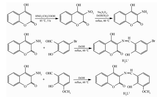
Scheme 1.
Synthetic routes of H2L1 and H2L2
Synthesis, Crystal Structure and Spectral Properties of Binuclear Ni(Ⅱ) and Cubane-like Cu4(μ3-O)4 Cored Tetranuclear Cu(Ⅱ) Complexes Based on Coumarin Schiff Base
Shu-Zhen ZHANG , Jian CHANG , Hong-Jia ZHANG , Ya WU , Yin-Xia SUN , Yan-Bin WANG

Schiff base compounds and their transition metal complexes are playing an important part in the development of coordination chemistry[1-5] because of their potential application in catalysis[6], bioscience[7-11], magnetic materials[12-17], luminescent[18-24], electrochem-ical systems[25-26] and constructing supramolecular stru-ctures building[27-33]. Schiff-base compounds and its derivatives are very important as versatile ligands, properties of interest in materials science. Also, the Schiff base ligands with N- and O- group are strong donors and therefore the oxime-containing ligands were found to efficiently stabilize high oxidation states of metal ions and prepare complexes with different structures and functionalities like Cu(Ⅱ) and Ni(Ⅱ) complexes[34-39]. In recent years, there has been enhanced interest in the synthesis and characterization of such complexes due to their interesting properties and other applications[40-47]. In order to further study the supramolecular of transition metal complexes and Schiff base ligands, we synthesized and analyzed two complexes, [Ni(L1)(DMF)(H2O)]2 (1) (H2L1=3-((5-bromo-2-hydroxy-benzylidene)-amino)-4-hydroxy-benzopyran-2-one) and [Cu4(L2)4]·DMF·CH3OH·2H2O (2) (H2L2=4-hydroxy-3-((2-hydroxy-3-methoxy-benzylidene)-amino)-benzopyran-2-one). Complex 1 is a dinuclear structure and is connected to a 1D supramolecular chain by intermolecular hydrogen bonding, C-H…π and π…π stacking interactions. Complex 2 is a cubane-like Cu4 (μ3-O)4 cored tetranuclear structure and is linked to a 3D network supramolecular structure by intramole-cular hydrogen bonding and π…π stacking interac-tions. The central metal Ni(Ⅱ) and Cu(Ⅱ) ions are all six-coordinated distorted octahedron geometries in complexes 1 and 2, respectively. In addition, the fluorescent properties of the ligands H2L1, H2L2 and their Ni(Ⅱ) complex 1 and Cu(Ⅱ) complex 2 are also studied.
4-Hydroxyl coumarin from Alfa Aesar was used without further purification. The other reagents and solvents were of analytical grade from Tianjin Chemical Reagent Factory, and were used without further purification.
C, H and N analyses were carried out with a GmbH Vario EL V3.00 automatic elemental analyzer. FT-IR spectra were recorded on a VERTEX70 FT-IR spectrophotometer, with samples prepared as KBr (400~4 000 cm-1) pellets. UV-Vis absorption spectra were recorded on a Hitachi UV-3900 spectrometer. Luminescence spectra in solution were recorded on a Hitachi F-7000 spectrometer. X-ray single crystal structure was determined on a Bruker Smart 1000 CCD area detector. Melting points were measured by an X-4 microscopic melting point apparatus made by Beijing Taike Instrument Limited Company and were uncorrected. 1H NMR spectra were recorded in DMSO-d6 solution at room temperature on a Bruker AV instrument operating at a frequency of 500 MHz and referenced to tetramethylsilane (δ=0.00) as an internal standard. Chemical shift multiplicities are reported as s=singlet, d=doublet, t=triplet and m=multiplet.
H2L1 and H2L2 were synthesized according to the following synthetic routes shown in Scheme 1.
3-Amino-4-hydroxycoumarin was synthesized according to an analogous method reported earlier[48]. Firstly, 0.9 mL concentrated HNO3 and 1.8 mL CH3COOH mixed solution was added dropwise into the 4-hydroxycoumarin (20.5 g, 12.6 mmol) solution containing 5 mL glacial acetic acid over 0.5 h at 85 ℃, and the mixture was subjected to heating at 80 ℃ for 4 h. The mixed solution was allowed to come to room temperature, and placed in an ice-water mixture until a pale yellow solid appeared, which was filtered to give the 3-nitro-4-hydroxycoumarin. Secondly, Na2S2O4 solid (33.0 g, 18.3 mmol) was added in batches to a solution of 3-nitro-4-hydroxycoumarin (10.0 g, 4.8 mmol) in EtOH/H2O (2:1, V/V) mixture solution (60 mL) at 87 ℃, and the reaction continued for 7 h. Then the ethanol was removed via vacuum distillation. The residual Na2S2O4 was removed by reaction with concentrated hydrochloric acid, and neutralized with saturated sodium bicarbonate solution. The product was suction filtered and dried to give a brown 3-amino-4-hydroxycoumarin powder. Yield: 67.8%. m.p. 209~211 ℃. Anal. Calcd. for C9H7NO3(%): C, 61.02; H, 3.98; N, 7.91. Found(%): C, 61.26; H, 3.67; N, 7.72.
3-Amino-4-hydroxycoumarin (177.0 mg, 1.0 mmol) and 2-hydroxy-5-bromobenzaldehyde (201.21 mg, 1.0 mmol) were placed in a 20 mL flask, and 7 mL of absolute ethanol was added to the flask. The mixture was subjected to reflux at 70 ℃ for 8 h, and allowed to come to room temperature. The light yellow precipitate was filtered and dried to obtain 267.79 mg H2L1. Yield: 61.07%. m.p. > 300 ℃. Anal. Calcd. for C16H10BrNO4(%): C, 53.36; H, 2.80; N, 3.89. Found (%): C, 53.43; H, 2.74; N, 3.79. 1H NMR (500 MHz, DMSO-d6): δ 6.99 (d, J=9.0 Hz, 1H, Ph), 7.24 (td, J=6.5, 1 Hz, 1H, Ph), 7.33 (m, 2H, Ph), 7.62 (m, 2H, Ph), 7.79 (dd, J=3.5 Hz, 1H, Ph), 8.07 (s, 1H, CH=N), 9.86 (s, 1H, OH), 10.99 (s, 1H, OH).
The ligand H2L2 was synthesized by a method similar to that of H2L1 except substituting 2-hydroxy-5-bromobenzaldehyde with 2-hydroxy-3-methoxybenzal-dehyde. H2L2: 193.21 mg, Yield: 62.23%. m.p. 247~249 ℃. Anal. Calcd. for C17H13NO5(%): C, 65.59; H, 4.21; N, 4.50. Found(%): C, 65.43; H, 4.35; N, 4.72. 1H NMR (500 MHz, DMSO-d6): δ 3.86 (t, J=14.5 Hz, 3H, OCH3), 6.97 (t, J=14.5 Hz, 1H, Ph), 7.08 (s, 1H, Ph), 7.00 (s, 1H, Ph), 7.22 (m, 2H, Ph), 7.4 (s, 1H, Ph), 7.69 (td, J=12.5, 6.5 Hz, 1H, Ph), 7.96 (dd, J=2, 0 Hz, 1H, CH=N), 9.91 (s, 1H, OH), 10.27 (s, 1H, OH).
A solution of Ni(Ⅱ) acetate monohydrate (2.48 mg, 0.01 mmol) in methanol (1 mL) was added dropwise to a solution of H2L1 (3.6 mg, 0.01 mmol) in acetone/DMF (7 mL, 6:1, V/V). The color of the mixture turned to yellow immediately, and then 2 drops of triethyla-mine were added in it. The mixture was stirred for 1 h at room temperature, filtered, and the filtrate was allowed to stand at room temperature for about two weeks. The solvent was partially evaporated, and pale yellow needle-like single crystals of complex 1 suitable for X-ray crystallographic analysis were obtained. Anal. Calcd. for C38H34Br2N4Ni2O12(%): C, 49.43; H, 3.37; N, 5.52. Found(%): C, 49.22; H, 3.75; N, 5.62.
The synthesis of Cu(Ⅱ) complex 2 was same as above to obtain yellow needle-like single crystals suitable for X-ray crystallographic analysis. Anal. Calcd. for C72H59Cu4N5O24(%): C, 52.97; H, 3.64; N, 4.29. Found(%): C, 53.20; H, 3.40; N, 4.51.
The single crystals with approximate dimensions of 0.22 mm×0.26 mm×0.28 mm (1) and 0.22 mm×0.25 mm×0.27 mm (2) were placed on a Bruker Smart 1000 CCD area detector. The reflections were collected using graphite-monochromatized Mo Kα radiation (λ=0.071 073 nm) at 296(1) K and 296(2) K, respectively. The Lp corrections were applied to the SAINT program[49]and semi-empirical correction were applied to the SADABS program[50]. The crystal structures were solved by the direct methods (SHELXS-2014)[51]. Details of the crystal parameters, data collection and refinements for complexes 1 and 2 are summarized in Table 1.
 下载:
导出CSV
下载:
导出CSV
| Complex | 1 | 2 |
| Empirical formula | C38H34Br2N4Ni2O12 | C72H59Cu4N5O24 |
| Formula weight | 1 015.87 | 1 491.27 |
| Crystal system | Monoclinic | Tetragonal |
| Space group | C2/c | I41/a |
| a/nm | 1.799 3(4) | 1.663 3(4) |
| b/nm | 0.921 0(2) | 1.663 3(4) |
| c/nm | 2.496 9(6) | 2.342 9(4) |
| β/(°) | 110.249(2) | |
| V/nm3 | 3.881 8(2) | 6.482(3) |
| Z | 4 | 4 |
| μ/mm-1 | 3.098 | 1.374 |
| F(000) | 2 048 | 3 024 |
| θ range/(°) | 1.70~25.0 | 2.5~25.0 |
| Limiting indices | -21 ≤ h ≤ 19, -10 ≤ k ≤10, -29 ≤ l ≤ 29 | -18 ≤ h ≤ 19, -19 ≤ k ≤ 18, -27 ≤ l ≤ 27 |
| Reflection collected, unique | 14 365, 3 413 (Rint=0.174) | 20 340, 2 862 (Rint=0.072) |
| Completeness to θ/% | 100 | 99.9 |
| Data, restraint, parameter | 3 413, 0, 263 | 2 862, 0, 218 |
| GOF on F2 | 1.07 | 1.06 |
| R1, wR2 [I>2σ(I)] | 0.044 4, 0.108 31 | 0.039 6, 0.100 2 |
| Largest diff. peak and hole/(e·nm-3) | 820 and -810 | 860 and -890 |
CCDC: 1922258, 1; 1922309, 2.
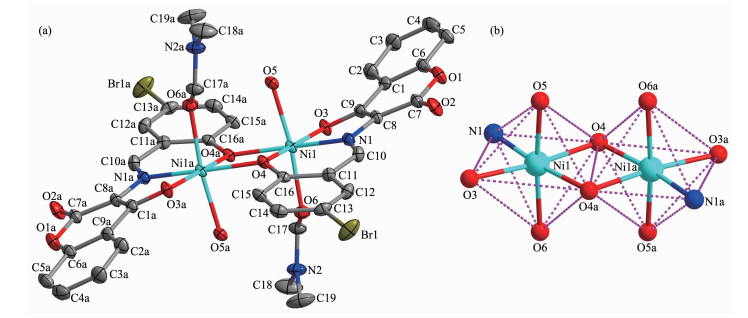
The molecular structure of complexes 1 and 2 are shown in Fig. 1 and 2, respectively, and selected bond lengths and angles are listed in Table 2. X-ray crystallographic analysis shows that complex 1 crystallizes in the monoclinic system, and the space group is C2/c. Complex 1 can be described as a binuclear Ni(Ⅱ) complex, consisting of two Ni(Ⅱ) ions, two (L1)2- units and two coordinated solvent molecules H2O and DMF. In complex 1, two deprotonated hydroxyl oxygen (O3, O4) atoms and one oxime nitrogen (N1) atoms come from the (L1)2- unit, as well as two oxygen (O5, O6) atoms of the coordinated solvent molecules H2O and DMF, respectively, which constitute the [Ni(L1)(H2O)(DMF)] moiety. And then the O4 and O4a atoms bridge the two [Ni(L1)(H2O)(DMF)] moieties to form the binuclear structure [Ni(L1)(H2O)(DMF)]2 (1). Thus, the central Ni(Ⅱ) ions are hexa-coordinated and their coordination sphere is best described as a slightly distorted octahedron.
Symmetry code: a: 3/2-x, 3/2-y, 1-z
Symmetry codes: a: 3/4+x, 3/4-y, 3/4-z; b: 3/4-x, -3/4+y, 3/4-z; c:-x, 3/2+y, z
下载:
导出CSV
| 1 | |||||
| Ni1-O3 | 0.205 0(2) | Ni1-O4 | 0.201 6(3) | Ni1-O5 | 0.208 4(2) |
| Ni1-O6 | 0.210 0(2) | Ni1-N1 | 0.201 2(3) | Ni1-O4a | 0.206 4(2) |
| O3-Ni1-O4 | 172.93(8) | O3-Ni1-O5 | 87.32(7) | O3-Ni1-O6 | 93.89(8) |
| O1-Ni1-N1 | 82.01(8) | O1-Ni1-O4a | 105.58(8) | O4-Ni1-O5 | 90.81(7) |
| O4-Ni1-O6 | 88.92(7) | O4-Ni1-N1 | 91.42(9) | O4-Ni1-O4a | 80.95(8) |
| O5-Ni1-O6 | 171.90(9) | O5-Ni1-N1 | 96.12(8) | O5-Ni1-O4a | 83.42(8) |
| O6-Ni1-N1 | 91.99(9) | O6-Ni1-O4a | 88.54(8) | N1-Ni1-O4a | 172.34(8) |
| 2 | |||||
| Cu1-O1 | 0.234 7(2) | Cu1-O2 | 0.198 5(2) | Cu1-O2a | 0.196 1(2) |
| Cu1-O3a | 0.194 5(2) | Cu1-N1a | 0.195 1(2) | Cu1b-O2 | 0.196 1(2) |
| Cu1b-O3 | 0.194 5(2) | Cu1b-N1 | 0.195 1(2) | Cu1-O2b | 0.268 2(2) |
| O1-Cu1-O2 | 75.51(7) | O1-Cu1-O2a | 100.98(7) | O2-Cu1-O2a | 88.76(8) |
| O1-Cu1-O3a | 84.41(7) | O2-Cu1-O3a | 91.75(7) | O2A-Cu1-O3a | 174.54(7) |
| O3A-Cu1-N1a | 85.16(8) | O1-Cu1-N1a | 114.44(7) | O2-Cu1-N1a | 169.13(8) |
| O2A-Cu1-N1a | 93.35(8) | Cu1-O2-Cu1b | 111.26(8) | ||
| Symmetry codes: a: 3/2-x, 3/2-y, 1-z for 1; a: 3/4+x, 3/4-y, 3/4-z; b: 3/4-x, -3/4+y, 3/4-z for 2. | |||||
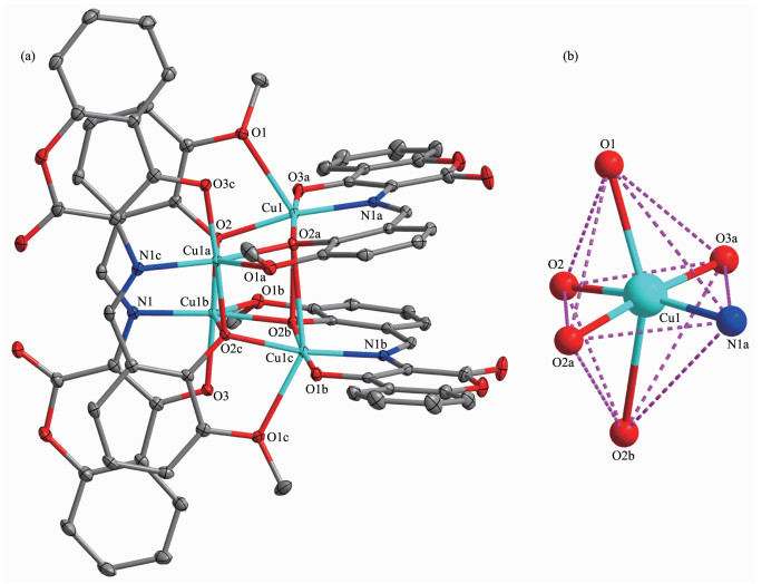
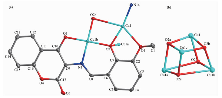
Complex 2 crystallizes in the tetragonal system, and the space group was I41/a. Complex 2 can be described as a cubane-like Cu4(μ3-O)4 cored tetranu-clear Cu(Ⅱ) complex, and consist of four Cu(Ⅱ) ions and four (L2)2- units, in which the ligand (L2)2- is both chelating and bridging after double deprotonation of the phenolic hydroxyls. A [Cu(L2)] moiety was constituted by two deprotonated hydroxyl oxygen (O2, O3) atoms, one oxime nitrogen (N1) atoms from one of the ligand units (L2)2-, and the oxygen (O1) atom of the methoxy group coming from this (L2)2- units coordinated to the adjacent [Cu(L2)] moiety (Fig. 3a). By self-assembly, four such monomeric [Cu(L2)] entities eventually are linked through alkoxo (O2 and O2a) bridges to produce the tetranuclear cubane Cu4(μ3-O)4 core. This Cu4(μ3-O)4 core consists of four alkoxo-bridged Cu(Ⅱ) centers approximately arranged in a cuboid geometry of an alternating array of Cu and O atoms that occupy the corners of the cube (Fig. 3b). This structure could alternatively be seen as two interpenetrated Cu4 and O4 tetrahedrons. The four pincer ligands (L2)2- are all in the binding mode μ3-η1:η1:η3, and all four bridging alkoxo oxygens are located at the four corners of the cube, each bridging three Cu(Ⅱ) ions. Four Cu(Ⅱ) ions are crystallographically equivalent and have the same coordination spheres, which is six coordinated by one nitrogen atom (N1a), five oxygen atoms (O1, O2, O2a, O2b, O3a) from the (L2)2- units, and all are in distorted [CuN1O5] octahedral geometries. Observed Cu-N bond distances lie in the normal range of 0.195 1(2) nm. The equatorial Cu-O bonds at each Cu center are shorter (ranging from 0.194 5(2) to 0.198 5(2) nm) than the axial Cu-O bond (in a range of 0.234 7(2)~0.268 2(2) nm)[52]. The metal-metal intramolecular bond distances range from 0.325 69(6) to 0.359 12(9) nm.
Symmetry codes: a: 3/4+x, 3/4-y, 3/4-z; b: 3/4-x, -3/4+y, 3/4-z; c:-x, 3/2+y, z
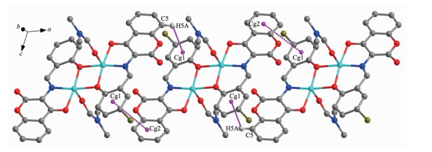
The intra- and intermolecular interactions data of complexes 1 and 2 are shown in Table 3 and 4. The structure of complex 1 was stabilized by three intra-molecular hydrogen bonds of O5-H5C…O6, C15-H15…O3, C10-H10…O2 (Fig. 4). An intermolecular hydrogen bonds O5-H5B…O2 (Fig. 5) and an inter-molecular non-classical C5-H5A…πcentroid(C11-C16) interac-tions (Fig. 6) link complex 1 molecules to form an infinite 1D supramolecular chain. Synchronously, this linkage is further stabilized via the intermolecular πcentroid(C1-C6)…πcentroid(C11-C16) stacking interactions between the benzene ring of adjacent complex 1 molecules with the distance of 0.428 1(2) nm (Fig. 6)[53-57]. Consequently, the intermolecular classical and non-classical hydrogen-bonding and π…π stacking interactions plays a very important role in the construction of supramolecular networks structure[58-64].
下载:
导出CSV
| D-H…A | d(D-H)/nm | d(H…A)/nm | d(D…A)/ nm | ∠(D-H…A)/(°) |
| 1 | ||||
| O5-H5C…O6a | 0.082 | 0.213 | 0.291 1(3) | 158 |
| C10-H10…O2 | 0.093 | 0.220 | 0.285 6(4) | 127 |
| C15-H15…O3a | 0.093 | 0.223 | 0.310 6(4) | 156 |
| O5-H5B…O2a | 0.086 | 0.183 | 0.267 9(3) | 172 |
| C5-H5A…Cg1a | 0.298 | 0.294 | 0.336 9(4) | 107 |
| 2 | ||||
| C8-H8…O5 | 0.93 | 2.17 | 2.856(3) | 130 |
| Symmetry code: 1-x, 2-y, 1-z; Cg1 is the centroids of C11~C16 in benzene ring. | ||||
下载:
导出CSV
| Ring (I) | Ring (J) | d(Cg…Cg)/nm | α/(°) | d(Cg(I)-perp)/nm | d(Cg(J)-perp)/nm | Slippage/nm |
| 1 | ||||||
| Cg2 | Cg1a | 0.428 1(2) | 26.58(2) | 0.283 5(1) | 0.394 4(1) | 0.961 0 |
| 2 | ||||||
| Cg3 | Cg4a | 0.329 8(2) | 13.77(1) | 0.275 51(8) | 0.307 81(9) | 0.118 3 |
| Cg3 | Cg5b | 0.329 8(2) | 13.77(1) | 0.275 51(8) | 0.307 81(9) | 0.118 3 |
| Cg4 | Cg6b | 0.357 3(2) | 13.08(9) | 0.300 81(9) | 0.309 50(8) | 0.178 5 |
| Cg4 | Cg7c | 0.357 3(2) | 13.08(9) | 0.300 81(9) | 0.309 50(8) | 0.178 5 |
| Cg5 | Cg6a | 0.357 3(2) | 13.08(9) | 0.300 81(9) | 0.309 50(8) | 0.178 5 |
| Cg5 | Cg7b | 0.357 3(2) | 13.08(9) | 0.300 81(9) | 0.309 50(8) | 0.178 5 |
| Cg7 | Cg6a | 0.312 5(1) | 15.47(8) | 0.303 72(8) | 0.303 71(8) | 0.073 7 |
| Cg6 | Cg7b | 0.312 5(1) | 15.47(8) | 0.303 72(8) | 0.303 71(8) | 0.073 7 |
| Symmetry codes: a: 1-x, 2-y, 1-z for 1; a: 3/4-y, 3/4+x, 3/4-z; b: -x, 3/2-y, z; c: -3/4+y, 3/4-x, 3/4-z for 2; α=dihedral angle between planes I and J; d(Cg…Cg)=distance between ring centroids; d(Cg(I)-perp)=perpendicular distance of Cg(I) on ring J; d(Cg(J)-perp)=perpendicular distance of Cg(J) on ring I; Slippage=distance between Cg(I) and perpendicular projection of Cg(J) on ring I; Cg1, Cg2 are the centroids of C11~C16 and C1~C6 in benzene ring, respectively; Cg3, Cg4, Cg5, Cg6, Cg7 are the centroids of ring Cu1-O1-C2-C7-O2, Cu1a-O3c-C10c-C9c-N1c, O3-C10-C9-N1-Cu1b, Cu1-O2a-C7a-C6a-C8a-N1a and O2b-C7b-C6b-C8b-N1b-Cu1c, respectively. | ||||||
Hydrogen atoms, except those forming hydrogen bonds, are omitted for clarity
Hydrogen atoms, except those forming hydrogen bonds, are omitted for clarity
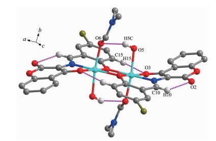
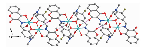
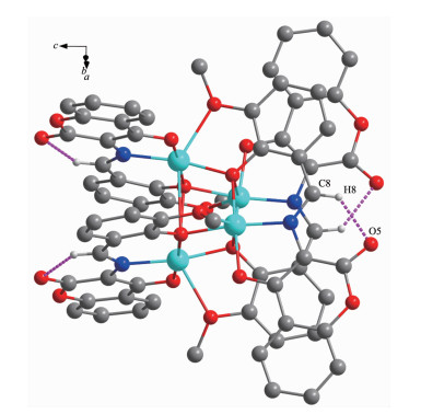
In complex 2, the structure were only stabilized by an intramolecular non-classical hydrogen bonds of C8-H8…O5 (Fig. 7 and Table 3), but there are eight complicated intermolecular π…π stacking interac-tions (Cg3…Cg4a, Cg3…Cg5b, Cg4…Cg6b, Cg4…Cg7c, Cg5…Cg6a, Cg5…Cg7b, Cg7…Cg6a, Cg6…Cg7b), with the distance of 0.329 7(7), 0.329 7(7), 0.357 3(11), 0.357 3(11), 0.357 3(0), 0.357 3(0), 0.312 5(4), 0.312 54(4) nm, respectively, which linked the neighboring molecules into a 3D network supramolecular structure (Table 4).
Hydrogen atoms, except those forming hydrogen bonds, are omitted for clarity
The FT-IR spectra of H2L1, H2L2, and their corresponding complexes 1 and 2 exhibited various bands in the 400~4 000 cm-1 region, and the most important FT-IR bands for H2L1, complex 1 and H2L2, complex 2 are given in Table 5. The free ligand H2L1 and H2L2 exhibited characteristic stretching bands of C=N group at 1 670 and 1 676 cm-1, respectively[65-73], while that of their corresponding complexes 1 and 2 were observed at 1 610 and 1 682 cm-1, respectively. Compared with the ligand, the C=N stretching frequency of complex 1 shifted to a lower frequency by ca. 60 cm-1, while that of complex 2 shifted to a higher frequency by ca. 6 cm-1. It is indicated that the C=N bond sequence is decreased or increased due to the coordination bond between the metal atom and the imino nitrogen lone pair[74-76]. In addition, the broad O-H group stretching bands at 3 403 and 3 438 cm-1 for the free ligands H2L1 and H2L2, disappeared for complexes 1 and 2, indicating the oxygen atoms in the phenolic hydroxyl groups were completely deprotonated and coordinated to the metal ions. Whereas, the stretching bands at 3 409 and 3 476 cm-1 in comp-lexes 1 and 2 are attributed to the stretching vibrations of the O-H group of coordinated water or methanol. The Ar-O stretching bands at 1 230 and 1 229 cm-1 of complexes 1 and 2 shifted toward lower frequencies by ca. 35 and 12 cm-1, respectively, compared with that of the free ligands H2L1 and H2L2 at 1 265 and 1 241 cm-1, respectively. The lower frequency of the Ar-O stretching shift indicates that M-O bond is formed between the metal ions and the oxygen atoms of the phenolic groups[77]. The FT-IR spectrum of complex 1 showed ν(M-N) and ν(M-O) vibration frequencies at 514 and 467 cm-1 (or 538 and 467 cm-1 for complex 2), respectively. These assignments are consistent with the frequency values in literature[78].
下载:
导出CSV
| Compound | ν(O-H) | ν(C=N) | ν(Ar-O) | ν(M-N) | ν(M-O) |
| H2L1 | 3 403 | 1 670 | 1 265 | — | — |
| [Ni(L1)(DMF)(H2O)]2 (1) | 3 409 | 1 610 | 1 230 | 514 | 467 |
| H2L2 | 3 438 | 1 676 | 1 241 | — | — |
| [Cu4(L2)4]·DMF·CH3OH·2H2O (2) | 3 476 | 1 682 | 1 229 | 538 | 467 |
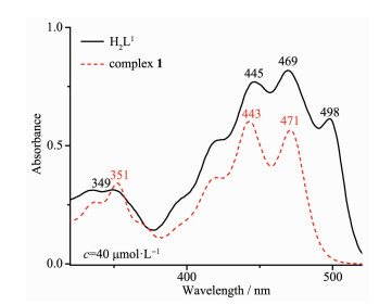
The absorption spectra of ligands H2L1, H2L2 and their corresponding Ni(Ⅱ) and Cu(Ⅱ) complexes 1 and 2 were determined in diluted DMSO solution, respectively. As shown in Fig. 8, compared with complex 1, an important feature of the absorption spectrum of H2L1 is shown that three absorption peaks were observed at 445, 469, 498 nm attributed to the intra-ligand π-π* transition of the C=N bonds and the conjugated aromatic chromophore, in which the absorption peak at 498 nm was absent in the spectrum of complex 1. The absorption peaks at 445, 469 nm were blue-shifted by 2 nm and red-shifted 2 nm, respectively, indicating that Ni(Ⅱ) ion coordinates with the O and N atoms of the deprotonated ligand units. And the absorption peak at 349 nm assigned to the π-π* transitions of the phenyl rings in H2L1 was shifted to 351 nm in complex 1, indicating the coordination of Ni(Ⅱ) atom with (L1)2-.
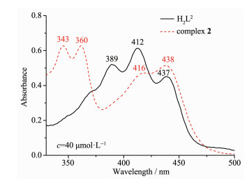
The electronic absorption spectrum of free ligand H2L2 exhibited three absorption peaks at approxi-mately 389, 412 and 437 nm (Fig. 9). The former absorption peaks at 389 nm can be assigned to the π-π* transition of benzene rings and the latter at 412 and 437 nm can be attributed to the intra-ligand π-π* transition of C=N group[79]. Upon coordination of the ligand, the absorption peaks at 412 and 437 nm were red-shifted to 416 and 438 nm, respectively, indicating that the amino nitrogen is involved in coordination with Cu(Ⅱ) ion[80]. The intraligand π-π* transitions of the benzene ring were bathochromically shifted to 343 and 360 nm in complex 2, indicating the coordination of Cu(Ⅱ) ion with deprotonated (L2)2- unit.
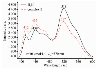
The fluorescence emission spectra of the ligands H2L1, H2L2 and their corresponding Ni(Ⅱ) and Cu(Ⅱ) complexes 1 and 2 were determined at room temp-erature in a diluted DMSO solution. As shown in Fig. 10, the free ligand H2L1 showed stronger fluorescence emission at 413, 439 and 518 nm with the excitation at 370 nm, respectively, which could be assigned to the intraligand π*-π transition. Compared with H2L1, the emission peaks of complex 1 was slightly blue-shifted 1~2 nm, and the fluorescence intensity at 412, 437 nm tended to increase and that at 518 nm tended to decrease significantly, indicating that the Ni(Ⅱ) ion coordinates with the N and O atoms and electron transition occurs.
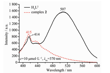
Meanwhile, the ligand H2L2 exhibited a relatively strong emission peak at ca. 507 nm and a weaker emission peak at 414 nm upon excitation at 370 nm (Fig. 11), which could be assigned to the intraligand π*-π transition. Compared with the H2L2, the emission intensity at 507 nm reduced obviously and that at 414 nm tended to increase for complex 2, indicating that the Cu(Ⅱ) ion coordinates with the N and O atoms and electron transition occurs. The fluorescence intensity of the free ligands H2L2 is probably enhanced via the occurrence of a photoinduced electron transfer process owing to the presence of a lone pair of nitrogen atoms. The process is prevented by the complexation of the free ligand H2L2 with the Cu(Ⅱ)ions. Therefore, the coordination of Cu(Ⅱ) ions can effectively reduce the fluorescence intensities[81].
Based on two Schiff base ligands, dinuclear complex 1 and cubane-like Cu4(μ3-O)4 core tetranuclear complex 2 were synthesized and their structural chara-cterization and fluorescence properties were carried out. The crystal structure analysis of complexes 1 and 2 shows that the atomic configurations of Ni(Ⅱ) and Cu(Ⅱ) are all six-coordinated distorted octahedrons. Complex 1 self-assembles into a 1D supramolecular chain through intermolecular hydrogen bonding, C-H…π and π…π stacking interactions, and complex 2 self-assembles into a 3D supramolecular network structure through intramolecular hydrogen bonding and π…π stacking interactions. Furthermore, the optical properties of complexes 1 and 2 indicate that the fluorescence variation of H2L1 and H2L2 is due to the coordination of the metal ions Ni(Ⅱ) and Cu(Ⅱ).
Liu Y A, Wang C Y, Zhang M, et al. Polyhedron, 2017, 127:278-286 doi: 10.1016/j.poly.2017.02.007
Dong W K, Ma J C, Dong Y J, et al. Polyhedron, 2016, 115:228-235 doi: 10.1016/j.poly.2016.05.017
Dong W K, Bai Y, Zhang L S, et al. Asian J. Chem., 2014, 26:2341-2343 doi: 10.14233/ajchem.2014.15903
Sun Y X, Zhang S T, Ren Z L, et al. Synth. React. Inorg. Met.-Org. Nano-Met. Chem., 2013, 43:995-1000 doi: 10.1080/15533174.2012.753614
杨玉华, 郝静, 董银娟, 等.无机化学学报, 2017, 33:1280-1292 doi: 10.11862/CJIC.2017.150YANG Yu-Hua, HAO Jing, DONG Yin-Juan, et al. Chinese J. Inorg. Chem., 2017, 33:1280-1292 doi: 10.11862/CJIC.2017.150
Wu H L, Pan G L, Bai Y C, et al. J. Chem. Res., 2014, 38:211-217 doi: 10.3184/174751914X13933417974082
Wu H L, Yuan J K, Bai Y, et al. Dalton Trans., 2012, 41:8829-8838 doi: 10.1039/c2dt30512g
Li X Y, Kang Q P, Liu L Z, et al. Crystals, 2018, 8:43 doi: 10.3390/cryst8010043
Wu H L, Pan G L, Bai Y C, et al. Res. Chem. Intermed., 2015, 41:3375-3388 doi: 10.1007/s11164-013-1440-5
Chen C Y, Zhang J W, Zhang Y H, et al. J. Coord. Chem., 2015, 68:1054-1071 doi: 10.1080/00958972.2015.1007965
Wu H L, Bai Y H, Zhang Y H, et al. Z. Anorg. Allg. Chem., 2014, 640:2062-2071 doi: 10.1002/zaac.201400109
Hao J, Li L L, Zhang J T, et al. Polyhedron, 2017, 134:1-10 doi: 10.1016/j.poly.2017.05.060
Wu H L, Wang C P, Wang F, et al. J. Chin. Chem. Soc., 2015, 62:1028-1034 doi: 10.1002/jccs.201500121
Song X Q, Liu P P, Xiao Z R, et al. Inorg. Chim. Acta, 2015, 438:232-244 doi: 10.1016/j.ica.2015.09.022
Dong W K, Li X L, Wang L, et al. Sens. Actuators B, 2016, 229:370-378 doi: 10.1016/j.snb.2016.01.139
Liu P P, Sheng L, Song X Q, et al. Inorg. Chim. Acta, 2015, 434:252-257 doi: 10.1016/j.ica.2015.05.026
Dong W K, Ma J C, Zhu L C, et al. New J. Chem., 2016, 40:6998-7010 doi: 10.1039/C6NJ00855K
Zhang H, Dong W K, Zhang Y, et al. Polyhedron, 2017, 133:279-293 doi: 10.1016/j.poly.2017.05.051
Dong X Y, Akogun S F, Zhou W M, et al. J. Chin. Chem. Soc., 2017, 64:412-419 doi: 10.1002/jccs.201600844
Li X Y, Kang Q P, Liu C, et al. New J. Chem., 2019, 43:4605-4619 doi: 10.1039/C9NJ00014C
Dong Y J, Dong X Y, Dong W K, et al. Polyhedron, 2017, 123:305-315 doi: 10.1016/j.poly.2016.12.010
Li G, Hao J, Liu L Z, et al. Crystals, 2017, 7:217 doi: 10.3390/cryst7070217
Liu L Z, Wang L, Yu M, et al. Spectrochim. Acta Part A, 2019, 222:117-209
Kang Q P, Li X Y, Wang L, et al. Appl. Organomet. Chem., 2019:e5013 doi: 10.1002/aoc.5013
Chai L Q, Tang L J, Chen L C, et al. Polyhedron, 2017, 122:228-240 doi: 10.1016/j.poly.2016.11.032
Chai L Q, Zhang K Y, Tang L J, et al. Polyhedron, 2017, 130:100-107 doi: 10.1016/j.poly.2017.04.010
Chen L, Dong W K, Zhang H, et al. Cryst. Growth Des., 2017, 17:3636-3648 doi: 10.1021/acs.cgd.6b01860
陆瑞娥, 李新然, 赵亚元, 等.无机化学学报, 2015, 31:1055-1062 http://www.wjhxxb.cn/wjhxxbcn/ch/reader/view_abstract.aspx?file_no=20150527&flag=1LU Rui-E, LI Xin-Ran, ZHAO Ya-Yuan, et al. Chinese J. Inog. Chem., 2015, 31:1055-1062 http://www.wjhxxb.cn/wjhxxbcn/ch/reader/view_abstract.aspx?file_no=20150527&flag=1
Wang P, Zhao L. Synth. React. Inorg. Met.-Org. Nano-Met. Chem., 2016, 46:1095-1101 doi: 10.1080/15533174.2015.1004416
Zhao L, Dang X T, Chen Q, et al. Synth. React. Inorg. Met.-Org. Nano-Met. Chem., 2013, 43:1241-1246 doi: 10.1080/15533174.2012.757236
Sun Y X, Wang L, Dong X Y, et al. Synth. React. Inorg. Met.-Org. Nano-Met. Chem., 2013, 43:599-603 doi: 10.1080/15533174.2012.751424
Dong W K, Ma J C, Zhu L C, et al. Cryst. Growth Des., 2016, 16:6903-6915 doi: 10.1021/acs.cgd.6b01067
董文魁, 王莉, 孙银霞, 等.无机化学学报, 2011, 27:372-376 http://www.wjhxxb.cn/wjhxxbcn/ch/reader/view_abstract.aspx?file_no=20110228DONG Wen-Kui, WANG Li, SUN Yin-Xia, et al. Chinese J. Inorg. Chem., 2011, 27:372-376 http://www.wjhxxb.cn/wjhxxbcn/ch/reader/view_abstract.aspx?file_no=20110228
Wu H L, Peng H P, Zhang Y H, et al. Appl. Organomet. Chem., 2015, 29:443-449 doi: 10.1002/aoc.3313
刘玲芝, 于萌, 李肖研, 等.无机化学学报, 2019, 35:1283-1294 doi: 10.11862/CJIC.2019.158LIU Ling-Zhi, YU Meng, LI Xiao-Yan, et al. Chinese J. Inorg. Chem., 2019, 35:1283-1294 doi: 10.11862/CJIC.2019.158
孙银霞, 李春宇, 杨成娟, 等.无机化学学报, 2016, 32:327-335 http://www.wjhxxb.cn/wjhxxbcn/ch/reader/view_abstract.aspx?file_no=20160218&flag=1SUN Yin-Xia, LI Chun-Yu, YANG Cheng-Juan, et al. Chinese J. Inorg. Chem., 2016, 32:327-335 http://www.wjhxxb.cn/wjhxxbcn/ch/reader/view_abstract.aspx?file_no=20160218&flag=1
Dong W K, Zhu L C, Ma J C, et al. Inorg. Chim. Acta, 2016, 453:402-408 doi: 10.1016/j.ica.2016.08.050
杨玉华, 郝静, 董银娟, 等.无机化学学报, 2017, 33:1280-1292 doi: 10.11862/CJIC.2017.150YANG Yu-Hua, HAO Jing, DONG Yin-Juan, et al. Chinese J. Inorg. Chem., 2017, 33:1280-1292 doi: 10.11862/CJIC.2017.150
Sun Y X, Gao X H. Synth. React. Inorg. Met.-Org. Nano-Met. Chem., 2011, 41:973-978 doi: 10.1080/15533174.2011.591329
Kang Q P, Li X Y, Wei Z L, et al. Polyhedron, 2019, 165:38-50 doi: 10.1016/j.poly.2019.03.008
Li L H, Dong W K, Zhang Y, et al. Appl. Organomet. Chem., 2017, 31:e3818 doi: 10.1002/aoc.3818
Li X Y, Chen L, Gao L, et al. RSC Adv., 2017, 7:35905-35916 doi: 10.1039/C7RA06796H
Zhao Q, An X X, Liu L Z, et al. Inorg. Chim. Acta, 2019, 490:6-15 doi: 10.1016/j.ica.2019.02.040
Hu J H, Sun Y, Qi J, et al. Spectrochim. Acta Part A, 2017, 175:125-133 doi: 10.1016/j.saa.2016.12.009
An X X, Zhao Qi, Mu H R, et al. Crystals, 2019, 9:101 doi: 10.3390/cryst9020101
Wu H L, Bai Y C, Zhang Y H, et al. J. Coord. Chem., 2014, 67:3054-3066 doi: 10.1080/00958972.2014.959507
Wu H L, Pan G L, Bai Y C, et al. J. Coord. Chem., 2013, 66:2634-2646 doi: 10.1080/00958972.2013.812725
Danis O, Yuce-Dursun B, Gunduz C, et al. Arzneim.-Forsch., 2010, 60:617-620
SAINT-Plus, Ver. 6.02, Bruker Analytical X-ray System, Madison, WI, 1999.
Sheldrick G M. SADABS, Program for Empirical Absorption Correction of Area Detector Data, University of Göttingen, Germany, 1996.
Sheldrick G M. SHELXS-97, Program for the Solution and the Refinement of Crystal Structures, University of Göttingen, Germany, 1997.
Wang F, Liu L Z, Gao L, et al. Spectrochim. Acta Part A, 2018, 203:56-64 doi: 10.1016/j.saa.2018.05.088
Dong W K, Wang Z K, Li G, et al. Z. Anorg. Allg. Chem., 2013, 639:2263-2268 doi: 10.1002/zaac.201300254
Chai L Q, Huang J J, Zhang J Y, et al. J. Coord. Chem., 2015, 68:1224-1237 doi: 10.1080/00958972.2015.1019875
Wang P, Zhao L. Asian J. Chem., 2015, 4:1424-1426
Chai L Q, Wang G, Sun Y X, et al. J. Coord. Chem., 2012, 65:1621-1631 doi: 10.1080/00958972.2012.677836
Wu H L, Bai Y, Yuan J K, et al. J. Coord. Chem., 2012, 65:2839-2851 doi: 10.1080/00958972.2012.707314
Dong W K, Zhang X Y, Sun Y X, et al. Synth. React. Inorg. Met.-Org. Nano-Met. Chem., 2015, 45:956-962 doi: 10.1080/15533174.2013.862814
Dong Y J, Li X L, Zhang Y, et al. Supramol. Chem., 2017, 29:518-527 doi: 10.1080/10610278.2017.1285031
Wang B J, Dong W K, Zhang Y, et al. Sens. Actuators B, 2017, 247:254-264 doi: 10.1016/j.snb.2017.02.154
Wang L, Hao J, Zhai L X, et al. Crystals, 2017, 7:277 doi: 10.3390/cryst7090277
Ma J C, Dong X Y, Dong W K, et al. J. Coord. Chem., 2016, 69:149-159 doi: 10.1080/00958972.2015.1108410
Dong W K, Zhu L C, Dong Y J, et al. Polyhedron, 2016, 117:148-154 doi: 10.1016/j.poly.2016.05.055
Xu L, Zhu L C, Ma J C, et al. Z. Anorg. Allg. Chem., 2015, 641:2520-2524 doi: 10.1002/zaac.201500619
Wu H L, Pan G L, Bai Y C, et al. J. Photochem. Photobiol. B, 2014, 135:33-43 doi: 10.1016/j.jphotobiol.2014.04.005
Song X Q, Peng Y J, Chen G Q, et al. Inorg. Chim. Acta, 2015, 427:13-21 doi: 10.1016/j.ica.2014.12.008
Hu J H, Li J B, Qi J, et al. New J. Chem., 2015, 39:843-848 doi: 10.1039/C4NJ01147C
Liu P P, Wang C Y, Zhang M, et al. Polyhedron, 2017, 129:133-140 doi: 10.1016/j.poly.2017.03.019
Chai L Q, Zhang H S, Huang J J, et al. Spectrochim. Acta Part A, 2015, 137:661-669 doi: 10.1016/j.saa.2014.08.084
Dong W K, Zhang F, Li N. Z. Anorg. Allg. Chem., 2016, 642:532-538 doi: 10.1002/zaac.201600010
Wang P, Zhao L. Spectrochim. Acta. Part A, 2015, 135:342-350 doi: 10.1016/j.saa.2014.06.129
Gao L, Wang F, Zhao Q, et al. Polyhedron, 2018, 139:7-16 doi: 10.1016/j.poly.2017.10.004
Dong W K, Ma J C, Dong Y J, et al. J. Coord. Chem., 2016, 69:3231-3241 doi: 10.1080/00958972.2016.1231302
Wu H L, Huang X C, Yuan J K, et al. Z. Naturforsch., 2011, 66b:1049-1055
Wu H L, Li K, Sun T, et al. Transition Met. Chem., 2011, 36:21-28 doi: 10.1007/s11243-010-9429-z
Wu H L, Wang K T, Kou F, et al. J. Coord. Chem., 2010, 64:2676-2687
Sun Y X, Xu L, Zhao T H, et al. Synth. React. Inorg. Met.-Org. Nano-Met. Chem., 2013, 43:509-513 doi: 10.1080/15533174.2012.740756
Sun Y X, Wang L, Dong X Y, et al. Synth. React. Inorg. Met.-Org. Nano-Met. Chem., 2013, 43:599-603 doi: 10.1080/15533174.2012.751424
Zhang Y G, Shi Z H, Yang L Z, et al. Inorg. Chem. Commun., 2014, 39:86-89 doi: 10.1016/j.inoche.2013.10.035
Wang F, Gao L, Zhao Q, et al. Spectrochim. Acta Part A, 2018, 190:111-115 doi: 10.1016/j.saa.2017.09.027
Chai L Q, Mao K H, Zhang J Y, et al. Inorg. Chim. Acta, 2017, 457:34-40 doi: 10.1016/j.ica.2016.12.004

Figure 1 (a) Molecular structure of complex 1 showing 30% probability displacement ellipsoids; (b) Coordination pattern diagram for Ni(Ⅱ) ions of complex 1
Symmetry code: a: 3/2-x, 3/2-y, 1-z

Figure 2 (a) Molecular structure of complex 2 showing 30% probability displacement ellipsoids; (b) Coordination pattern diagram for Cu(Ⅱ) ions of complex 2
Symmetry codes: a: 3/4+x, 3/4-y, 3/4-z; b: 3/4-x, -3/4+y, 3/4-z; c:-x, 3/2+y, z

Figure 3 (a) Structure of [Cu(L2)] moiety of complex 2 showing 30% probability displacement ellipsoids; (b) Distorted Cu4O4 core of the cubane-like isomer (30% probability ellipsoids) with no crystallographic constraint
Symmetry codes: a: 3/4+x, 3/4-y, 3/4-z; b: 3/4-x, -3/4+y, 3/4-z; c:-x, 3/2+y, z

Figure 4 Intramolecular hydrogen bonding of complex 1
Hydrogen atoms, except those forming hydrogen bonds, are omitted for clarity

Figure 5 Intermolecular hydrogen bonding of complex 1
Hydrogen atoms, except those forming hydrogen bonds, are omitted for clarity

Figure 6 View of 1D supramolecular chain linked by C-H…π and π…π stacking interaction of complex 1

Figure 7 Intramolecular hydrogen bonding of complex 2
Hydrogen atoms, except those forming hydrogen bonds, are omitted for clarity

Figure 8 UV-Vis absorption spectra of H2L1 and complex 1 in diluted DMSO solution at room temperature

Figure 9 UV-Vis absorption spectra of H2L2 and complex 2 in diluted DMSO solution at room temperature
Table 1. Crystal data and structure refinement for complexes 1 and 2
| Complex | 1 | 2 |
| Empirical formula | C38H34Br2N4Ni2O12 | C72H59Cu4N5O24 |
| Formula weight | 1 015.87 | 1 491.27 |
| Crystal system | Monoclinic | Tetragonal |
| Space group | C2/c | I41/a |
| a/nm | 1.799 3(4) | 1.663 3(4) |
| b/nm | 0.921 0(2) | 1.663 3(4) |
| c/nm | 2.496 9(6) | 2.342 9(4) |
| β/(°) | 110.249(2) | |
| V/nm3 | 3.881 8(2) | 6.482(3) |
| Z | 4 | 4 |
| μ/mm-1 | 3.098 | 1.374 |
| F(000) | 2 048 | 3 024 |
| θ range/(°) | 1.70~25.0 | 2.5~25.0 |
| Limiting indices | -21 ≤ h ≤ 19, -10 ≤ k ≤10, -29 ≤ l ≤ 29 | -18 ≤ h ≤ 19, -19 ≤ k ≤ 18, -27 ≤ l ≤ 27 |
| Reflection collected, unique | 14 365, 3 413 (Rint=0.174) | 20 340, 2 862 (Rint=0.072) |
| Completeness to θ/% | 100 | 99.9 |
| Data, restraint, parameter | 3 413, 0, 263 | 2 862, 0, 218 |
| GOF on F2 | 1.07 | 1.06 |
| R1, wR2 [I>2σ(I)] | 0.044 4, 0.108 31 | 0.039 6, 0.100 2 |
| Largest diff. peak and hole/(e·nm-3) | 820 and -810 | 860 and -890 |
 下载: 导出CSV
Table 2. Selected bond lengths(nm) and bond angles(°) for complexes 1 and 2
| 1 | |||||
| Ni1-O3 | 0.205 0(2) | Ni1-O4 | 0.201 6(3) | Ni1-O5 | 0.208 4(2) |
| Ni1-O6 | 0.210 0(2) | Ni1-N1 | 0.201 2(3) | Ni1-O4a | 0.206 4(2) |
| O3-Ni1-O4 | 172.93(8) | O3-Ni1-O5 | 87.32(7) | O3-Ni1-O6 | 93.89(8) |
| O1-Ni1-N1 | 82.01(8) | O1-Ni1-O4a | 105.58(8) | O4-Ni1-O5 | 90.81(7) |
| O4-Ni1-O6 | 88.92(7) | O4-Ni1-N1 | 91.42(9) | O4-Ni1-O4a | 80.95(8) |
| O5-Ni1-O6 | 171.90(9) | O5-Ni1-N1 | 96.12(8) | O5-Ni1-O4a | 83.42(8) |
| O6-Ni1-N1 | 91.99(9) | O6-Ni1-O4a | 88.54(8) | N1-Ni1-O4a | 172.34(8) |
| 2 | |||||
| Cu1-O1 | 0.234 7(2) | Cu1-O2 | 0.198 5(2) | Cu1-O2a | 0.196 1(2) |
| Cu1-O3a | 0.194 5(2) | Cu1-N1a | 0.195 1(2) | Cu1b-O2 | 0.196 1(2) |
| Cu1b-O3 | 0.194 5(2) | Cu1b-N1 | 0.195 1(2) | Cu1-O2b | 0.268 2(2) |
| O1-Cu1-O2 | 75.51(7) | O1-Cu1-O2a | 100.98(7) | O2-Cu1-O2a | 88.76(8) |
| O1-Cu1-O3a | 84.41(7) | O2-Cu1-O3a | 91.75(7) | O2A-Cu1-O3a | 174.54(7) |
| O3A-Cu1-N1a | 85.16(8) | O1-Cu1-N1a | 114.44(7) | O2-Cu1-N1a | 169.13(8) |
| O2A-Cu1-N1a | 93.35(8) | Cu1-O2-Cu1b | 111.26(8) | ||
| Symmetry codes: a: 3/2-x, 3/2-y, 1-z for 1; a: 3/4+x, 3/4-y, 3/4-z; b: 3/4-x, -3/4+y, 3/4-z for 2. | |||||
下载: 导出CSV
Table 3. Putative hydrogen-bonding interactions for complexes 1 and 2
| D-H…A | d(D-H)/nm | d(H…A)/nm | d(D…A)/ nm | ∠(D-H…A)/(°) |
| 1 | ||||
| O5-H5C…O6a | 0.082 | 0.213 | 0.291 1(3) | 158 |
| C10-H10…O2 | 0.093 | 0.220 | 0.285 6(4) | 127 |
| C15-H15…O3a | 0.093 | 0.223 | 0.310 6(4) | 156 |
| O5-H5B…O2a | 0.086 | 0.183 | 0.267 9(3) | 172 |
| C5-H5A…Cg1a | 0.298 | 0.294 | 0.336 9(4) | 107 |
| 2 | ||||
| C8-H8…O5 | 0.93 | 2.17 | 2.856(3) | 130 |
| Symmetry code: 1-x, 2-y, 1-z; Cg1 is the centroids of C11~C16 in benzene ring. | ||||
下载: 导出CSV
Table 4. π…π stacking interactions for complexes 1 and 2
| Ring (I) | Ring (J) | d(Cg…Cg)/nm | α/(°) | d(Cg(I)-perp)/nm | d(Cg(J)-perp)/nm | Slippage/nm |
| 1 | ||||||
| Cg2 | Cg1a | 0.428 1(2) | 26.58(2) | 0.283 5(1) | 0.394 4(1) | 0.961 0 |
| 2 | ||||||
| Cg3 | Cg4a | 0.329 8(2) | 13.77(1) | 0.275 51(8) | 0.307 81(9) | 0.118 3 |
| Cg3 | Cg5b | 0.329 8(2) | 13.77(1) | 0.275 51(8) | 0.307 81(9) | 0.118 3 |
| Cg4 | Cg6b | 0.357 3(2) | 13.08(9) | 0.300 81(9) | 0.309 50(8) | 0.178 5 |
| Cg4 | Cg7c | 0.357 3(2) | 13.08(9) | 0.300 81(9) | 0.309 50(8) | 0.178 5 |
| Cg5 | Cg6a | 0.357 3(2) | 13.08(9) | 0.300 81(9) | 0.309 50(8) | 0.178 5 |
| Cg5 | Cg7b | 0.357 3(2) | 13.08(9) | 0.300 81(9) | 0.309 50(8) | 0.178 5 |
| Cg7 | Cg6a | 0.312 5(1) | 15.47(8) | 0.303 72(8) | 0.303 71(8) | 0.073 7 |
| Cg6 | Cg7b | 0.312 5(1) | 15.47(8) | 0.303 72(8) | 0.303 71(8) | 0.073 7 |
| Symmetry codes: a: 1-x, 2-y, 1-z for 1; a: 3/4-y, 3/4+x, 3/4-z; b: -x, 3/2-y, z; c: -3/4+y, 3/4-x, 3/4-z for 2; α=dihedral angle between planes I and J; d(Cg…Cg)=distance between ring centroids; d(Cg(I)-perp)=perpendicular distance of Cg(I) on ring J; d(Cg(J)-perp)=perpendicular distance of Cg(J) on ring I; Slippage=distance between Cg(I) and perpendicular projection of Cg(J) on ring I; Cg1, Cg2 are the centroids of C11~C16 and C1~C6 in benzene ring, respectively; Cg3, Cg4, Cg5, Cg6, Cg7 are the centroids of ring Cu1-O1-C2-C7-O2, Cu1a-O3c-C10c-C9c-N1c, O3-C10-C9-N1-Cu1b, Cu1-O2a-C7a-C6a-C8a-N1a and O2b-C7b-C6b-C8b-N1b-Cu1c, respectively. | ||||||
下载: 导出CSV
Table 5.
Main bands in IR spectra of H2L1, H2L2 and complexes 1 and 2
| Compound | ν(O-H) | ν(C=N) | ν(Ar-O) | ν(M-N) | ν(M-O) |
| H2L1 | 3 403 | 1 670 | 1 265 | — | — |
| [Ni(L1)(DMF)(H2O)]2 (1) | 3 409 | 1 610 | 1 230 | 514 | 467 |
| H2L2 | 3 438 | 1 676 | 1 241 | — | — |
| [Cu4(L2)4]·DMF·CH3OH·2H2O (2) | 3 476 | 1 682 | 1 229 | 538 | 467 |
下载: 导出CSV

 扫一扫看文章

扫一扫关注我们