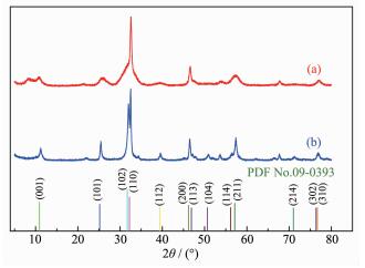
Figure 1.
XRD patterns of persimmon-like BiOBr (a) and BiOBr nanosheets (b)
Facile Solvothermal Synthesis of Porous Persimmon-like BiOBr Photocatalyst
Huang-Gen YANG , Yuan CHEN , Zhi-Wei WANG , Jia-Tian YANG , Li-Gang ZHU , Li-Qin QIN , Hong-Xing DAI

Photocatalysis has been an effective pathway to address the urgent energy crisis and environmental problems since Fujishima and Honda reported the photocatalytic splitting of water over a TiO2 electrode in 1972[1-2]. However, the wide bandgap of TiO2 (ca. 3.2 eV) hinders its widespread applications due to its only ultraviolet-light (4% of the solar spectrum) response[3]. Therefore, it is of significance to develop novel high-efficiency, good-stability and visible-light-driven photocatalysts for pollutant decomposition, water splitting, and carbon dioxide reduction[4-5]. Bismuth oxyhalides (BiOX; X=Cl, Br, I) have attracted much attention because of their good physicochemical properties[6-7]. Among the family of BiOX, BiOBr with a layered structure, in which there are an alternating arrangement of [Bi2O2]2+ slabs and double Br slabs, can absorb a part of visible light owing to its narrow bandgap (ca. 2.7 eV)[8-9]. The unique structure endows BiOBr with high carrier mobility and efficient separa-tion of photogenerated electron-hole pairs, which would be beneficial for enhancing its photocatlytic activity and stability[10-12]. Nonetheless, it is necessary to further improve the photocatalytic activity of BiOBr for the practical applications by tailor-making its morphology and microstructure.
It is well known that controlling the particle size, surface area, and morphology of a photocatalyst is an effective strategy to optimize its photocatalytic perfor-mance. Hence, controllable synthesis of BiOBr with specific morphologies is important. In the past years, the BiOBr photocatalysts with various morphologies and structures, including nanobelts[13], nanosheets[14], and microflowers[15], and microsperes[16], have been developed. For example, Sin et al.[17] prepared the flower-like BiOBr hierarchical structures by the hydrothermal method, which exhibits much higher photocatalytic activity as compared to the commercial TiO2 (P25) for the degradation of sunset yellow. The two-dimensional BiOBr ultrathin nanosheets synthes-ized via a hydrothermal route were used as an oxygen reduction reaction catalyst in the air electrode of aluminum-air batteries[18]. Flower-structured BiOBr photocatalysts with excellent photocatalytic activity and high stability in the degradation of rhodamine B (RhB) was fabricated adopting the chemical precipita-tion procedures[19]. The photocatalytic activity of the ultrathin BiOBr nanosheets is better than the BiOBr microspheres for the degradation of RhB due to the unique micromorphology and microstructure of the former[20]. Hollow BiOBr microspheres with nanoslices were synthesized via a simple one-pot solvothermal method, which exhibits high photocatalytic activity for MB and RhB degradation under visible-light irradia-tion[21]. Flower-like BiOBr microspheres with a controlled morphology were fabricated in ethanol using Bi(NO3)3·5H2O and NaBr as precursors, and the obtained sample shows a high photocatalytic activity due to its abundance of exposed (110) facets for RhB degradation[22].
BiOBr with three-dimensional hierarchical structures assembled from low-dimensional nanoscale building blocks has become one of attractive materials due to peculiar structures and unique properties. As previous studies have demonstrated, BiOBr with hierarchical structures is more structurally stable and could provide a large number of surface active sites for the diffusion and mass transportation of organic molecules and reactive radicals, hence exhibiting good photocatalytic activity in photocatalytic dye degrada-tion[23]. Recently, persimmon-like nanostructures have attracted considerable attention. For example, Cao et al.[24] fabricated persimmon-like (BiO)2CO3, and found that it is able to efficiently degrade RhB and eosin sodium salt under the simulated solar-light irradiation. Dong et al.[25] synthesized persimmon-like N-doped (BiO)2CO3 with a hierarchical superstructure, and observed that such samples show enhanced visible-light-driven catalytic activity as compared to pure (BiO)2CO3 and TiO2-based visible-light photocatalysts for NO removal under visible- or solar-light illumina-tion. Nearly monodisperse YVO4 with a persimmon-like architecture was synthesized on a large scale via a complexing agent-assisted solution route, the ultraviolet absorption and photoluminescence spectra of the samples showed that the optical properties of YVO4 nanopersimmons are relevant to their sizes and shapes[26]. Hexagonal ZnIn2S4 photocatalysts with a three-dimensional (3D)-hierarchical persimmon-like shape were fabricated using the oleylamine-assisted solvothermal method, and hydrogen evolution experiments revealed that the obtained hierarchical ZnIn2S4 possesses good photocatalytic activity[27]. Persimmon-like SrMoO4 with a uniform structure was obtained via a microwave radiation route with ethyl-enediamine tetraacetic acid as the chelating agent[28]. Using the rapid and high-efficient microwave radiation method, CaMoO4 with an interpenetrated persimmon-like structure was generated in a high yield[29]. Uniform and highly hierarchical CaMoO4 microstru-ctures with a persimmon-like morphology were obtained on a large scale by the green solution-based synthesis method without adding any surfactant[30]. Employing N(C4H9)4BF4 as fluoride source, multilayer-stacked hierarchical persimmon-shaped CeF3 was prepared via a template-free hydrothermal route[31]. Persimmon-like NaLa(WO4)2 with a microarchitecture was obtained through a hydrothermal process using trisodium citrate as chelated reagent[32]. To the best of our knowledge, however, preparation of porous nanosheet-aggregated persimmon-like BiOBr and its influence on photocatalytic activity has not yet been investigated systematically.
In this work, we report synthesis of the porous nanosheet-aggregated persimmon-like BiOBr with hierarchical microstructures for the first time utilizing a facile polyvinyl pyrrolidone (PVP)-assisted solvo-thermal approach. Their physicochemical properties were characterized, and their photocatalytic perfor-mance for MB degradation under visible-light irradia-tion was evaluated.
The persimmon-like BiOBr with hierarchical microstructures and the BiOBr nanosheets were fabricated using (or without) the PVP-assisted or solvothermal method. In a typical preparation process, 5.0 mmol of Bi(NO3)3·5H2O and 7.5 mmol of cetyltrie-thylammnonium bromide (CTAB) were dissolved in 30 mL of ethylene glycol (EG) under stirring until a homogeneous solution was formed. Then, 0, 0.2, 0.5, 0.7, 1.0 and 1.5 g of PVP was dissolved in the above mixed solution under stirring for 0.5 h, respectively. The obtained mixed solution was transferred to a 50 mL Teflon-lined stainless autoclave for the solvo-thermal treatment at 120 ℃ for 12 h. The resulting product was filtered and washed with pure ethanol and deionized water several times until the pH value of the filtrate reached 7, followed by drying in an oven at 60 ℃ for 24 h, thus obtaining the persimmon-like BiOBr with hierarchical microstructures and the BiOBr nanosheets. The preparation of the BiOBr contrast samples was same as described above except that the time was changed at a solvothermal temper-ature of 120 ℃ in the presence of 0.7 g PVP. The commercial TiO2 (which was denoted as P25) was purchased from Degussa company.
The X-ray diffraction (XRD) patterns of the samples were recorded on an X-ray diffractometer (Bruker D8-advance, Germany) operated at 40 kV and 35 mA using Cu Kα radiation and nickel filter (λ=0.154 178 nm) within a range of 5°~80°(2θ). The N2 adsorption of the samples were investigated at 196 ℃ on a Quadrasorb S1-3MP analyzer with the samples being degassed at 250 ℃ for 3 h in vacuum before measurement. The surface areas of the samples were calculated by Brunauer-Emmett-Teller (BET) method. The surface morphologies of the samples were observed by a scanning electron microscopy (SEM, Hitachi S-4800) operated at 15 kV. Transmission electron microscopic (TEM) images and selected-area electron diffraction (SAED) patterns of the samples were obtained using a JEM-2100F equipment (operating at 200 kV). Ultraviolet-visible diffuse reflectance spectra (UV-Vis DRS) was recorded with a UV-Vis spectro-photometer (UV-2550, Shimadzu) using BaSO4 as the reference, and the scanning range was 200~700 nm.
Photocatalytic activities of the samples for MB degradation were measured in a quartz reactor (AK6522, Lianyungang, Jiangsu, Guibaoda Customized Quartz Processing Sci. & Technol. Co. Ltd.) under visible-light irradiation using a 300 W Xe lamp with a 400 nm cutoff filter (PLS-SXE300UV, Beijing, Perfect Light Sci. & Technol. Co. Ltd.). 0.2 g BiOBr or commercial TiO2 (Degussa P25) samples were added to 100 mL of aqueous solution containing MB (initial MB concentration C0=10 mg·L-1). Prior to visible-light irradiation, the suspension was ultrasonicated for 0.5 h and magnetically stirred for 2 h in the dark to ensure that the MB could reach an adsorption-desorption equilibrium on the photocatalyst surface. Then, the suspension was magnetically stirred and exposed to visible light. The temperature of the reaction solution was kept at ca. 25 ℃ using flowing cool water. At regular intervals, 5 mL of the suspension was taken out and then centrifuged to remove the photocatalyst particles for the analysis of MB concentration. The concentration (Ct) of the remaining MB was measured by its absorbance (At) at 664 nm with an Agilent Cary 5000 spectrophotometer. The MB degradation efficiency (X) was calculated according to the following equation: X=(C0-Ct)/C0×100%=(A0-At)/A0×100%.
Fig. 1 shows the XRD patterns of the BiOBr samples prepared without and with 0.7 g PVP after solvothermal treatment at 120 ℃ for 12 h. By referencing to the XRD pattern of the standard BiOBr sample (PDF No.09-0393), one can realize that both samples with good crystallinity contain no other impurity phases, and all of the diffraction peaks are characteristic of a tetragonal bismuth oxybromide crystal structure. Similar results were also reported by other researchers[22]. According to the Scherrer formula, the broad diffraction peak in the BiOBr sample synthesized with 0.7 g PVP (Fig. 1a) indicates a small size of the BiOBr crystal. As shown in Fig. 1b, the XRD pattern of the BiOBr sample synthesized without PVP possessed the stronger intensity of the (102) and (110) crystal planes, indicating that this BiOBr crystal had a preferential growth in the two directions. However, the (102) crystal plane (Fig. 1a) of the sample synthesized with 0.7 g PVP was significantly inhibited, and the (101), (112), (104), and (214) crystal planes were also suppressed to certain degree. This might be due to the selective adsorption of PVP on the crystal surface, which could induce generation of the samples with the anisotropic structure and morphology[33].
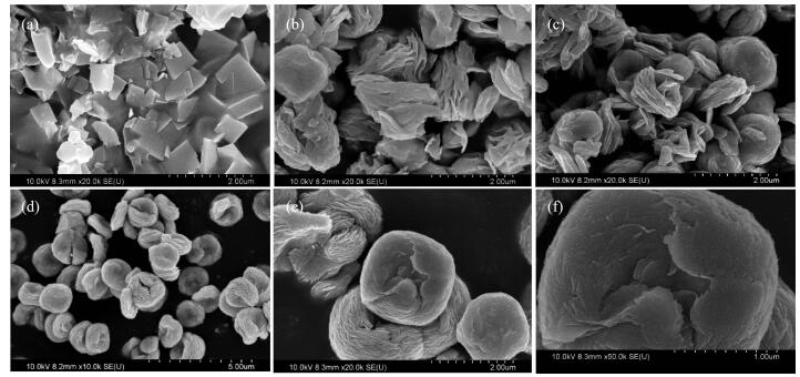
Morphologies and nanostructures of the BiOBr samples were analyzed using SEM and HRTEM techniques, as shown in Fig. 2. The sample prepared with 0.7 g PVP after solvothermal treatment at 120 ℃ for 12 h was composed of a large number of persimmon-like structures with a diameter of 15~20 μm (Fig. 2a), and these persimmon-like microstructures were formed by the aggregation of a number of nanosheets (Fig. 2b and 2c). These nanosheets were intersected and grown together to form a persimmon-like shape that would facilitate separation of the photogenerated electron-hole pairs and hence improve the photocatalytic performance for dye degradation[24-25]. Two complete persimmon-like BiOBr micronstructures displayed a nearly circular morphology with a serrated edge and a layered structure (Fig. 2d and 2e). The central part of the persimmon-like BiOBr was lighter than the marginal part, suggesting that the actual thickness of the middle section is smaller than the edge part. Formation of such a morphology (Fig. 2d) might be a cause of the persimmon-like BiOBr shaping hierarchical structure with cavity. This void made the central portion of the persimmon-like BiOBr hierarchical structure loose, forming a structure similar to a hollow sphere. The recording of multiple bright electron diffraction rings in the SAED patterns (inset of Fig. 2d) of the BiOBr sample suggests that the obtained persimmon-like BiOBr is polycrystalline. The persimmon-like BiOBr micronstructures were composed of many small square nanosheets (Fig. 2e). The intraplanar spacings (d values) were measured to be ca. 0.254 8 and 0.257 1 nm (Fig. 2f), in good consistence with those of the (200) and (113) crystal planes of standard BiOBr (PDF No.09-0393), respectively. It can be seen from Fig. 2g that the persimmon-like BiOBr sample was mainly composed of Bi, O and Br. Most of the sample derived without PVP after solvothermal treatment at 120 ℃ for 12 h were irregular sheet particles with poor dispersi-bility and serious agglomeration (Fig. 2h and 2i). This result indicates the importance of PVP for the formation of persimmon-like BiOBr hierarchical microstructures.
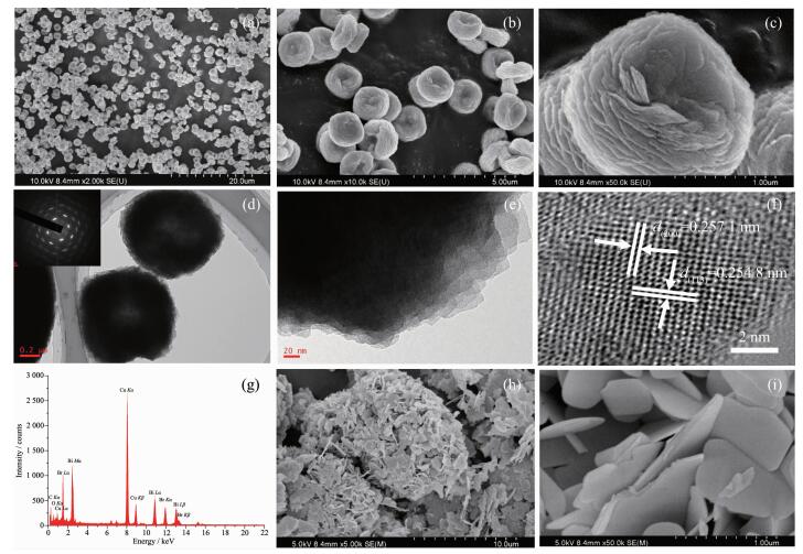
Inset of Fig. 2d: SAED patterns
Fig. 3 shows the N2 adsorption-desorption isotherms and pore-size distributions of the persimmon-like and nanosheet-like BiOBr samples. According to the BDDT classification[34], the isotherms were type Ⅳ. In the range (P/P0=0.8~1.0) of high relative pressures, there was a hysteresis loop for the BiOBr nanosheets, indicating formation of a mesoporous slit structure. Compared with the BiOBr nonasheets, the hysteresis loop of the persimmon-like BiOBr sample moved to a lower relative pressure (P/P0=0.1), suggesting existence of a bimodal mesoporous structure. This was reflected more clearly in the corresponding pore-size distributions (inset), which contained small mesopores of ca. 3.9 and 9.8 nm in diameter and big mesopores of ca. 39.2 nm in diameter. The small mesopores are formed within the nanosheets[35], while the big mesopores are generated between two-dimensional nanolayers. BET surface areas of the persimmon- and nanosheet-like BiOBr samples were 4 and 2 m2·g-1, and the pore volumes were 0.011 and 0.004 mL·g-1, respectively. Obviously, such a 3D hierarchical structure could be conducive to increasing the adsorption efficiency of reactants[36].
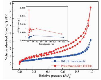
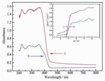
Fig. 4 shows the UV-Vis DRS and (αhν)1/2 versus hν plots (inset) of the persimmon- and nanosheet-like BiOBr samples. Both samples displayed a sharp increase in absorption below 450 nm. Compared with the BiOBr nonasheets, absorption of the persimmon-like BiOBr sample was significantly enhanced in the ultraviolet- and visible-light regions. As a semicon-ductor material, BiOBr obeys the following formula: αhν=A(hν-Eg)n/2, where A is the optical absorption coefficient, ν is the optical frequency, h is Planck constant, n is related to carrier transition, and Eg is the bandgap energy. Since BiOBr is the indirect transitive semiconductor[37-38], n is equal to 4. According to the above formula, we can draw the (αhν)1/2 versus hν plots of the samples. When αhν=0, the corres-ponding tangent value is the Eg of the sample. The Eg values of the persimmon- and nonasheet-like BiOBr samples were 2.64 and 2.73 eV, respectively. The narrower bandgap energy can be excited by the lower-energy visible-light to produce electron-hole pairs. Therefore, the persimmon-like BiOBr hierarchical microstructures would more effectively absorb the incident light, thus improving the catalytic activity.
In order to explore the formation mechanism and evolution process of the persimmon-like BiOBr, we synthesized the BiOBr samples by changing the solvothermal time at 120 ℃ in the presence of 0.7 g PVP, and their SEM images are shown in Fig. 5. As can be seen from Fig. 5a, a large number of square nanosheet entities were formed after 1 h of solvo-thermal treatment, and a few square nanosheets were stacked together. This result suggests that the initial stage of the reaction leads to first generation of small square nanoparticles and then assembling to larger ones. After 4 h of solvothermal treatment, the flaky particles gradually decreased, and the persimmon-like structure began to appear (Fig. 5b). After 6 h of solvothermal treatment, accumulation of the thin-nanosheet particles basically disappeared, and the skeleton structure of primary persimmon-like microstructures were formed (Fig. 5c). After 8 h of solvothermal treatment, a skeletal structure of the persimmon-like stratified BiOBr sample was generated (Fig. 5d). As shown in Fig. 2a~c, the perfect porous sheet-aggregated persimmon-like BiOBr sample were obtained after 12 h of solvothermal treatment. When the solvothermal time was 18 h, part of the persimmon-like structure was damaged (Fig. 5e and f).
To further investigate the crystallization process of BiOBr, we recorded the XRD patterns of the BiOBr samples derived from different solvothermal time, as shown in Fig. 6. When the solvothermal time was 1 h, the product was amorphous. With the extension of solvothermal time to 4 h, crystallinity of the product increased. When the solvothermal time was 6, 8, 12 and 18 h, the diffraction peaks became sharper and increased in intensity. Therefore, formation of the persimmon-like BiOBr phase is a slow crystallization process, and the oriented growth of BiOBr is in agreement with the SEM observations.
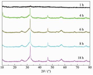
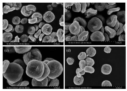
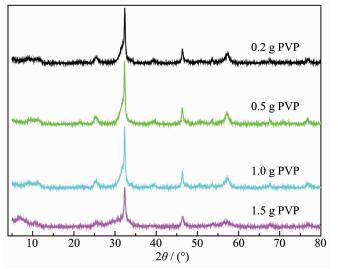
Fig. 7 shows SEM photographs of the BiOBr samples fabricated with different amounts of PVP after solvothermal treatment at 120 ℃ for 12 h. When 0.2 g of PVP was used, some persimmon-like particles of BiOBr were preliminarily generated (Fig. 7a). When the amount of PVP was 0.5 g, the persimmon-like morphological BiOBr particles were basically formed (Fig. 7b). Addition of 0.7 g PVP led to formation of a microstructure of BiOBr with a diameter of 15~20 μm (Fig. 2a~c). When the amount of PVP increased to 1.0 g, the morphology of the product did not change significantly (Fig. 7c). Further rise in amount of PVP to 1.5 g obtained the BiOBr sample with a ball-shaped structure and a diameter of ca. 25 μm (Fig. 7d). The above results indicate that an appropriate amount (0.7 g) of PVP could allow the nanosheets to form a persimmon-like BiOBr hierarchical nanostructure. Fig. 8 shows the XRD patterns of the BiOBr samples derived with different amounts of PVP. The crystallinity of the sample first increased and then decreased with the rise in amount of PVP, confirming the presence of an appropriate amount of PVP.
Based on the above results and combined with the crystal structure and morphology analyses, formation processes of the persimmon-like BiOBr hierarchical microstructures are illustrated in Fig. 9. In the initial stage of the persimmon-like BiOBr growth, Bi(NO3)3·5H2O and CTAB reacted to form the BiOBr nanoparticles in the ethylene glycol solution, and PVP molecules were adsorbed on the surface of the nanoparticles[39]. When the solution was thermally treated for 1 h, the surface of the particles adsorbed by PVP molecules began to crystallize, forming the tiny square nanosheet particles. As the solvothermal treatment time was prolonged, rectangular nano-particles were aligned to generate big two-dimensional nanosheets via the side-side binding. With the aid of PVP, these two-dimensional nanosheets were arranged in a certain way to form a three-dimensional micro-structure. Formation of the primary BiOBr square nanosheets was a typical Ostwald ripening process. The crystal nuclei of BiOBr were formed and grown in the supersaturated solution. According to Gibbs-Thomson′s law, the solubility of big and small particles is different, and the big nanoparticles are grown from the small nanoparticles. Further growth of BiOBr crystals into two-dimensional nanostructures is directly related to the intrinsic crystal structure of the substance itself. BiOBr (200) and (113) crystal facets have higher chemical activity than other crystal facets. In this way, the growth rate of (200) and (113) crystal facets is faster than that of other crystal facets. Therefore, the growth of crystals is preferentially carried out along the layered plane, and the square nanosheet BiOBr was formed after 1 h of reaction. In the subsequent self-assembly process, PVP played an important role in forming big layered nanosheets and multilayered structures[40]. Selective adsorption of PVP on multiple crystal planes of square nanosheet BiOBr was an important process in the initial stage of crystal growth. TEM results show that self-assembled layered nanosheet BiOBr is composed of a large number of small square nanosheet BiOBr, and these primary small nanosheets tend to gradually expand their two-dimensional surface areas via the side-by-side self-assembly. With the help of PVP, these small nanosheets rotated to make the two-dimensional crystal surface of the adjacent nanosheets aligned, and then these small nanosheets fused to form a complete layered nanosheet. As observed in the SEM image (Fig. 5c), newly formed layered nanosheets contained flat and multilayered disks. This process could further reduce the surface energy of the layered nanosheets, and the two-dimensional geometry was particularly conducive to such an accumulation. As the reaction time increased, the layered nanosheets did not continue to accumulate into a cylindrical structure, but was slightly warped to form a concave surface around the flat disks, possibly due to lattice tension or surface interaction at the edge of the layered nanosheets. After the assembly process was carried out in this concave range, more warped layered nanosheets were added to this concave surface, causing the edges of the entire structure to gradually thicken and become denser, while the central part of the disk was almost stagnant in the process[41]. In this way, BiOBr evolved from a flat two-dimensional disk into a three-dimensional persimmon-like BiOBr hierar-chical microstructure with small cavities in the middle. Therefore, the phenomenon that the edge of BiOBr hierarchical microstructure was thicker than the central part is observed in the TEM image (Fig. 2d).
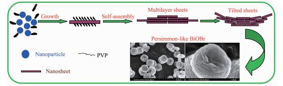
Fig. 10 shows the photocatalytic activities of the samples for MB degradation under visible-light illumination. The MB solution was relatively stable in the absence of a catalyst and only under visible-light irradiation, since almost no decolorization (about 1% degradation efficiency) was observed after 50 min of visible-light illumination. When the MB solution was irradiated by visible light for 10~15 min, MB degradation efficiencies of P25 and BiOBr nonasheets were 11%~15% and 49%~61%, respectively. However, when the MB solution with persimmon-like BiOBr was exposed to visible light for 5 min, MB degradation efficiency reached 99%. After 15 min of visible-light irradiation, the MB solution was almost completely decolorized (ca. 100% MB degradation efficiency), probably because the concentration of MB is lower and more inactive molecules or ions are adsorbed on the photocatalyst surface as the reaction proceeds[42]. The results indicate that the persimmon-like BiOBr sample shows excellent photocatalytic performance under visible-light irradiation.
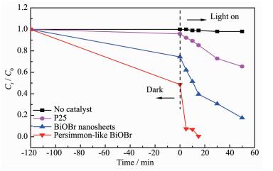
Fig. 11 shows the temporal absorption spectra of MB during the photodegradation processes over persimmon- and nanosheet-like BiOBr. As can be seen from Fig. 11a, the intensity of the MB absorption peaks at 246, 290 and 664 nm continuously decreased under the irradiation of visible light, and the color of the solution became lighter. After 15 min of visible-light illumination, the MB absorption peaks almost disappeared. Over the nanosheet-like BiOBr sample, however, the intensity of the MB absorption peaks at 246, 290 and 664 nm decreased with the light illumination time, but the absorbance at 664 nm was still ca. 0.4 after 50 min of visible-light illumination (Fig. 11b), indicating that MB solution was not completely degraded. Therefore, the visible-light-driven catalytic activity of the persimmon-like BiOBr sample was much higher than that of the nanosheet-like BiOBr sample.
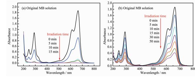
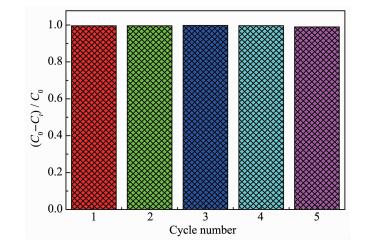
Fig. 12 shows the reusability of the persimmon-like BiOBr sample. It can be clearly seen that the photocatalytic activity of this sample remained basically unchanged after 5 recycles. This result demonstrates that the persimmon-like BiOBr sample is highly catalytically stable under the irradiation of visible light.
It is well known that a photocatalytic process involves the reductive and oxidative reactions of the photo-induced charge carries (e-/h+ pairs) in the degra-dation of organic pollutants, in which the ·OH radicals and active oxygen species participate[43]. Under visible-light irradiation, the porous sheet-aggregated persimmon-like BiOBr is inspired to generate photo-induced e-/h+ pairs. The electrons from the valence band (VB) are transferred to the conduction band (CB), leaving lots of holes in the VB[44]. The photo-induced holes react with H2O on the surface of persimmon-like BiOBr to form ·OH radicals. The photo-induced electrons react with adsorbed O2 on the surface of persimmon-like BiOBr or dissolved O2 in water to generate ·O2- radicals. Therefore, the surface of persimmon-like BiOBr could generate a large number of ·OH and ·O2- radicals with strong oxidation ability which would be beneficial for the degradation of MB[42].
In general, the photocatalytic activity of a semiconductor oxide is mainly determined by the factors, such as crystal structure, bandgap energy, crystallinity, grain size, specific surface area, and pore structure[4]. The main reason for the higher visible-light-driven catalytic activity of the persimmon-like BiOBr sample might be due to the following factors: firstly, the crystallinity of persimmon-like BiOBr is good and this sample possesses a special layered structure, which leads to its wider absorption band edges (corresponding to the forbidden band width of less than BiOBr nonasheets). The improvements in light absorption range and light utilization efficiency of the persimmon-like BiOBr sample render it to show enhanced photocatalytic activity. Secondly, the persimmon-like BiOBr sample possesses a larger surface area, which would be helpful for the adsorption of MB molecules and transfer between different active sites. Consequently, the MB molecules adsorbed on the surface of the material could easily undergo redox reactions with the photogenerated electrons or holes, thus showing higher photocatalytic performance[42]. Thirdly, the three-dimensional hierar-chical structures have a large number of three-dimensional channels, which allow MB and degrada-tion intermediates to be diffused in the channels. These structures would favor the interactions of MB and intermediates with photogenerated electrons or holes, hence enabling more effective and thorough degradation of the organic dyes [39-40].
With bismuth nitrate as Bi source, CTAB as Br source, and EG as solvent in the presence of PVP, a novel sheet-aggregated persimmon-like BiOBr photocatalyst was successfully synthesized via a facile solvothermal route. The solvothermal time and PVP addition greatly influence the hierarchical microstruc-ture, porous structure, and crystallinity of the BiOBr product. After solvothermal treatment at 120 ℃ for 12 h in the presence of 0.7 g PVP, a porous sheet-aggregated persimmon-like BiOBr microstructure (Diameter=15~20 μm) was obtained. The porous persimmon-like BiOBr sample with a surface area of 4 m2·g-1 and a bandgap energy of 2.64 eV exhibited good optical absorption behaviors in visible-light regions and excellent photocatalytic activity and stability for the removal of MB under visible-light illumination. It is concluded that the factors, such as high surface area, low bandgap energy, porous structure and unique morphology, are responsible for the excellent photocatalytic performance of porous persimmon-like BiOBr for MB degradation under visible-light irradiation.
Fujishima A, Honda K. Nature, 1972, 238:37-38 doi: 10.1038/238037a0
Kisch H. Angew. Chem. Int. Ed., 2013, 52:812-847 doi: 10.1002/anie.201201200
Yao Y, Guan L X, Ma Y, et al. J. Mater. Sci., 2017, 28:3013-3019
Ma Y, Wang X, Jia Y, et al. Chem. Rev., 2014, 114:9987-10043 doi: 10.1021/cr500008u
Zheng L X, Han S C, Liu H, et al. Small, 2016, 12:1527-1536 doi: 10.1002/smll.201503441
Bhachu D S, Moniz S J A, Sathasivam S, et al. Chem. Sci., 2016, 7:4832-4841 doi: 10.1039/C6SC00389C
Si Y J, Zhong J B, Li J Z, et al. Mater. Lett., 2016, 163:175-178 doi: 10.1016/j.matlet.2015.10.095
Lin H P, Lee W W, Huang S T, et al. J. Mol. Catal. A, 2016, 417:168-183 doi: 10.1016/j.molcata.2016.03.021
Jiang Y R, Chou S Y, Chang J L, et al. RSC Adv., 2015, 5:30851-30860 doi: 10.1039/C5RA01702E
Fan Z, Zhao Y B, Zhai W, et al. RSC Adv., 2016, 6:2028-2031 doi: 10.1039/C5RA18768K
Lin H P, Lee W W, Huang S T, et al. J. Mol. Catal. A, 2016, 417:168-183 doi: 10.1016/j.molcata.2016.03.021
Li K, Zhang H B, Tang Y P, et al. Appl. Catal. B, 2015, 164:82-91 doi: 10.1016/j.apcatb.2014.09.017
Li H P, Li J Y, Hu T X, et al. Mater. Res. Bull., 2016, 77:171-177 doi: 10.1016/j.materresbull.2016.01.039
Lei F, Sun Y, Liu K, et al. J. Am. Chem. Soc., 2014, 136:6826-6829 doi: 10.1021/ja501866r
Xiao P P, Zhu L L, Zhu Y C, et al. J. Solid State Chem., 2011, 184:1459-1464 doi: 10.1016/j.jssc.2011.04.015
Zhao Y, Tan X, Yu T, et al. Mater. Lett., 2016, 164:243-247 doi: 10.1016/j.matlet.2015.10.155
Sin J C, Lim C A, Lam S M, et al. J. Mater. Sci., 2017, 28:13236-13246
Li M C, Yuan J L, Nan B, et al. Electrochim. Acta, 2017, 249:413-420 doi: 10.1016/j.electacta.2017.08.005
Mao X M, Li M, Li H. J. Adv. Oxid. Technol., 2017, 20:2371-1175
Cao G, Liu Z, Feng P, et al. Mater. Chem. Phys., 2017, 199:131-137 doi: 10.1016/j.matchemphys.2017.06.056
Wang H T, Shi M S, Yang H F, et al. Mater. Lett., 2018, 222:164-167 doi: 10.1016/j.matlet.2018.03.179
Li R P, Ren H J, Ma W H, et al. Catal. Comm., 2018, 106:1-5 doi: 10.1016/j.catcom.2017.11.015
Xiong J Y, Dong Q S, Wang T, et al. RSC Adv., 2014, 4:583-586 doi: 10.1039/C3RA46088F
Cao X F, Zhang L, Chen X T, et al. CrystEngComm, 2011, 13:1939-1945 doi: 10.1039/C0CE00324G
Dong F, Wang R, Li X W, et al. Appl. Surf. Sci., 2014, 319:256-264 doi: 10.1016/j.apsusc.2014.07.062
Qian L W, Zhu J, Chen Z, et al. Chem. Eur. J., 2009, 15:1233-1240 doi: 10.1002/chem.200801724
Shen J, Zai J T, Yuan Y P, et al. Int. J. Hydrogen Energy, 2012, 37:16986-16993 doi: 10.1016/j.ijhydene.2012.08.038
Sun Y, Li C S, Wang L N, et al. Cryst. Res. Technol., 2011, 46:973-978
Sun Y, Li C S, Zhang Z J, et al. Solid State Sci., 2012, 14:219-224 doi: 10.1016/j.solidstatesciences.2011.11.015
Zhou Y X, Chen X B, Zeng X H. Micro Nano Lett., 2011, 6:988-991 doi: 10.1049/mnl.2011.0535
Tang Y F, Zhu J L, Sun T M, et al. Mater. Lett., 2013, 107:20-22 doi: 10.1016/j.matlet.2013.05.121
Tian Y, Chen B J, Tian B N, et al. J. Colloid Interface Sci., 2013, 393:44-52 doi: 10.1016/j.jcis.2012.10.031
Shi X J, Chen X, Chen X L, et al. Chem. Eng. J., 2013, 222:120-127 doi: 10.1016/j.cej.2013.02.034
Sing K S W, Everett D H, Haul R A W, et al. Pure Appl. Chem., 1985, 57:603-619 doi: 10.1351/pac198557040603
Liu S W, Yu J G. J. Solid State Chem., 2008, 181:1048-1055 doi: 10.1016/j.jssc.2008.01.049
Yu X X, Yu J G, Cheng B, et al. J. Phys. Chem. C, 2009, 113:17527-17535 doi: 10.1021/jp906992r
Deng Z G, Chen D, Peng B, et al. Cryst. Growth Des., 2008, 8:2995-3003 doi: 10.1021/cg800116m
Xia J X, Yin S, Li H M, et al. Dalton Trans., 2011, 40:5249-5258 doi: 10.1039/c0dt01511c
Zhang C, Zhu Y F. Chem. Mater., 2005, 17:3537-3545 doi: 10.1021/cm0501517
Wu J, Duan F, Zheng Y, et al. J. Phys. Chem. C, 2007, 111:12866-12871 doi: 10.1021/jp073877u
Li Y Y, Liu J P, Huang X T, et al. Cryst. Growth Des., 2007, 7:1350-1355 doi: 10.1021/cg070343+
安伟佳, 刘利, 李炘檑, 等.无机化学学报, 2015, 31(2):329-337 doi: 10.11862/CJIC.2015.042AN Wei-Jia, LIU Li, LI Xin-Lei, et al. Chinese J. Inorg. Chem., 2015, 31(2):329-337 doi: 10.11862/CJIC.2015.042
潘金波, 刘建军, 马贺成, 等.无机化学学报, 2018, 34(8):1421-1429 doi: 10.11862/CJIC.2018.197PAN Jin-Bo, LIU Jian-Jun, MA He-Cheng, et al. Chinese J. Inorg. Chem., 2018, 34(8):1421-1429 doi: 10.11862/CJIC.2018.197
王鹏, 李昭, 周颖梅, 等.无机化学学报, 2019, 35(2):217-224 doi: 10.11862/CJIC.2019.022WANG Peng, LI Zhao, ZHOU Ying-Mei, et al. Chinese J. Inorg. Chem., 2019, 35(2):217-224 doi: 10.11862/CJIC.2019.022
Zhang J, Shi F J, Lin J, et al. Chem. Mater., 2008, 20:2937-2941 doi: 10.1021/cm7031898

Figure 2 SEM (a~c, h, i) of persimmon-like BiOBr (a~f) and BiOBr nanosheets (h, i); TEM (d~f) images and EDS spectra (g) of persimmon-like BiOBr
Inset of Fig. 2d: SAED patterns

Figure 3 Nitrogen adsorption-desorption isotherms and pore- size distributions (inset) of persimmon-like BiOBr and BiOBr nanosheets

Figure 4 UV-Vis DRS and plots of the (αhν)1/2 vs hν (inset) of persimmon-like BiOBr (a) and BiOBr nanosheets (b)

Figure 5 SEM images of BiOBr samples synthesized after solvothermal treatment for 1 h (a), 4 h (b), 6 h (c), 8 h (d), and 18 h (e, f)

Figure 6 XRD patterns of BiOBr samples synthesized after solvothermal treatment for different times

Figure 7 SEM images of BiOBr samples synthesized with PVP addition of 0.2 g (a), 0.5 g (b), 1.0 g (c) and 1.5 g (d)

Figure 9 Schematic illustration of morphological evolution process of persimmon-like BiOBr

Figure 10 Photocatalytic activities of the samples for MB degradation under visible-light illumination

Figure 11 Temporal absorption spectra of MB during the photodegradation process over persimmon-like BiOBr (a) and BiOBr nanosheets (b)

 扫一扫看文章

扫一扫关注我们
 下载:
下载:

 下载:
下载:

