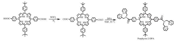
Scheme 1.
Synthesis of Porphyrin-2-DPA

Fluorescent molecular sensors, in particular those for detecting the heavy and transition metal (HTM) ions, have attracted increasing attention due to their high sensitivity, rapid responsiveness, and low-detection-limit[1-6]. However, most fluorescent sensors focus just on single-target detection with few dealing with "single sensor for multiple-analytes"[7-12]. Towards future practical applications, it is necessary to develop the single fluorescent sensors with different responses for multiple-analytes since they could shorten the preparation process to multiple sensors and facilitate to detect multiple analytes with high efficiency. For the purpose of realizing the versatile molecular optical detection for multiple-analytes, usually more than one receptors and/or fluorophores have been incorporated into the multiple-detective single fluorescent sensors[13-15]. In 2005, a three chromogenic units-containing chemosensor was prepared by Suzuki and co-workers for Fe3+/Pb2+/Al3+/Cu2+ on the basis of different coordination mode between metal ion and multi-dentate single receptor[16]. Akkaya synthesized a three-receptor-consisting styryl-Bodipy sensor, which is able to detect Zn2+, Ca2+, and Hg2+ [17]. Subsequently, one-receptor and two-fluorophores composed Porphyrin-BODIPY FRET ratiometric sensor for Fe2+/Hg2+ was synthesized by our group[18]. Meanwhile, there is less study on single-fluorophore-receptor-constructing chem-osensors for di/tri-analytes associated with the different sensing mechanism[19-20]. Notably, the studies on the multiple-analyte sensors have thus far focused mainly on its multiple-detecting function, and there still exists less investigation over the detecting effect of each receptor for multi-receptor-consisting sensors.
Porphyrin chromophore has been one of the most promising signaling units in constructing fluorescent sensors due to its advantageous photophysical charact-eristics such as pronounced photostability, high extinc-tion coefficient, and tunable fluorescence emission[21-27]. Moreover, the central tetrapyrrole macrocyclic moiety of porphyrin derivatives are also able to act as excellent functional receptor for various metal ions due to the strong binding ability attributed to four-pyrrole-nitrogen atoms[28]. As a consequence, extensive investigations have been carried out over the porphyrin-based fluorescent sensors for Cu2+, Hg2+, Cu2+/Pb2+, etc[29-31]. Our group also prepared a series of N, N-di(2-pyridylmethyl)amino(DPA)-based porphyrinato zinc complexes to investigate the number effect of DPA receptor on Fe3+-detecting function[32]. Herein, a di-DPA-based metal-free porphyrin, namely 5, 15-di(p-N, N-bis(2-pyridylmethyl)amino-phenyl)-10, 20-di(4-tert-butylphenyl)-porphyrin (Porphyrin-2-DPA) was synth-esized, as shown in Scheme 1[32]. The tetrapyrrole macrocyclic moiety in Porphyrin-2-DPA as a rigid π-conjugated plane is almost perpendicular to the phenyl plane attached to DPA moiety, indicating the non-electronic coupling nature between porphyrin core and DPA unit. Accordingly, the rigid porphyrin core in Porphyrin-2-DPA is utilized not only as sensitive signaling fluorophore but also as the primary binding receptor with excellent affinity and distinctive selectivity to metal ion, which together with the flexible DPA auxiliary receptor endows Porphyrin-2-DPA the excellent sensing function to Pb2+/Cu2+ on the basis of versatile optical-signals.
Column chromatography was carried out on silica gel (Merck, Kieselgel 60, 70-230 mesh) with the indicated eluents. The tetrahydrofuran (THF) and methanol used for spectral experiments were purified via the standard methods. Other reagents and solvents were used as received. The reference compounds of 5, 10, 15, 20-tetra(4-tert-butylphenyl)porphyrin (Porphyrin-0-DPA), 5-(p-N, N-bis(2-pyridylmethyl)aminophenyl) 10, 15, 20-tri(4-tert-butylphenyl)porphyrin (Porphyrin-1-DPA), 5, 10, 15, 20-tetra(p-N, N-bis(2-pyridylmethyl) aminophenyl)porphyrin (Porphyrin-4-DPA), 5, 15-di(p-N, N-bis(2-pyridylmethyl)aminophenyl)10, 20-di(4-tert-butylphenyl)porphyrinato zinc complex (Porphyrin-2-DPA-Zn) and 5, 10-di(4-carboxylphenyl)-10, 20-di(4-tert-butylphenyl)porphyrin were prepared according to the reported procedures[31-34].
1H NMR spectra was recorded on a Bruker DPX 400 MHz spectrometer in CDCl3 with shifts referenced to SiMe4 (0.00 ppm). Electronic absorption spectra were recorded on a U-4100 spectrophotometer. Steady-state fluorescence spectroscopic studies were performed on an F 4500 (Hitachi). The slit width was 5 nm for emission, and the photon multiplier voltage was 700 V.
According to the reference[32], the mixture of 5, 10-di(4-carboxylphenyl)-10, 20-di(4-tert-butylphenyl)porphyrin (81 mg, 0.1 mmol) and thionyl chloride (15 mL) was refluxed for 2 h under N2 atmosphere and evaporated by atmospheric distillation. The residue obtained was dissolved in anhydrous THF (10 mL) and then added into the solution of DPA (60 mg, 0.3 mmol), followed by adding two drops of anhydrous triethylamine. After stirring for another 3 h at 55 ℃, the resulting black-green mixture was evaporated under reduced pressure, and the residue was chromatographed on a silica gel column using CHCl3/MeOH (98:2, V/V) as eluent. Repeated chromatography followed by recrystallization from CHCl3 and MeOH gave Porphyrin-2-DPA, 82 mg in the yield of 70%. 1H NMR (CDCl3, 400 MHz): δ 8.89 (d, 2H, J=8 Hz), 8.78(d, 4H, J=8 Hz), 8.69 (d, 4H, J=4 Hz), 8.61 (d, 4H, J=8 Hz), 8.23 (d, 4H, J=8 Hz), 8.12 (d, 4H, J=8 Hz), 7.98 (d, 6H, J=8 Hz), 7.77 (d, 8H, J=8 Hz), 7.62 (d, 2H, J=12 Hz), 7.42 (d, 2H, J=12 Hz), 5.07 (s, 8H), 1.32 (s, 18H), -2.83 (d, 2H).
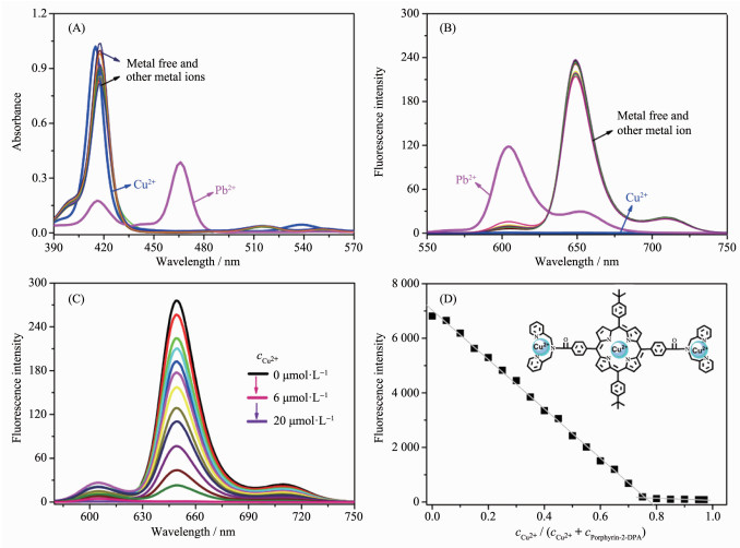
To investigate the sensing function of Porphyrin-2-DPA to metal ions, the photophysical properties of this compound (2 μmol·L-1) upon addition of different metal ion was studied in a mixed in CH2Cl2/MeOH (1:1, V/V). As shown in Fig. 1A and 1B, the electronic absorption and fluorescent emission spectra of metal free Porphyrin-2-DPA kept almost unchanged upon addition of different metal ion such as Fe2+, Co2+, Hg2+, Mn2+, Zn2+, Ni2+, Cd2+, Ca2+, Ba2+, Mg2+, Li+, Na+, or K+, except for Cu2+ and Pb2+ (20 μmol·L-1). After adding Cu2+, though the maximum absorption of Porphyrin-2-DPA was slightly blue-shifted (from 417 to 415 nm), the Q bands of this compound at 515, 551, and 590 nm disappeared synchronously accompanied by the appearance of a new absorption around 538 nm, leading to the varying ratio of A538/A515 from 0.25 to 5.42. Meanwhile a remarkable change also occured in the fluorescence emission of Porphyrin-2-DPA after adding Cu2+: the fluorescence emission of this compound centered at 650 nm was obviously weakened even to be completely quenched. Moreover, the fluore-scence titration experiments for Porphyrin-2-DPA (2 μmol·L-1) with increasing amount of Cu2+ (0~20 μmol·L-1) show that the fluorescence decrease mainly occured in the concentration range of Cu2+ from 0 to 6 μmol·L-1, suggesting the possible 1:3 binding stoichiometry between Porphyrin-2-DPA and Cu2+ in Fig. 1C. The fluorescence Job′s plot gave additional support for this point, revealing the possible binding mode between Porphyrin-2-DPA and Cu2+ with both DPA moiety and Porphyrin core in Porphyrin-2-DPA as the receptor for Cu2+ in Fig. 1D. The detection limit for Cu2+ ion with Porphyrin-2-DPA was determined to be 1.6×10⁻⁷ mol·L-1 under the present condition, revealing the sensitive dual-optical detecting nature of porphyrin-2-DPA for Cu2+ [35-37].
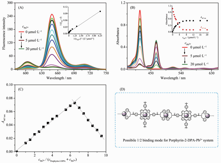
More notably, addition of Pb2+ into Porphyrin-2-DPA not only resulted in the quenching of the fluorescent emission of this compound at 650 nm but also induced the remarkable change in its electronic absorption spectrum. As shown in Fig. 1, upon addition of Pb2+, the maximum absorption of Porphyrin-2-DPA centered at 417 nm as well as Q bands at 515, 551 and 590 nm was diminished synchronously accomp-anied the appearance of a new strong absorption band at 467 nm, leading to the multi-ratiometric changes including the intensity radio of A467/A417 from 0.01 to 3.38, A467/A515 from 0.23 to 42 and A467/A551 from 0.42 to 47.5 (Ai is the absorbance at the wavelength i and Amax is the maximum intensity). Meanwhile the fluorescent emission of this compound at 650 nm was obviously decreased while the fluorescent emission at 604 nm was increased after adding Pb2+, with a changing ratio of I604/I650 from approximate 0.03 to 22. Furthermore, the quantitative fluorescence titration experiments for Porphyrin-2-DPA (at 650 nm) with the increasing amount of Pb2+ (0~20 μmol·L-1) showed that the change in fluorescent emission intensity mainly occursed in the concentration range (0~5 μmol·L-1) of Pb2+, and the emission intensity 1/(Imax-I) increased linearly against the change in 1/(CPb2+)2 according to the Benesi-Hildebrand equation, suggesting the possible 1:2 bind-ing stoichiometry between Porphyrin-2-DPA and Pb2+, as shown in Fig. 2A. This is confirmed by the absorption titration experiments as well as fluorescent Job′s plot in Fig. 2B and 2C, suggesting the most possible binding mode between Porphyrin-2-DPA and Pb2+ (Fig. 2D).
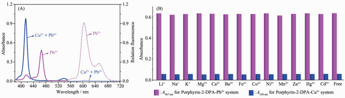
It is worth noting that upon subsequent addition of Cu2+ (20 μmol·L-1), the absorption of Porphyrin-2-DPA-Pb2+ system centered at 467 nm got disappeared synchronously accompanied by the appearance of the maximum absorption at 415 nm obviously attributed to the Porphyrin-2-DPA-Cu2+ system, indicating the displa-cement of Pb2+ in Porphyrin-2-DPA-Pb2+ complex by Cu2+. This is further validated by the disappearance of the fluorescence emission band at 604 nm attributed to Porphyrin-2-DPA-Pb2+ complex after addition of Cu2+ ion (Fig. 3A). In contrast, upon addition of other metal ion such as Fe2+, Co2+, Hg2+, Mn2+, Zn2+, Ni2+, Cd2+, Ca2+, Ba2+, Mg2+, Li+, Na+, or K+, the electronic absorption and fluorescent emission spectra of Porphyrin-2-DPA-Pb2+ system kept almost unchanged, which was also true for the Porphyrin-2-DPA-Cu2+ system (Fig. 3B), clearly indicating the excellent selectivity of Porphyrin-2-DPA to Cu2+ or Pb2+ ions among all the tested metal ions. As a consequence, Porphyrin-2-DPA can work as a dual-mode Cu2+-selective sensor via porphyrin fluor-escence ON-OFF mechanism as well as dual-signal (the ratio of A467/A415 and fluorescence ON-OFF) metal displacement from the Porphyrin-2-DPA-Pb2+ complex.
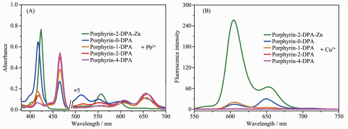
To understand the respective sensing role of porphyrin and DPA receptors to Cu2+/Pb2+, the control experiments of the reference porphyrin derivatives including Porphyrin-0-DPA and Porphyrin-X-DPA (X=1 and 4) as well as Porphyrin-2-DPA-Zn upon addition of Pb2+/Cu2+ have been carried out in a just same manner as for Porphyrin-2-DPA[31-32]. As shown in Fig. 4, upon addition of Pb2+, the electronic absorption and fluorescence emission spectra of Porphyrin-2-DPA-Zn kept almost unchanged. By contrast, addition of Pb2+ into Porphyrin-0-DPA induced the decrease in its maximum absorption at 417 nm synchronously with the appearance of a new strong absorption band at 467 nm. Moreover, the absorption at 417 nm was more weakened while the one at 467 nm obviously increased along with increasing the peripheral DPA number of Porphyrin-X-DPA derivatives from 1 to 2 and 4. Similarly, the fluorescence emission of Porphyrin-DPA derivatives at 650 nm was gradually decreased while the one at 604 nm ncreased along with the increase of the DPA number from 0→1→2→4 upon addition of Pb2+. These results clearly suggest that the rigid Porphyrin core in Porphyrin-DPA derivatives is employed not only as fluorophore signal unit but also as primary binding ligand, which combined with the flexible DPA auxiliary receptor endows Porphyrin-DPA derivatives the excellent detecting potential to Pb2+. This is also true for Cu2+ in Fig. 4B. However, the optical change degree for Porphyrin-DPA derivatives along with increasing the DPA number from 0 to 4 was slightly less after adding Cu2+ than Pb2+, implying that porphyrin core as the firstly binding ligand plays more important role in detecting the former. This may be attributed to the slightly smaller atomic radius together with the distinctive characteristic outermost electronic structure of Cu2+, which can induce the more strong binding affinity of the rigid porphyrin core to Cu2+ than Pb2+, thus gives Porphyrin-2-DPA the diverse optical-detecting-signal to Cu2+/Pb2+.
Briefly summarizing above, a two-DPA-modified metal free porphyrin compound has been synthesized and characterized. Systemic studies show that the rigid π-conjugated porphyrin core was utilized not only as signaling fluorophore but also as the primary metal ion receptor with the excellent binding affinity and distinctive selectivity, which together with the no-conjugated flexible DPA receptor endows DPA-modified porphyrin compound the excellent sensing function to Pb2+/Cu2+ on the basis of versatile optical-signals.
Zhu H, Fan J L, Wang B H, et al. Chem. Soc. Rev., 2015, 44:4337-4366 doi: 10.1039/C4CS00285G
Qin M, Huang Y, Li F Y, et al. J. Mater. Chem. C, 2015, 3:9265-9275 doi: 10.1039/C5TC01939G
Tang J, Yin H Y, Zhang J L. Chem. Sci., 2018, 9:1931-1939 doi: 10.1039/C7SC04498D
Chen Y T, Pan H H, Jiang J Z, et al. Eur. J. Inorg. Chem., 2017, 19:5254-5259
Su D D, Teoh C L, Wang L, et al. Chem. Soc. Rev., 2017, 46:4833-4844 doi: 10.1039/C7CS00018A
Yao Y W, Wang F, Zhang X L, et al. J. Porphyrins Phthalo-cyanines, 2018, 22:1-7 doi: 10.1142/S1088424617500699
Chen Y T, Wang K L, Jiang J Z. Photochem. Photobiol. Sci., 2013, 12:2001-2007 doi: 10.1039/c3pp50126d
Kotani H, Ohkubo K, Crossley M J, et al. J. Am. Chem. Soc., 2011, 133:11092-11095 doi: 10.1021/ja204161j
Diana R, Caruso U, Concilio S, et al. Dyes Pigm., 2018, 155:249-257 doi: 10.1016/j.dyepig.2018.03.055
Aprahamian I. Chem. Commun., 2017, 53:6674-6684 doi: 10.1039/C7CC02879B
Wu W N, Wu H, Wang Y, et al. RSC Adv., 2018, 8:5640-5646 doi: 10.1039/C7RA10219D
Yang M, Chae J B, Kim C, et al. Photochem. Photobiol. Sci., 2019, doi. org/10.1039/C8PP00545A
He L W, Dong B L, Liu Y, et al. Chem. Soc. Rev., 2016, 45:6449-6461 doi: 10.1039/C6CS00413J
Chen Y T, Pan H H, Wang F, et al. Dyes Pigm., 2019, 165:279-286 doi: 10.1016/j.dyepig.2019.02.034
Kolanowski J L, Liu F, New E J. Chem. Soc. Rev., 2018, 47:195-208 doi: 10.1039/C7CS00528H
Bozdemir O A, Guliyev R, Buyukcakir O, et al. J. Am. Chem. Soc., 2010, 132:8029-8036 doi: 10.1021/ja1008163
Komatsu H, Citterio D, Fujiwara Y, et al. Org. Lett., 2005, 7:2857-2859 doi: 10.1021/ol0507219
Chen Y T, Wan L, Li W J, et al. Org. Lett., 2011, 13:5774-5777 doi: 10.1021/ol202338c
Huang C B, Li H R, Luo Y Y, et al. Dalton. Trans., 2014, 43:8102-8108 doi: 10.1039/c4dt00014e
Zhang X L, Guo X H, Yuan H H, et al. Dyes Pigm., 2018, 155:100-106 doi: 10.1016/j.dyepig.2018.03.037
Zhang J F, Zhou Y, Yoon J, et al. Org. Lett., 2010, 12:3852-3855 doi: 10.1021/ol101535s
Zhu M L, Zhang J H, Zhou Y B, et al. Inorg. Chem., 2018, 57:11537-11542 doi: 10.1021/acs.inorgchem.8b01581
Chen J D, Zhou Z X, Chen Z X, et al. New J. Chem., 2017, 41:10272-10280 doi: 10.1039/C7NJ01263B
Zhang L, Chen Y T, Jiang J Z. Dyes Pigm., 2016, 124:110-119 doi: 10.1016/j.dyepig.2015.09.005
Weng Y Q, Yue F, Zhong Y R, et al. Inorg. Chem., 2006, 46:7749-7755
Zhu X J, Fu S T, Wong W K, et al. Angew. Chem. Int. Ed., 2006, 45:150-3153 doi: 10.1002/anie.200502706
Abdullah K, Chen Y L, Jiang J Z. J. Porphyrins Phthalocy-anines, 2017, 21:893-899 doi: 10.1142/S1088424617500985
Fang Z, Pu K Y, Liu B. Macromolecules, 2008, 41:8380-8387 doi: 10.1021/ma801874z
Li C Y, Zhang X B, Qiao L, et al. Anal. Chem., 2009, 81:9993-10001 doi: 10.1021/ac9018445
Brittle S A, Richardson T H, Dunbar A D F, et al. J. Mater. Chem., 2011, 21:4882-4887 doi: 10.1039/c0jm03670f
Chen Y T, Jiang J Z. Org. Biomol. Chem., 2012, 10:4782-4787 doi: 10.1039/c2ob25313e

Figure 1 (A) Electronic absorption spectra and (B) fluorescence emission spectra of Porphyrin-2-DPA at the concentration of 2 μmol·L-1 in CH2Cl2/MeOH (1:1, V/V) upon addition of different metal ions such as Cu2+, Fe2+, Co2+, Hg2+, Mn2+, Zn2+, Ni2+, Cd2+, Pb2+, Ca2+, Ba2+, Mg2+, Li+, Na+, or K+, respectively; (C) Fluorescence emission spectra of Porphyrin-2-DPA upon addition of increasing amount of Cu2+ (0~20 μmol·L-1); (D) Fluorescence Job′s plot indicating the 1:3 binding stoichiometry between Porphyrin-2-DPA and Cu2+ in Porphyrin-2-DPA-Cu2+ system. The inset shows the binding mode between Porphyrin-2-DPA and Cu2+

Figure 2 (A) Fluorescence emission and (B) electronic absorption spectra of Porphyrin-2-DPA upon addition of increasing amount of Pb2+ (0~20 μmol·L-1); (C) Absorption Job′s plot (at 467 nm) indicating the 1:2 binding stoichiometry between Porphyrin-2-DPA and Pb2+ in Porphyrin-2-DPA-Pb2+ system; (D) Possible binding mode between Porphyrin-2-DPA and Pb2+

Figure 3 (A) Electronic absorption and fluorescence emission spectra of Porphyrin-2-DPA (2 μmol·L-1) upon sequential addition of Pb2+ (20 μmol·L-1) and Cu2+ ion (20 μmol·L-1) in CH2Cl2/MeOH (1:1, V/V); (B) Change in the electronic absorption of 467 nm for Porphyrin-2-DPA-Pb2+ system and 538 nm for Porphyrin-2-DPA-Cu2+ system upon addition of other metal ions such as Fe2+, Co2+, Hg2+, Mn2+, Zn2+, Ni2+, Cd2+, Ca2+, Ba2+, Mg2+, Li+, Na+, or K+, respectively

 扫一扫看文章

扫一扫关注我们
 下载:
下载:

 下载:
下载:
