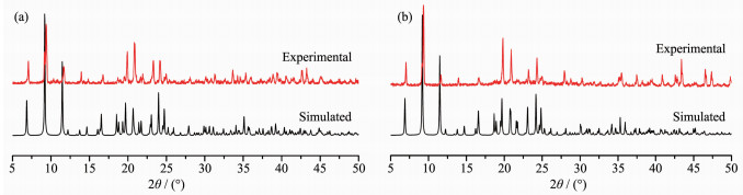
Figure 1.
PXRD patterns of 1 (a) and 2 (b)

Bi-functional materials combining luminescence with magnetic properties have become a research focus due to their various applications, such as information storage, sensing (optical detection of magnetic compounds) and bio-imaging (combining for instance magnetic relaxation imaging (MRI) and fluorescence labelling)[1-3]. However, because of the quenching effect of paramagnetic transition metal (TM) ions on the luminescence of organic dyes, it is difficult to obtain magneto-optical materials from TM-based complexes[4]. On the contrary, lanthanide-based complexes are well known for their magnetic properties with large angular momentum and crystal-field effect[5], as well as their exceptional luminescent properties with narrow band and long-lived emission, large Stokes shifts, high luminescence efficiency, and ligand-dependent lumin-escence sensitization[6-7]. In this regard, lanthanide ions have been incrementally used to design bi-functional magneto-optical materials by the rational choice of suitable ligands, which should hold not only efficient energy-transfer properties for the luminescent materials but also the proper orbitals for the construc-tion of lanthanide-based magnetic materials[8-11].
Aromatic carboxylate ligands are usually used to construct lanthanide-based materials because carboxylic groups can clip fit well with the oxytropic lanthanide ions, and mediate strong magnetic interactions between paramagnetic centers[9]. Simultaneously, aromatic subs-tituent groups can act as sensitive antennas to promote the luminescence of lanthanide ions and lead to π stacking[12-13]. Previously, we chose the N-succinylpyri-dine ligand to achieve two d10-based compounds[14] and several lanthanide-based compounds[15], revealing the flexible coordination modes, rich fluorescent properties, and well magnetic mediums of N-succinylpyridine ligand. These results are very helpful for our further design and investigation on the bi-functional magneto-optical lanthanide-based materials.
In this work, we reported the preparation, structures, magnetic and fluorescent properties of two new complexes, [Ln(HL)2(H2O)4]Cl3 (Ln=Pr (1), Eu (2), HL=N-succinopyridine).
N-succinopyridine was obtained according to the reported procedure[16], and other chemicals were comm-ercially available analytical grade and used without further purification. The elemental analyses were carried out on an Elementar Vario EL Ⅲ microanalyzer 9. Powdered X-ray diffraction (PXRD) patterns were tested on a Rigaku MiniFlex Ⅱ diffractometer using Cu Kα radiation (λ=0.154 2 nm) at 30 kV and 15 mA in a range of 5°~50°. The simulated patterns were finished by the Mercury software. The FT-IR spectra were measured on a Perkin-Elmer spectrum using KBr pellets as the matrices within 4 000~400 cm-1. Optical diffuse reflectance spectra were achieved at room temperature on a PE Lambda 900 UV-Vis spectro-photometer used BaSO4 plate as a standard (100% reflectance). Thermogravimetric analysis (TGA) experiments were recorded on a Netzsch STA449C-QMS403C thermogravimetric analyzer under N2 atmosphere with the sample heated in an Al2O3 crucible at a heating rate of 10 K·min-1. The tempera-ture dependence of magnetic susceptibility was done with a Quantum Design MPMS-XL superconducting quantum interference device (SQUID) magnetometer in a temperature range of 2~300 K at a field of 1 kOe and diamagnetic corrections were made using Pascal′s constants. The determination of PL was conducted on a double excitation monochromator Edinburgh FL920 fluorescence spectrometer equipped with a R928P PMT detector.
The preparation procedures and methods of the two complexes are similar. The lanthanide halide (2 mmol) and N-succinylpyridine (195.2 mg, 1 mmol) were dissolved in 10.0 mL of distilled water and stirred under an atmosphere for 3 hours. Then the resulting solution was filtered into a glass beaker and slowly evaporated to crystallize. After about half a month, prismatic crystals which can be used for X-ray analysis were obtained. The crystal samples used for all testing and characterization were carefully hand-picked with the help of a microscope. The purity of the obtained crystalline samples was measured PXRD (Fig. 1). Elemental analysis data of C, H, N was also used to prove the purity of test samples. Anal. Calcd. for C18H26Cl3N2O12Pr (1)(%): C, 30.44; H, 3.66; N, 3.95. Found(%): C, 29.31; H, 3.84; N, 3.74. Anal. Calcd. for C18H26Cl3N2O12Eu (2)(%): C, 29.97; H, 3.61; N, 3.89. Found(%): C, 29.39; H, 3.88; N, 3.60.
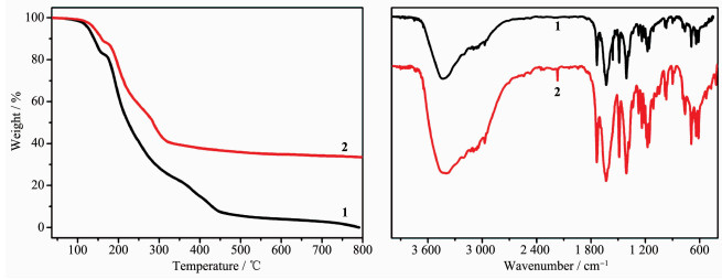
TG analysis data (Fig. 2a) indicates that 1 and 2 are stable up to 109 and 114 ℃, then show a weight loss between 155 and 162 ℃, in accordance with the removal of four coordinated water molecules (Obsd. 13.86% and 9.52%; Calcd. 10.15% and 10.00% for 1 and 2, respectively). Yield based on HL: 49% for 1; 18% for 2. IR (KBr, cm-1) (Fig. 2b) for 1: 3 430 (s), 1 735 (s), 1 632 (s), 1 559 (s), 1 488 (s), 1 409 (s), 1 272 (w), 1 238 (w), 1 210 (s), 1 175 (m), 1 159 (m), 1 109 (w), 984 (w), 971 (w), 896 (m), 785 (w), 764 (w), 694 (m), 637 (m), 613 (m); 2: 3 447~3 393 (s), 1 737 (s), 1 630 (s), 1 489 (s), 1 409 (s), 1 338 (m), 1 274 (m), 1 238 (m), 1 177 (s), 1 156 (s), 1 111 (m), 1 082 (m), 1 050 (m), 984 (m), 969 (m), 899 (w), 764 (m), 693 (s), 640 (m), 613 (s), 529 (m), 477 (w), 420 (w).
The single-crystal X-ray diffraction measurement was collected on a Rigaku AFC7R diffractometer for 1 and Rigaku SCX mini for 2, respectively, which were equipped with Mo Kα radiation (λ=0.071 07 nm). Evidence during data collection indicated that the crystal did not decay, indicating that the obtained polymers were stable at ambient temperature. The intensity data sets were collected with the ω scan technique and corrected for Lp effects. The primitive structures were solved by the direct method and reduced by the CrystalClear software[17]. The subsequent successive difference Fourier syntheses yielded the other non-hydrogen atoms. The final structure was refined using a fullmatrix least-squares refinement on F2. All non-hydrogen atoms were refined anisotropically. The hydrogen atoms of HL molecule were added geometrically and refined using the riding model. The hydrogen atoms of all water molecules were located in the idealized positions and refined with O-H distances restrained to a target value of 0.085 nm, the H-H distance to 0.134 nm, and Uiso(H)=1.5Ueq(O). All of the calculations were performed by the Siemens SHELXTL version 5 package of crystallographic software[18]. The crystal data and structure refinement results for 1 and 2 are given in Table 1. The selected bond lengths (nm) and angles (°) are given in Table 2 and Table 3.
 下载:
导出CSV
下载:
导出CSV
| Compound | 1 | 2 |
| Formula | C18H26Cl3N2O12Pr | C18H26Cl3N2O12Eu |
| Formula weight | 709.67 | 720.72 |
| Crystal system | Monoclinic | Monoclinic |
| Space group | P2/c | P2/c |
| a / nm | 1.308 2(6) | 1.301 2(5) |
| b / nm | 0.961 9(5) | 0.962 7(4) |
| c / nm | 1.119 2(5) | l.l08 2(4) |
| β / (°) | 99.73(4) | 99.563(6) |
| V / nm3 | 1.388 0(11) | 1.368 8(9) |
| Z | 2 | 2 |
| Dc / (g·cm-3) | 1.698 | 1.749 |
| μ / mm-1 | 2.102 | 2.643 |
| Parameter, restraint, data | 187, 7, 2 187 | 179, 0, 2 163 |
| R1a [I > 2σ(I)] | 0.031 8 | 0.042 8 |
| wR2b [I > 2σ(I)] | 0.082 5 | 0.112 1 |
| Goodness of fit | 1.007 | 1.069 |
下载:
导出CSV
| Pr(1)-O(1) | 0.242 7(3) | Pr(1)-O(2)#1 | 0.237 8(3) | Pr(1)-O(1W) | 0.251 4(3) |
| Pr(1)-O(2W) | 0.252 5(4) | ||||
| O(2)#1-Pr(1)-O(1)#3 | 86.52(11) | O(1)#3-Pr(1)-O(1) | 147.12(18) | O(1W)#3-Pr(1)-O(1W) | 68.65(17) |
| O(1)-Pr(1)-O(1W) | 72.67(12) | O(2W)-Pr(1)-O(2W)#3 | 79.56(18) | O(2)#2-Pr(1)-O(1W)#3 | 75.14(13) |
| Symmetry codes: #1: x, -y+1, z+1/2; #2: -x+1, -y+1, -z+2; #3: -x+1, y, -z+5/2. | |||||
下载:
导出CSV
| Eu(1)-O(2) | 0.233 1(4) | Eu(1)-O(1W) | 0.247 5(4) | Eu(1)-O(2W) | 0.247 6(4) |
| Eu(1)-O(1)#2 | 0.237 5(4) | ||||
| O(2)-Eu(1)-O(1)#3 | 86.21(15) | O(1)#2-Eu(1)-O(2W) | 140.28(15) | O(1)#2-Eu(1)-O(1)#3 | 146.8(2) |
| O(2)#1-Eu(1)-O(2W)#1 | 77.59(15) | O(1W)-Eu(1)-O(1W)#1 | 78.O(2) | O(1)#3-Eu(1)-O(2W) | 72.67(15) |
| Symmetry codes: #1: -x+1, y, -z+3/2; #2: x, -y+1, z-1/2; #3: -x+1, -y+1, -z+2. | |||||
CCDC: 885100, 1; 902937, 2.
The single-crystal X-ray diffraction analyses indicated that 1 and 2 belong to isostructural phases. Hence, compound 1 is taken as an example to depict the crystal structure. Compound 1 crystallizes in the monoclinic system P2/c. (No.147), and the asymmetric unit consists of one Pr(Ⅲ) ion, one neutral HL ligand, two coordinated water molecules, one and half a Cl- anions.
As shown in Fig. 3a, every Pr(Ⅲ) ion coordinates eight oxygen atoms and exhibits distorted square anti-prismatic geometry. One basal aspect of the reverse prism composes of O1, O1W, O2#2 and O2W atoms, and the other base builds up with O2W#3, O2#1, O1#3 and O1W#3 atoms. The Pr-O bond distances ranging from 0.237 8(3) to 0.252 5(4) nm are normal as compared with Pr carboxylates[15, 19-21]. Note that Cl2 atom splits up into Cl2 and Cl21 with the SOF value of 0.35(0) and 0.15(0). Adjacent Pr(Ⅲ) ions are doubly bridged by syn-syn carboxylate groups forming linear 1D chains. Neighboring chains form 2D layered structure by O3…Cl1…O1W hydrogen bonds along b axis (Fig. 3b). Finally, these layers are further expanded into a 3D network through the hydrogen bonding of O1W…Cl2…O2W (O3…Cl1 0.304 0(4) nm, Cl1…O1W 0.306 3(5) nm, O1W…Cl2 0.314 5(5) nm, Cl2…O2W 0.313 5(5) nm, Fig. 3c). The coordination mode of ligand in 1 is different to that in our previous work[15], revealing the flexible coordination modes of HL. The intrachain distance of Pr…Pr is 0.564 0(2) nm and the nearest interchain distance of Pr…Pr is 0.961 9(5) nm. It is worth noting that although the lanthanide ions has a good affinity for oxygen, only one carboxylate unit is coordinated to Pr(Ⅲ) ions in the HL ligand.

Symmetry codes: #1: x, -y+1, z+1/2; #2:-x+1, -y+1, -z+2; #3: -x+1, y, -z+5/2
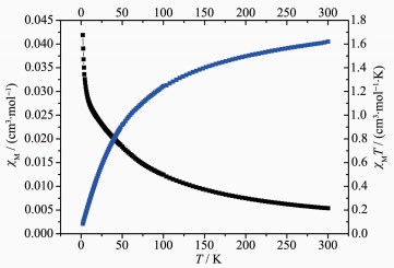
The temperature dependence of magnetic susceptibility for 1 is investigated and presented in Fig. 4. Compound 1 exhibited a χMT value of 1.62 cm3·K·mol-1 at room temperature, which is in excellent agreement with the theoretical value of 1.60 cm3·K·mol-1 for an uncoupled Pr(Ⅲ) ion (3H4, g=4/5) in the ground state. As the temperature was lowered from 300 to 2 K, the χMT value decreased gradually and reached a minimum of 0.08 cm3·K·mol-1, which could be caused by a selective depopulation of the excited crystal field state and antiferromagnetic interaction between Pr(Ⅲ) ions. There are no available expressions to estimate the magnetic susceptibility because of the large anisotropy in this 1D system. In order to get a rough quantitative estimation of the magnetic interaction between Pr(Ⅲ) ions, it can be assumed that the Pr(Ⅲ) ion exhibits a splitting of the mj energy levels (
|
$ \begin{array}{l} {\chi _{{\rm{Pr}}}} = N{g^2}{\beta ^2}\left[ {2{{\rm{e}}^{ - \mathit{\Delta }/(kT)}} + 8{{\rm{e}}^{ - 4\mathit{\Delta }/(kT)}} + 18{{\rm{e}}^{ - 9\mathit{\Delta }/(kT)}} + 32{{\rm{e}}^{ - 16\mathit{\Delta }/(kT)}}} \right]/\\ \;\;\;\;\;\;\;\;\left\{ {kT\left[ {1 + 2{{\rm{e}}^{ - \mathit{\Delta }/(kT)}} + 2{{\rm{e}}^{ - 4\mathit{\Delta }/(kT)}} + 2{{\rm{e}}^{ - 9\mathit{\Delta }/(kT)}} + 2{{\rm{e}}^{ - 16\mathit{\Delta }/(kT)}}} \right]} \right\} \end{array} $ |
(1) |
|
$ {\chi _{\rm{M}}} = {\chi _{{\rm{Pr}}}}/\left\{ {1 - \left[ {2z{J^\prime }/\left( {N{g^2}{\beta ^2}} \right)} \right]{\chi _{{\rm{Pr}}}}} \right\} $ |
(2) |
In these formulas, Δ is the zero-field splitting parameter, and N, g, β, and k represent their basic meanings. By using the above equation and considering the molecular field approximation with zJ′ as the total exchange parameter between Pr(Ⅲ) ions, we can fit our experimental data with Eq.(2). The best fitting of the susceptibility data in a temperature range of 8~300 K gave zJ′=-2.71 cm-1, Δ=0.99 cm-1, g=0.89, and R= ∑(χobsd-χ′cacld)2/∑(χobsd)2=1.82×10-4. The negative value of zJ′ demonstrates that an overall antiferromagnetic interaction between Pr(Ⅲ) ions is operative.
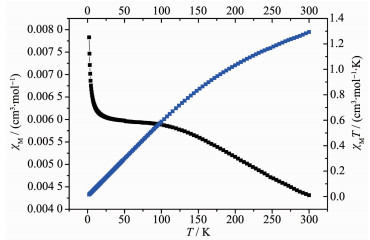
About Eu(Ⅲ), due to the weak energy separation and spin-orbit coupling, the crystal field effects and the possible thermal population of higher states should be accounted. The χMT and χM vs T plots for 2 are shown in Fig. 5. 2 had a χMT value of 1.29 cm3·K·mol-1, which is much higher than the expected value of 0 cm3·K·mol-1 for one isolated Eu(Ⅲ) ion in the ground state. Upon cooling, χMT gradually reached a value close to zero (0.016 cm3·K·mol-1) at 2 K because of the depopulation of Stake levels, which corresponds to a nonmagnetic ground state of 7F0 for Eu(Ⅲ) ions. The shape of the curve is a typical characteristic occurrence of thermally populated excited states. As for Eu(Ⅲ), the 7F ground term is split by the spin-orbit coupling (
|
$ {\chi _{\rm{M}}} = AN{\beta ^2}/[3Bkx(T - \theta )] $ |
(3) |
with A=24+[(27x-3)/2]e-x+[(135x-5)/2]e-3x+(189x-7/2)e-6x+(405x-9/2)e-10x+[(1 485x-11)/2]e-15x+[(2 457x-13)/2]e-21x and B=1+3e-x+5e-3x+7e-6x+9e-10x+11e-15x+13e-21x and where N stands for Avogadro′s number, β for the Bohr magneton, k the Boltzmann constant, T the temperature, θ the Weiss constant, x=λ/(kT) and λ the spin-orbit coupling parameter. The best fitting of the χMT vs T curve in a temperature range of 20~300 K gave the spin-coupling parameter, λ=373 cm-1, a value in the expected range, and θ=3.17 K, R2=0.999 41. The positive θ value reveals the presence of weak ferromagnetic interactions between Eu(Ⅲ) ions.
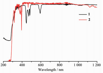
The reflectance diffusion spectra (Fig. 6) reveals that 1 and 2 display intense π-π* transition absorption bands at 274 and 285 nm, respectively. In addition, the characteristic f-f electronic transition bands at 444, 470, 482, and 594 nm attributed to 3H4→3PJ (J=2, 1, 0) and 1D2 for Pr(Ⅲ) ion in 1, 318 nm (7F0→5H5), 363 nm (7F0→5D4), 375 nm (7F0→5G4), 383 nm (7F0→5G3), 393 nm (7F0→5L6), 465 nm (7F0→5D2), 525 nm (7F0→5D1), 534 nm (7F1→5D1) for Eu(Ⅲ) ion in 2 were also observed.
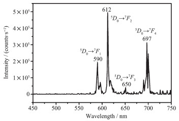
The solid-state luminescence properties of these two complexes are also investigated at room temperature. Upon excitation at 396 nm, 2 emitted intense red luminescence (Fig. 7) and exhibited four characteristic peaks at 590, 612, 650, and 697 nm, respectively, corresponding to the transitions of Eu(Ⅲ) from 5D0 to 7FJ (J=1, 2, 3 and 4). The quite weak emission band for 5D0→7F0 in the position of 575 nm is due to the symmetry-forbidden transition of the Eu(Ⅲ) ion in 2. It is well known that the 5D0→7F1 transition is magnetic dipole in nature and less sensitive to its environment, while 5D0→7F2 is electric dipole in origin and its intensity is strongly influenced by the crystal field[22-23]. The intensity ratio of D0→7F2 to 5D0→7F1 is about 2.5, implying relatively low-symmetric crystal field for the center Eu(Ⅲ) in 2, which is consistent with the single-crystal structure. Luminescence lifetime measurements in solid monitored at 614 nm of 2 was 0.233 ms. Regrettably, because of the large energy gap of the Pr(Ⅲ) ion, no characteristic peak for Pr(Ⅲ) ion was observed in the luminescent spectra of 1. These results suggest that energy transfer from the ligands to the resonance level of lanthanide ions is efficient for Eu(Ⅲ) ion but not for Pr(Ⅲ) ion.
In conclusion, two new 3D isostructural polymers based on N-succinopyridine ligand have been synthesized in aqueous solution at room temperature and characterized in terms of structure, magnetic properties and luminescence. The magnetic studies show that the χMT value of 1 decreases as the temperature drops, which could arise from a selective depopulation of the excited crystal field state and antiferromagnetic interaction between Pr(Ⅲ) ions. And 2 is a typical characteristic occurrence of thermally populated excited states because of the weak energy separation and spin-orbit coupling. The efficient energy transfer from the HL ligand to the resonance level of lanthanide ions results in that 2 exhibited intense characteristic luminescent property of Eu(Ⅲ).
Bi Y, Wang X T, Liao W, et al. Inorg. Chem., 2009, 48(24):11743-11747 doi: 10.1021/ic9017807
Cucinotta G, Perfetti M, Luzon J, et al. Angew. Chem. Int. Ed., 2012, 51(7):1606-1610 doi: 10.1002/anie.201107453
Bi Y, Chen C, Zhao Y F, et al. Chem. Sci., 2016, 7(8):5020-5031 doi: 10.1039/C6SC01157H
Long J, Guari Y, Ferreira R A S, et al. Coord. Chem. Rev., 2018, 363:57-70 doi: 10.1016/j.ccr.2018.02.019
Liddle S T, Slageren J J. Chem. Soc. Rev., 2015, 44(19):6655-6669 doi: 10.1039/C5CS00222B
Eliseeva S V, Bunzli J C. Chem. Soc. Rev., 2010, 39(1):189-227 doi: 10.1039/B905604C
Bunzli J C. Chem. Rev., 2010, 110(5):2729-2755 doi: 10.1021/cr900362e
Pointillart F, Guennic B L, Cador O, et al. Acc. Chem. Res., 2015, 48(11):2834-2842 doi: 10.1021/acs.accounts.5b00296
Pointillart F, Cador O, Guennic B L, et al. Coord. Chem. Rev., 2017, 346:150-175 doi: 10.1016/j.ccr.2016.12.017
Hareri M A, Gavey E L, Regier J, et al. Chem. Commun., 2016, 52(76):11335-11338 doi: 10.1039/C6CC03578G
Long J, Rouquette J, Thibaud J M, et al. Angew. Chem. Int. Ed., 2015, 54(7):2236-2240 doi: 10.1002/anie.201410523
Guillou O, Daiguebonne C, Calvez G, et al. Acc. Chem. Res., 2016, 49(5):844-856 doi: 10.1021/acs.accounts.6b00058
Wu J W, Zhang H B, Du S W. J. Mater. Chem. C, 2016, 4(16):3364-3374 doi: 10.1039/C5TC04432D
Cai L Z, Wang M S, Zhang M J, et al. CrystEngComm, 2012, 14(19):6196-6200 doi: 10.1039/c2ce25755f
Cai L Z, Wang M S, Wang S H, et al. CrystEngComm, 2013, 15(38):7670-7679 doi: 10.1039/c3ce41050a
Kotov V Y, Gorbunova Y G, Kostina S A, et al. Mendeleev Commun., 2001, 11(5):181-182 doi: 10.1070/MC2001v011n05ABEH001464
CrystalClear, Ver. 1.35, Software User's Guide for the Rigaku R-Axis, and Mercury and Jupiter CCD Automated X-ray Imaging System, Rigaku Molecular Structure Corporation: UT, 2002.
SHELXTL Reference Manual, Ver. 5, Siemens Energy & Automation Inc.: Madison, WI, 1994.
Chen F, Wang J, Dong M W, et al. J. Mol. Struct., 2019, 1177:117-123 doi: 10.1016/j.molstruc.2018.09.032
Yuan G, Zhang C, Shao K Z, et al. Inorg. Chem. Commun., 2019, 99:126-130 doi: 10.1016/j.inoche.2018.11.017
Wang Y, Xing S H, Bai F Y, et al. Inorg. Chem., 2018, 57(20):12850-12859 doi: 10.1021/acs.inorgchem.8b02050
Zhou X, Wang H, Jiang S, et al. Inorg. Chem., 2019, 58(6):3780-3788 doi: 10.1021/acs.inorgchem.8b03319
Wang Z X, Wu Q F, Liu H J, et al. CrystEngComm, 2010, 12(4):1139-1146 doi: 10.1039/B910701K

Figure 3 (a) Coordination environment of Pr(Ⅲ) in 1; (b) View of the hydrogen bonding 2D framework of 1 along b axis; (c) View of the hydrogen bonding 3D framework of 1 along b axis
Symmetry codes: #1: x, -y+1, z+1/2; #2:-x+1, -y+1, -z+2; #3: -x+1, y, -z+5/2
Table 1. Crystal and structure refinement data for 1 and 2
| Compound | 1 | 2 |
| Formula | C18H26Cl3N2O12Pr | C18H26Cl3N2O12Eu |
| Formula weight | 709.67 | 720.72 |
| Crystal system | Monoclinic | Monoclinic |
| Space group | P2/c | P2/c |
| a / nm | 1.308 2(6) | 1.301 2(5) |
| b / nm | 0.961 9(5) | 0.962 7(4) |
| c / nm | 1.119 2(5) | l.l08 2(4) |
| β / (°) | 99.73(4) | 99.563(6) |
| V / nm3 | 1.388 0(11) | 1.368 8(9) |
| Z | 2 | 2 |
| Dc / (g·cm-3) | 1.698 | 1.749 |
| μ / mm-1 | 2.102 | 2.643 |
| Parameter, restraint, data | 187, 7, 2 187 | 179, 0, 2 163 |
| R1a [I > 2σ(I)] | 0.031 8 | 0.042 8 |
| wR2b [I > 2σ(I)] | 0.082 5 | 0.112 1 |
| Goodness of fit | 1.007 | 1.069 |
 下载: 导出CSV
Table 2. Selected bond lengths (nm) and angles (°) for 1
| Pr(1)-O(1) | 0.242 7(3) | Pr(1)-O(2)#1 | 0.237 8(3) | Pr(1)-O(1W) | 0.251 4(3) |
| Pr(1)-O(2W) | 0.252 5(4) | ||||
| O(2)#1-Pr(1)-O(1)#3 | 86.52(11) | O(1)#3-Pr(1)-O(1) | 147.12(18) | O(1W)#3-Pr(1)-O(1W) | 68.65(17) |
| O(1)-Pr(1)-O(1W) | 72.67(12) | O(2W)-Pr(1)-O(2W)#3 | 79.56(18) | O(2)#2-Pr(1)-O(1W)#3 | 75.14(13) |
| Symmetry codes: #1: x, -y+1, z+1/2; #2: -x+1, -y+1, -z+2; #3: -x+1, y, -z+5/2. | |||||
下载: 导出CSV
Table 3. Selected bond lengths (nm) and angles (°) for 2
| Eu(1)-O(2) | 0.233 1(4) | Eu(1)-O(1W) | 0.247 5(4) | Eu(1)-O(2W) | 0.247 6(4) |
| Eu(1)-O(1)#2 | 0.237 5(4) | ||||
| O(2)-Eu(1)-O(1)#3 | 86.21(15) | O(1)#2-Eu(1)-O(2W) | 140.28(15) | O(1)#2-Eu(1)-O(1)#3 | 146.8(2) |
| O(2)#1-Eu(1)-O(2W)#1 | 77.59(15) | O(1W)-Eu(1)-O(1W)#1 | 78.O(2) | O(1)#3-Eu(1)-O(2W) | 72.67(15) |
| Symmetry codes: #1: -x+1, y, -z+3/2; #2: x, -y+1, z-1/2; #3: -x+1, -y+1, -z+2. | |||||
下载: 导出CSV

 扫一扫看文章

扫一扫关注我们