Scheme1.
Synthesis route of H2L1
Syntheses, Structures and Hirshfeld Analyses of Trinuclear Ni(Ⅱ) Salamo-Type Complexes
Meng YU , Hao-Ran MU , Ling-Zhi LIU , Na LI , Yang BAI , Xiu-Yan DONG

As we known, Salen and its derivatives[1-4] are extremely important N2O2 chelating ligands in organometallic and coordination chemistry. Not only their transition metal complexes[5-9] are widely studied in the biological fields[10-17], but also there are certain advantages in luminescent[18-25] and magnetic[26-31] materials, electrochemical fields[32-33], supramolecular constructions[34-38], and so on.
As derivatives of Salen, in recent years, our research mostly concentrated on the syntheses of Salamo-type ligands[39-46]. These compounds have been investigated in forming metal complexes with interesting properties[47-48], and some works have been devoted to synthesize and characterize mono-[49], di- and multi-nuclear metal(Ⅱ) complexes bearing Salamo ligand and its derivatives[50-51]. Herein, as part of our ongoing interest in Salamo-type complexes, we have described the syntheses, crystal structures, and Hir-shfeld surfaces analyses of two trinuclear Ni(Ⅱ) compl-exes, which were constructed from Salamo-type bisoxime ligands 5-methoxy-2, 2′-(ethylenedioxybis(azomethine))diphenol (H2L1) and 4, 4′-dinitro-2, 2′-(ethylenediyldio-xybis(nitrilomethylidyne))diphenol (H2L2).
All chemical reagents are analytical pure reagents, which have not been purified before used. C, H and N analyses were obtained using a GmbH Vario EL V3.00 automatic elemental analyzer. Elemental analysis for Ni(Ⅱ) was conducted using an IRIS ER/S·WP-1 ICP atomic emission spectrometer. 1H NMR spectra were recorded using a Bruker AVANCE DRX-400 spectrometer. The melting points were determined by microscopic melting point instrument made in Beijing Tektronix Instruments Limited Company. IR spectra were recorded on a VERTEX70 FT-IR spectrophotometer, with samples prepared as KBr (500~4 000 cm-1) and CsI (100~500 cm-1) pellets. UV-Vis absorption spectra were recorded on a Shimadzu UV-2550 spectrometer.
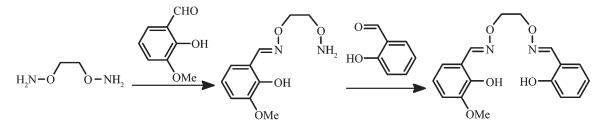
The asymmetric Salamo-type ligand H2L1 was synthesized by a modified method reported in the earlier literature (Scheme 1)[24].
To an ethanol solution (30 mL) of 4-methoxy-2-hydroxybenzaldehyde (316.62 mg, 2.08 mmol) was added an ethanol solution (20 mL) of 1, 2-bis(aminooxy)ethane (289.32 mg, 3.14 mmol). The mixture solution was heated at 50~55 ℃ for 5 h. The solution was concentrated in reduced pressure to obtain 210.37 mg of yellow solid, 2-(O-(1-ethyloxyamide))oxime-5-metho-phenol. Yield: 44.68%, m.p. 96~98 ℃. Anal. Calcd. for C10H14N2O4(%): C 53.09, H 6.24, N 12.38; Found(%): C 53.08, H 6.25, N 12.36.
A solution of above-obtained 2-(O-(1-ethyloxya-mide))oxime-5-methophenol (210.37 mg, 0.93 mmol) in ethanol (20 mL) was added dropwise to a solution of salicylaldehyde (107.03 mg, 0.88 mmol) in ethanol (15 mL), and the mixture was heated at 50~55 ℃ for 3 h. After cooling to room temperature, the solution was concentrated in reduced pressure. The residue was purified by column chromatography (SiO2, chloroform/ethyl acetate, 10:1, V/V) to afford 139.26 mg colorless flocculent crystalline solid (H2L1). Yield: 48.1%. m.p. 69~71 ℃. 1H NMR (400 MHz, CDCl3): δ 3.81 (s, 3H, CH3), 4.44 (m, 4H, CH2), 6.46~6.49 (m, 2H, ArH), 6.89 ~6.92 (d, J=12 Hz, 1H, ArH), 6.97~6.99 (d, J=8 Hz, 1H, ArH), 7.04~7.06 (d, J=8 Hz, 1H, ArH), 7.15~7.17 (d, J=8 Hz, 1H, ArH), 8.18 (s, 1H, CH=N), 8.24 (s, 1H, CH=N), 8.25 (s, 1H), 9.75 (s, 1H, OH), 9.92 (s, 1H, OH). Anal. Calcd. for C17H18N2O5(%): C 61.81, H 5.49, N 8.48; Found(%): C 61.80, H 5.50, N 8.49.
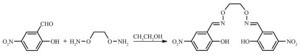
The reaction route is shown in Scheme 2. 4, 4′-Dinitro-2, 2′-(ethylenediyldioxybis(nitrilomethylidyne))diphenol (H2L2) was synthesized according to the method reported earlier[39]. Yield: 80.0%, m.p. 202~203 ℃, Anal. Calcd. for C16H14N4O8(%): C 49.24, H 3.62, N 14.35. Found(%): C 49.33, H 3.58, N 14.04. 1H NMR (400 MHz, DMSO-d6): δ 4.44 (s, 4H), 7.03 (d, J=9.2 Hz, 2H), 8.09 (dd, J=9.2, 2.6 Hz, 2H), 8.35 (d, J=2.6 Hz, 2H), 8.37 (s, 2H), 10.13 (s, 2H, OH).
A n-propanol solution (3.0 mL) of nickel(Ⅱ) acetate tetrahydrate (2.46 mg, 0.01 mmol) was added to a dichloromethane solution (2.0 mL) of H2L1: 5-methoxy-2, 2′-(ethylenedioxybis(azomethine))diphenol (3.31 mg, 0.01 mmol) at room temperature. After the mixture was stirred for 2 h, the mixture was filtered off. The resulting filtrate was left undisturbed for about one week to form block-like crystals suitable for X-ray crystallographic analysis. Yield: 40.5%. Anal. Calcd. for C50H70Ni3N4O18(%): C 50.41, H 5.92, N 4.70; Ni 14.78; Found(%): C 50.60; H 5.98; N 4.52; Ni 14.62.
A solution of nickel(Ⅱ) acetate tetrahydrate (2.48 mg, 0.01 mmol) in n-butanol (3 mL) was added dropwise to a solution of H2L2 (3.90 mg, 0.01 mmol) in acetoni-trile (3 mL) at room temperature. The color of the mixing solution turned yellow immediately, and was stirring continually for 1h at room temperature. The mixture was filtered and the filtrate was allowed to stand at room temperature for about three weeks. The solvent was partially evaporated and several green prismatic single crystals suitable for X-ray crystallo-graphic analysis were obtained. Yield: 47.8%. Anal. Calcd. for C52H70Ni3N8O24(%): C 45.68, H 5.16, N 8.20, Ni 12.88; Found(%): C 45.90, H 5.22, N 8.02, Ni 12.63.
X-ray single crystal diffraction data of complexes 1 and 2 were recorded using a Super Nova Dual (Cu at zero) diffractometer with a monochromated Mo Kα radiation (λ=0.710 73 nm) at 294.29(10) and 293.84(19) K, respectively. The Lp factor semi-empirical absorp-tion corrections were applied using the SADABS program[52a]. The structures were solved by the direct methods (SHELXS-2014[52b]). All hydrogen atoms were added theoretically and difference-Fourier map revealed the positions of the remaining atoms. All non-hydrogen atoms were refined anisotropically using a full-matrix least-squares procedure on F2 with SHELXL -2014[52b]. The structure contained large void, and the solvent molecules and the positive or negative ions locating in the void couldn′t be identified because they were highly disordered and had so small residual peak. Therefore, SQUEEZE in PLATON program was performed to remove the highly disordered solvent and ions. The structures were then refined again using the data generated. The crystal data and experimental parameters relevant to the structure determinations are listed in Table 1.
 下载:
导出CSV
下载:
导出CSV
| Complex | 1 | 2 |
| Formula | C50H70Ni3N4O18 | C52H70Ni3N8O24 |
| Formula weight | 1 191.23 | 1 367.29 |
| Crystal system | Trigonal | Triclinic |
| Space group | ||
| a / nm | 3.651 4(2) | 0.980 80(10) |
| b / nm | 3.651 4(2) | 1.088 56(12) |
| c / nm | 1.213 53(7) | 1.564 37(9) |
| α/ (°) | 97.265(6) | |
| β/ (°) | 90.468(6) | |
| γ / (°) | 95.428(9) | |
| V / nm3 | 14.012(2) | 1.649 0(3) |
| Z | 9 | 1 |
| Dc / (g·cm-3) | 1.271 | 1.377 |
| μ / mm-1 | 0.963 | 0.927 |
| F(000) | 5 634 | 714 |
| Crystal size / mm | 0.33×0.31×0.27 | 0.11×0.13×0.21 |
| θ range / (°) | 3.597~26.022 | 3.344~26.022 |
| Index ranges | -45 ≤ h ≤ 29, -45 ≤ k ≤ 44, -14 ≤ l ≤ 14 | -12 ≤ h ≤ 12, -13 ≤ k ≤ 11, -19 ≤ l ≤ 17 |
| Reflection collected | 13 730 | 10 927 |
| Independent reflection | 5 942 | 6 409 |
| Rint | 0.042 6 | 0.063 |
| Completeness / % | 97.3 | 98.8 |
| Data, restraint, parameter | 5 942, 105, 355 | 6 409, 72, 417 |
| GOF | 1.043 | 1.031 |
| Final R1, wR2 [I>2σ(I)] | 0.079 1, 0.223 9 | 0.083 7, 0.194 9 |
| R1, wR2 indices (all data) | 0.121 6, 0.261 9 | 0.152 4, 0.255 8 |
CCDC: 1589650, 1; 1589813, 2.
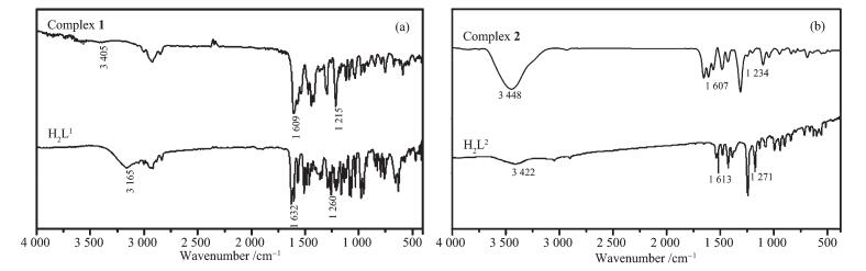
The FT-IR spectra of H2L1 and H2L2 with their corresponding complexes 1 and 2 exhibited various bands in the 4 000~400 cm-1 region (Fig. 1 and Table 2). The infrared spectra of the ligand H2L1 and its complex 1 are shown in Fig. 1(a). The ligand H2L1 had a strong absorption peak at 3 165 cm-1, which belongs to the stretching vibration of the phenolic hydroxyl groups, while complex 1 exhibited a new vibration peak at 3 405 cm-1, indicating the presence of n-butanol molecules. This result is consistent with the previous element analysis results. It can be seen from Fig. 1 and Table 2 that the stretching vibration absorption peak of the νC=N bond in the ligand H2L2 appeared at 1 632 cm-1, and that of complex 1 appeared at 1 609 cm-1. The peak-to-low wavenumber is shifted by 23 cm-1 [64-65], indicating that the Ni(Ⅱ) ions coordinate to the oxime nitrogen atoms, giving rise to reduction in the bond energy of the C=N bond.
下载:
导出CSV
| cm-1 | |||||
| Compound | νO-H | νC=N | νAr-O | νNi-N | νNi-O |
| H2L1 | 3 165 | 1 632 | 1 260 | — | — |
| Complex 1 | 3 405 | 1 609 | 1 215 | 590 | 419 |
| H2L2 | 3 422 | 1 613 | 1 271 | — | — |
| Complex 2 | 3 448 | 1 607 | 1 234 | 552 | 422 |
As shown in Fig. 1(b), the stretching vibration absorption peaks of νC=N and νAr-O in the ligand H2L2 appeared near 1 613 and 1 271 cm-1, whereas those of complex 2 appeared at 1 607 and 1 234 cm-1, respe-ctively[66-67], and moving toward the direction of low wave number.
The far-IR spectra (550~100 cm-1) of both complexes 1 and 2 were also obtained so as to identify the bonds of Ni-O and Ni-N frequencies. The bands at 419 and 422 cm-1 of complexes 1 and 2 can be attributed to νNi-O, while the bands at 590 and 552 cm-1 are assigned to νNi-N, respectively[34].
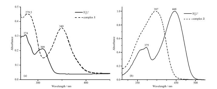
The UV-Vis absorption spectra of the free ligands H2L1 and H2L2 with their corresponding complexes 1 and 2 in ethanol solutions (10 μmol·L-1) at 298 K are shown in Fig. 2.
Obviously, the absorption peaks of the ligands H2L1 and H2L2 differ from those of complexes 1 and 2. From Fig. 2(a), we can see that the ligand H2L1 showed two strong absorption peaks appear near 274 and 309 nm. The former belongs to the π-π* transition of the benzene rings in the ligand molecule. The latter is assigned to the transition absorption peak of n-π* on the C=N bonds[68-69]. Compared with the π-π* transition absorption peak of the benzene rings at 274 nm of the ligand H2L1, it was red-shifted to 279.5 nm in complex 1, indicating that the Ni(Ⅱ) ions have coordinated to the ligand H2L1. At the same time, the absorption peak of the ligand H2L1 appearing near 309 nm disappeared in complex 1, indicating that the oxime nitrogen atoms have coordinated with the Ni(Ⅱ) ions. In addition, complex 1 exhibited a new absorption peak at 349 nm. It can be seen that the Ni(Ⅱ) ions chelate with two oxime nitrogen atoms and two phenol oxygen atoms to produce a charge transfer effect.
As shown in Fig. 2(b), the ligand H2L2 showed two absorption peaks appearing around 375 and 448 nm. The absorption peak near 375 nm is attributed to the π-π* transition of the aromatic rings in the ligand H2L2, and the absorption peak at 448 nm is attributed to the π-π* transition of the C=N bonds in H2L2. However, compared with the ligand H2L2, the positions of the absorption peaks of complex 2 were blue-shifted, and the absorption peak at 448 nm was blue-shifted by about 51 nm, while the absorption peak of the ligand H2L2 near 375 nm disappeared. It is indicated that there are strong coordination bonds between the ligand H2L2 and the Ni(Ⅱ) ions in complex 2.
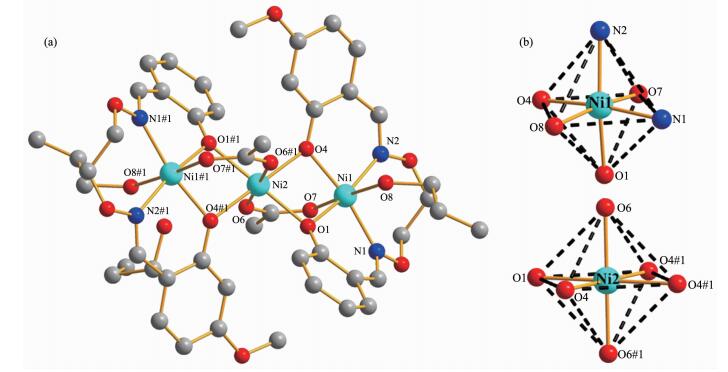
As shown in Fig. 3, in the crystal structure of complex 1, the terminal hexa-coordinated Ni(Ⅱ) ion (Ni1) is located in the cis-N2O2 coordination cavity (O1, O4, N1 and N2) of the completely deprotonated (L1)2- unit, and one μ-acetate oxygen atom (O7) and one coordinated n-propanol oxygen atom (O8) occupy together the axial positions. Meanwhile, the coordina-tion environment of the central hexa-coordinated Ni(Ⅱ) ion (Ni2) is completed by quadruple μ-phenoxo oxygen atoms from the two (L1)2- moieties and two μ-acetate oxygen atoms (O6 and O6#1). All of the six oxygen atoms coordinate to Ni2 constituting an octahedral geometry. The μ-acetate ligand serves as bridging group for Ni1 and Ni2 and another coordinates to Ni2 and Ni1#1, in both cases via Ni-O-C-O-Ni bridges. Then, all of the hexa-coordinated Ni(Ⅱ) ions of complex 1 have slightly distorted octahedral geometries[23].
Hydrogen atoms and solvent molecules are omitted for clarity; Symmetry codes: #1: 1-x, -y, 1-z
下载:
导出CSV
| Ni1-O1 | 0.201 1(5) | Ni1-N1 | 0.206 9(5) | Ni2-O1 | 0.208 2(4) |
| Ni1-O4 | 0.201 7(4) | Ni1-N2 | 0.205 1(7) | Ni2-O4 | 0.206 0(5) |
| Ni1-O8 | 0.216 2(5) | Ni1-O7 | 0.203 5(4) | Ni2-O6 | 0.209 5(4) |
| O7#1-Ni1-N2 | 92.7(2) | O4-Ni1-N2 | 88.5(2) | O1-Ni2-O6 | 92.11(15) |
| O1-Ni1-O4 | 81.17(18) | O4-Ni1-O7#1 | 90.27(18) | O1-Ni2-O1#1 | 180.00 |
| O1-Ni1-O8 | 88.1(2) | O8-Ni1-N1 | 87.3(2) | O1-Ni2-O4#1 | 101.54(17) |
| O1-Ni1-N1 | 86.2(2) | O8-Ni1-N2 | 86.6(2) | O1-Ni2-O6#1 | 87.89(15) |
| O1-Ni1-N2 | 168.11(18) | O7#1-Ni1-O8 | 176.45(19) | O4-Ni2-O6 | 89.59(18) |
| O1-Ni1-O7#1 | 93.3(2) | O7#1-Ni1-N1 | 89.5(2) | O4-Ni2-O4#1 | 180.00 |
| O4-Ni1-O8 | 93.19(18) | N1-Ni1-N2 | 104.1(2) | O4-Ni2-O6#1 | 90.41(18) |
| O4-Ni1-N1 | 167.4(2) | O1-Ni2-O4 | 78.47(17) | O6-Ni2-O6#1 | 180.00 |
| Symmetry codes: #1: 1-x, -y, 1-z. | |||||
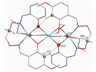
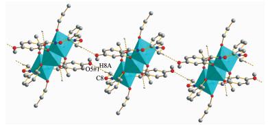
In the crystal structure of complex 1, there are two pairs of intra-molecular hydrogen bonding interactions (C8-H8B…O7#2 and C13-H13…O1#2) (Fig. 4)[53-57]. Table 4 summarizes the intra-molecular hydrogen bonding interactions in complex 1. As shown in Fig. 5 and Table 4, the supramolecular structure of complex 1 was linked by inter-molecular hydrogen bonding interactions (O8-H8…O9, O9-H9…O6 and C8-H8A…O5#1), which performs a crucial role in constructing and stabilizing 1D supramolecular chain structure[58].
Hydrogen atoms are omitted except those forming hydrogen bonds; Symmetry codes: #2: 1-x, -y, 1-z
下载:
导出CSV
| D-H…A | d(D-H) / nm | d(H…A) / nm | d(D…A) / nm | ∠D-H…A / (°) |
| O8-H8…O9 | 0.086(6) | 0.177(6) | 0.262 7(10) | 172(12) |
| O9-H9…O6 | 0.082 | 0.206 | 0.277 9(9) | 146 |
| C8-H8A…O5#1 | 0.097 | 0.253 | 0.317 7(9) | 124 |
| C8-H8B…O7#2 | 0.097 | 0.229 | 0.314 3(11) | 146 |
| C13-H13…O1#2 | 0.093 | 0.256 | 0.327 7(8) | 134 |
| Symmetry codes: #1: x, y, 1+z; #2: 1-x, -y, 1-z. | ||||
Symmetry codes: #1: x, y, 1+z
X-ray crystallographic analysis of complex 2 revealed symmetric trinuclear structure. It crystallizes in the triclinic system, space group
下载:
导出CSV
| Ni1-O1 | 0.202 7(5) | Ni1-O11 | 0.210 0(6) | Ni2-O1 | 0.208 0(4) |
| Ni1-O8 | 0.202 9(4) | Ni1-N3 | 0.207 9(6) | Ni2-O8 | 0.206 8(5) |
| Ni1-O9 | 0.203 3(5) | Ni1-N2 | 0.208 7(6) | Ni2-O10 | 0.201 1(5) |
| O1-Ni1-O8 | 80.67(18) | O8-Ni1-N3 | 85.9(2) | O8-Ni2-O10 | 87.92(19) |
| O1-Ni1-O9 | 89.40(19) | O9-Ni1-O11 | 176.9(2) | O8-Ni2-O8#1 | 180.00 |
| O1-Ni1-O11 | 92.4(2) | O9-Ni1-N2 | 90.0(2) | O8-Ni2-O10#1 | 92.08(19) |
| O1-Ni1-N2 | 87.7(2) | O9-Ni1-N3 | 91.4(2) | O1#1-Ni2-O8#1 | 78.53(18) |
| O1-Ni1-N3 | 166.6(2) | O1-Ni2-O10 | 89.98(18) | O11-Ni1-N2 | 87.6(2) |
| O8-Ni1-O9 | 94.4(2) | O1-Ni2-O1#1 | 180.00 | O11-Ni1-N3 | 87.4(2) |
| O8-Ni1-O11 | 88.4(2) | O1-Ni2-O8#1 | 101.48(18) | N2-Ni1-N3 | 105.7(2) |
| O8-Ni1-N2 | 167.5(2) | O1-Ni2-O10#1 | 90.02(18) | O1-Ni2-O8 | 78.53(18) |
| Symmetry codes: #1: 2-x, 1-y, 2-z | |||||
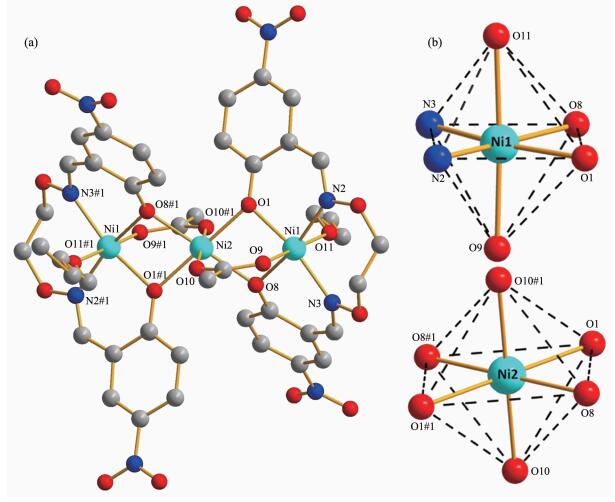
As shown in Fig. 6, the hexa-coordinated terminal Ni(Ⅱ) ion (Ni1 or Ni#1) lies in the N2O2 coordination sphere (O1, O8, N2 and N3 or O1#1, O8#1, N2#1 and N3#1) of (L2)2-, and coordinates with one μ-acetate oxygen atom (O9 or O9#1) and one coordinated n-butanol oxygen atom (O11 or O11#1).
Solvent molecules and hydrogen atoms are omitted for clarity; Symmetry codes: #1: 2-x, 1-y, 2-z
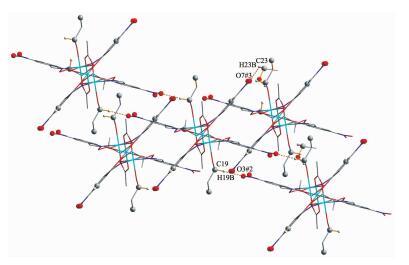
As illustrated in Fig. 7, in the crystal structure of complex 2, there are six pairs of intra-molecular hydrogen bonding interactions(C0AA-H0AA…O3, C2-H2…O8#1, C2-H2…O10#1, C9-H9A…O9, C13-H13…O10#1 and C19-H19A…O10#1)[59-63]. As shown in Fig. 8 and Table 6, complex 2 molecules are linked into an infinite 2D supramolecular structure by inter-molecular hydrogen bonding interactions (C19-H19B…O3#2, C23-H23B…O7#3).
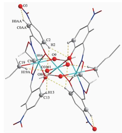
Hydrogen atoms, except those forming hydrogen bonds, are omitted for clarity; Symmetry codes: #1: 2-x, 1-y, 2-z
Symmetry codes: #2: 1-x, -y, 2-z; #3: x, -1+y, z
下载:
导出CSV
| D-H…A | d(D-H) / nm | d(H…A) / nm | d(D…A) / nm | ∠D-H…A / (°) |
| C0AA-H0AA…O3 | 0.093 | 0.237 | 0.269 0(11) | 100 |
| C2-H2…O8#1 | 0.093 | 0.257 | 0.329 2(9) | 135 |
| C2-H2…O10#1 | 0.093 | 0.257 | 0.216 5(9) | 122 |
| C9-H9A…O9 | 0.097 | 0.221 | 0.309 5(12) | 150 |
| C13-H13…O10#1 | 0.093 | 0.254 | 0.313 5(8) | 122 |
| C19-H19A…O10#1 | 0.097 | 0.248 | 0.343 2(11) | 167 |
| C19-H19B…3#2 | 0.097 | 0.252 | 0.330 1(12) | 137 |
| C23-H23B…O7#3 | 0.097 | 0.246 | 0.320(2) | 133 |
| Symmetry codes: #1: 2-x, 1-y, 2-z; #2: 1-x, -y, 2-z; #3: x, -1+y, z. | ||||
The Hirshfeld surfaces[70] of complexes 1 and 2 are illustrated in Fig. 9 showing surfaces that have been mapped over dnorm and the corresponding location in shape index exists the complementary region of red concave surface surrounded by receptors and the blue convex surface surrounding receptors, further proving that such hydrogen bonding exists. As for the large amount of white region in dnorm surfaces, it is suggested that there is a weaker and farther contact between molecules, rather than hydrogen bonding.
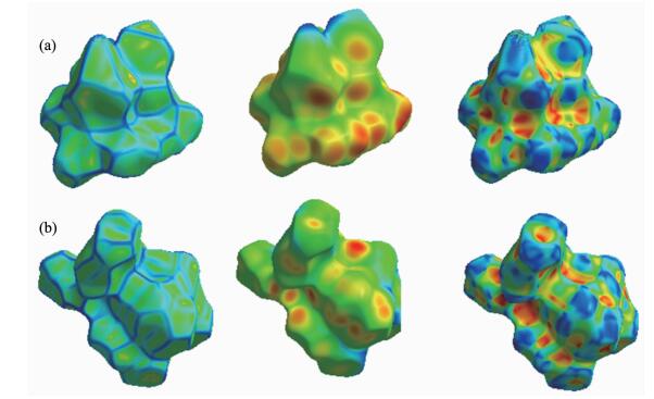
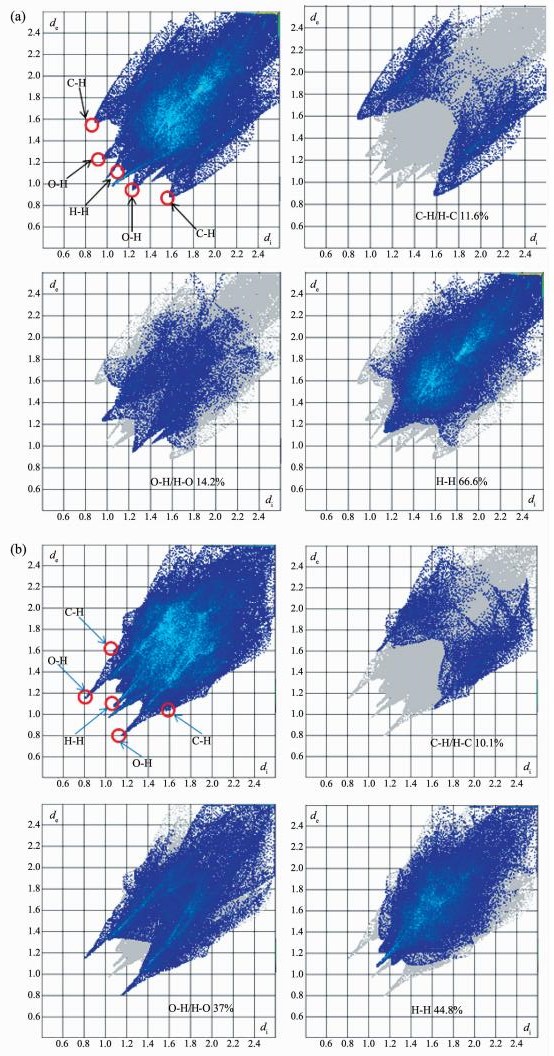
Fig. 10 shows the 2D plots generated[71] from the Hirshfeld surfaces of complexes 1 and 2 corresponding to the O…H, C…H and H…H interactions. As shown in Fig. 10(a), the H…H interactions appeared at (0.115 nm, 0.115 nm) accounting for 66.6% of the total area of Hirshfeld surfaces for complex 1. The C…H/H…C interactions in a range of (0.160 nm, 0.09 nm) appeared as a pair of symmetrical wings, accounting for 11.6% of the total area of Hirshfeld surfaces. The proportions of O…H/H…O interactions comprised 14.2% of the total Hirshfeld surfaces for each molecule of complex 1. As shown in Fig. 10(b), for complex 2, the interactions of H…H appeared at (0.115 nm, 0.115nm) accounting for 44.8% of the total area of Hirshfeld surfaces. The C…H/H…C interactions in a range of (0.160 nm, 0.105 nm) accounted for 10.1% of the total area of Hirshfeld surfaces. The proportions of O…H/H…O interactions comprised 37% of the total Hirshfeld surfaces for each molecule of complex 2. It is because of the existence of these weaker hydrogen bonds that complexes 1 and 2 can be stable.
In summary, we have reported the successful syntheses and characterizations of two newly designed trinuclear Ni(Ⅱ) Salamo-type complexes, {[Ni(L1)(n-propanol)]2(OAc)2Ni}·2(n-propanol) (1) and {[Ni(L2)(n-butanol)]2(OAc)2Ni}·2(n-butanol) (2). All of the hexa-coordinated Ni(Ⅱ) ions in complexes 1 and 2 are slightly distorted octahedral configurations, and complex 1 forms an infinite 1D chain-like supramolecular structure through inter-molecular hydrogen bonding interactions. The supramolecular structure of complex 2 is formed by inter-molecular hydrogen bonding interactions, resulting in a self-assembled infinite 2D supramolecular network.
Chai L Q, Tang L J, Chen L C, et al. Polyhedron, 2017, 122:228-240 doi: 10.1016/j.poly.2016.11.032
Wu H L, Wang C P, Wang F, et al. J. Chin. Chem. Soc., 2015, 62:1028-1034 doi: 10.1002/jccs.v62.11
Song X Q, Liu P P, Xiao Z R, et al. Inorg. Chim. Acta, 2015, 438:232-244 doi: 10.1016/j.ica.2015.09.022
Sun Y X, Wang L, Dong X Y, et al. Synth. React. Inorg. Met.-Org. Nano-Met. Chem., 2013, 43:599-603 doi: 10.1080/15533174.2012.751424
Chai L Q, Mao K H, Zhang J Y, et al. Inorg. Chim. Acta, 2017, 457:34-40 doi: 10.1016/j.ica.2016.12.004
Zhao L, Wang L, Sun Y X, et al. Synth. React. Inorg. Met.-Org. Nano-Met. Chem., 2012, 42:1303-1308 doi: 10.1080/15533174.2012.684235
Wang P, Zhao L. Synth. React. Inorg. Met-Org. Nano-Met. Chem., 2016, 46:1095-1101 doi: 10.1080/15533174.2015.1004416
Zhao L, Dang X T, Chen Q, et al. Synth. React. Inorg. Met.-Org. Nano-Met. Chem., 2013, 43:1-6 doi: 10.1080/15533174.2011.652275
Dong X Y, Kang Q P, Li X Y, et al. Crystals, 2018, 8:139 doi: 10.3390/cryst8030139
Wu H L, Pan G L, Bai Y C, et al. Res. Chem. Intermed., 2015, 41:3375-3388 doi: 10.1007/s11164-013-1440-5
Wu H L, Pan G L, Bai Y C, et al. J. Coord. Chem., 2013, 66:2634-2646 doi: 10.1080/00958972.2013.812725
Wu H L, Pan G L, Bai Y C, et al. J. Chem. Res., 2014, 38:211-217 doi: 10.3184/174751914X13933417974082
Wang F, Xu Y L, Aderinto S O, et al. J. Photochem. Photobiol. A, 2017, 332:273-282 doi: 10.1016/j.jphotochem.2016.09.004
Chen C Y, Zhang J W, Zhang Y H, et al. J. Coord. Chem., 2015, 68:1054-1071 doi: 10.1080/00958972.2015.1007965
Li X Y, Kang Q P, Liu L Z, et al. Crystals, 2018, 8:43 doi: 10.3390/cryst8010043
Wu H L, Pan G L, Wang H, et al. J. Photochem. Photobiol. B, 2014, 135:33-43 doi: 10.1016/j.jphotobiol.2014.04.005
Sun S S, Stern C L, Nguyen S T, et al. J. Am. Chem. Soc., 2004, 126:6314-6326 doi: 10.1021/ja037378s
Ren Z L, Hao J, Hao P, et al. Z. Naturforsch., 2018, 73:203-210 doi: 10.1515/znb-2017-0191
Song X Q, Cheng G Q, Liu Y A, et al. Inorg. Chim. Acta, 2016, 450:386-394 doi: 10.1016/j.ica.2016.06.028
Dong X Y, Akogun S F, Zhou W M, et al. J. Chin. Chem. Soc., 2017, 64:412-419 doi: 10.1002/jccs.2017.64.issue-4
Song X Q, Peng Y J, Chen G Q, et al. Inorg. Chim. Acta, 2015, 427:13-21 doi: 10.1016/j.ica.2014.12.008
Zhang L W, Li X Y, Kang Q P. Crystals, 2018, 8:173 doi: 10.3390/cryst8040173
Kang Q P, Li X Y, Zhao Q, et al. Appl. Organomet. Chem., 2018, 32:e4379 doi: 10.1002/aoc.v32.7
Dong W K, Zhang J, Zhang Y, et al. Inorg. Chim. Acta, 2016, 444:95-102 doi: 10.1016/j.ica.2016.01.034
Wang L, Ma J C, Dong W K, et al. Z. Anorg. Allg. Chem., 2016, 642:834-839 doi: 10.1002/zaac.v642.15
Dong W K, Zhu L C, Dong Y J, et al. Polyhedron, 2016, 117:148-154 doi: 10.1016/j.poly.2016.05.055
Li J, Zhang H J, Chang J, et al. Crystals, 2018, 8:176 doi: 10.3390/cryst8040176
Dong W K, Ma J C, Dong Y J, et al. Polyhedron, 2016, 115:228-235 doi: 10.1016/j.poly.2016.05.017
Dong W K, Ma J C, Zhu L C, et al. New J. Chem., 2016, 40:6998-7010 doi: 10.1039/C6NJ00855K
Song X Q, Liu P P, Liu Y A, et al. Dalton Trans., 2016, 45:8154-8163 doi: 10.1039/C6DT00212A
Liu Y A, Wang C Y, Zhang M, et al. Polyhedron, 2017, 127:278-286 doi: 10.1016/j.poly.2017.02.007
Ömer S, ÜmmühanÖ Ö, Nurgul S, et al. Tetrahedron, 2016, 72:5843-5852 doi: 10.1016/j.tet.2016.08.004
Chai L Q, Zhou L, Zhang K Y, et al. Appl. Organomet. Chem., 2018:e4576
Dong W K, Ma J C, Zhu L C, et al. Inorg. Chim. Acta, 2016, 445:140-148 doi: 10.1016/j.ica.2016.02.043
Dong X Y, Li X Y, Liu L Z, et al. RSC Adv., 2017, 7:48394-48403 doi: 10.1039/C7RA07826A
Chen L, Dong W K, Zhang H, et al. Cryst. Growth Des., 2017, 17:3636-3648 doi: 10.1021/acs.cgd.6b01860
Chai L Q, Wang G, Sun Y X, et al. J. Coord. Chem., 2012, 65:1621-1631 doi: 10.1080/00958972.2012.677836
Zhou L, Hu Q, Chai L Q, et al. Polyhedron, 2019, 158:102-116 doi: 10.1016/j.poly.2018.10.052
Akine S, Taniguchi T, Dong W K, et al. J. Org. Chem., 2005, 70:1704-1711 doi: 10.1021/jo048030y
Peng Y D, Li X Y, Kang Q P, et al. Crystals, 2018, 8:107 doi: 10.3390/cryst8020107
Wang F, Liu L Z, Gao L, et al. Spectrochim. Acta Part A, 2018, 203:56-64 doi: 10.1016/j.saa.2018.05.088
Peng Y D, Wang F, Gao L, et al. J. Chin. Chem. Soc., 2018, 65:893-899 doi: 10.1002/jccs.2018.65.issue-7
Dong W K, Lan P F, Zhou W M, et al. J. Coord. Chem., 2016, 69:1-22 doi: 10.1080/00958972.2015.1118469
Zhang L W, Liu L Z, Wang F, et al. Molecules, 2018, 23:1141 doi: 10.3390/molecules23051141
Dong X Y, Zhao Q, Kang Q P, et al. Crystals, 2018, 8:230 doi: 10.3390/cryst8050230
Wang F, Gao L, Zhao Q, et al. Spectrochim. Acta Part A, 2018, 190:111-115 doi: 10.1016/j.saa.2017.09.027
王莉, 康全鹏, 郝静, 等.无机化学学报, 2018, 34:525-533 doi: 10.11862/CJIC.2018.035WANG Li, KANG Quan-Peng, HAO Jing, et al. Chinese J. Inorg. Chem., 2018, 34:525-533 doi: 10.11862/CJIC.2018.035
Dong X Y, Zhao Q, Wei Z L, et al. Molecules, 2018, 23:1006 doi: 10.3390/molecules23051006
Gao L, Liu C, Wang F, et al. Crystals, 2018, 8:77 doi: 10.3390/cryst8020077
Hao J, Li X Y, Zhang Y, et al. Materials, 2018, 11:523 doi: 10.3390/ma11040523
Jia H R, Li J, Sun Y X, et al. Crystals, 2017, 7:247 doi: 10.3390/cryst7080247
(a) Sheldrick G M. SADABS, University of Göttingen, Germany, 1996.
(b)Sheldrick G M. Acta Crystallogr., Sect. C: Cryst. Struct. Commun., 2015, C71: 3-8
Dong W K, Zheng S S, Zhang J T, et al. Spectrochim. Acta Part A, 2017, 184:141-150 doi: 10.1016/j.saa.2017.04.061
Chai L Q, Huang J J, Zhang H S, et al. Spectrochim. Acta Part A, 2014, 131:526-533 doi: 10.1016/j.saa.2014.04.127
孙银霞, 赵亚元, 李春宇, 等.无机化学学报, 2016, 32:913-920 doi: 10.11862/CJIC.2016.118SUN Yin-Xia, ZHAO Ya-Yuan, LI Chun-Yu, et al. Chinese J. Inorg. Chem., 2016, 32:913-920 doi: 10.11862/CJIC.2016.118
Chai L Q, Liu G, Zhang J Y, et al. J. Coord. Chem., 2013, 66:3926-3938 doi: 10.1080/00958972.2013.857016
Chai L Q, Zhang K Y, Tang L J, et al. Polyhedron, 2017, 130:100-107 doi: 10.1016/j.poly.2017.04.010
Guo W T, Li X Y, Kang Q P, et al. Crystals, 2018, 8:154 doi: 10.3390/cryst8040154
Dong W K, Zhang X Y, Sun Y X, et al. Synth. React. Inorg. Met.-Org. Nano-Met. Chem., 2015, 45:956-962 doi: 10.1080/15533174.2013.862814
Sun Y X, Xu L, Zhao T H, et al. Synth. React. Inorg. Met.-Org. Nano-Met. Chem., 2013, 43:509-513 doi: 10.1080/15533174.2012.740756
张宏佳, 常健, 贾浩然, 等.无机化学学报, 2018, 34(12):2261-2270 doi: 10.11862/CJIC.2018.261ZHANG Hong-Jia, CHANG Jian, JIA Hao-Ran, et al. Chinese J. Inorg. Chem., 2018, 34(12):2261-2270 doi: 10.11862/CJIC.2018.261
常健, 张宏佳, 贾浩然, 等.无机化学学报, 2018, 34(11):2097-2107 doi: 10.11862/CJIC.2018.256CHANG Jian, ZHANG Hong-Jia, JIA Hao-Ran, et al. Chinese J. Inorg. Chem., 2018, 34(11):2097-2107 doi: 10.11862/CJIC.2018.256
Chai L Q, Huang J J, Zhang J Y, et al. J. Coord. Chem., 2015, 68:1224-1237 doi: 10.1080/00958972.2015.1019875
Zhang H, Dong W K, Zhang Y, et al. Polyhedron, 2017, 133:279-293 doi: 10.1016/j.poly.2017.05.051
杨玉华, 郝静, 董银娟, 等.无机化学学报, 2017, 33:1280-1292 doi: 10.11862/CJIC.2017.150YANG Yu-Hua, HAO Jing, DONG Yin-Juan, et al. Chinese J. Inorg. Chem., 2017, 33:1280-1292 doi: 10.11862/CJIC.2017.150
Zhao L, Dang X T, Chen Q, et al. Synth. React. Inorg. Met.-Org. Nano-Met. Chem., 2013, 43:1241-1246 doi: 10.1080/15533174.2012.757236
Dong X Y, Gao L, Wang F, et al. Crystals, 2017, 7:267 doi: 10.3390/cryst7090267
Sun Y X, Gao X H. Synth. React. Inorg. Met.-Org. Nano-Met. Chem., 2011, 41:973-978 doi: 10.1080/15533174.2011.591329
Li G, Hao J, Liu L Z, et al. Crystals, 2017, 7:217 doi: 10.3390/cryst7070217
Rohl A L, Moret M, Kaminsky W, et al. Cryst. Growth Des., 2012, 8:4517-4525
Chattopadhyay B, Mukherjee A K, Narendra N, et al. Cryst. Growth Des., 2010, 10:65-68

Figure 3 (a) Molecular structure and atom numberings of complex 1 with 30% probability displacement ellipsoids; (b) Coordination polyhedrons for Ni(Ⅱ) ions of complex 1
Hydrogen atoms and solvent molecules are omitted for clarity; Symmetry codes: #1: 1-x, -y, 1-z

Figure 4 View of the intra-molecular hydrogen bonding interaction of complex 1
Hydrogen atoms are omitted except those forming hydrogen bonds; Symmetry codes: #2: 1-x, -y, 1-z

Figure 5 View of inter-molecular hydrogen bonding interactions of complex 1
Symmetry codes: #1: x, y, 1+z

Figure 6 (a) Molecular structure and atom numberings of complex 2 with 30% probability displacement ellipsoids; (b) Coordination polyhedrons for Ni(Ⅱ) ions of complex 2
Solvent molecules and hydrogen atoms are omitted for clarity; Symmetry codes: #1: 2-x, 1-y, 2-z

Figure 7 Intra-molecular C-H…O hydrogen bonding interactions of complex 2
Hydrogen atoms, except those forming hydrogen bonds, are omitted for clarity; Symmetry codes: #1: 2-x, 1-y, 2-z

Figure 8 Two dimensional supramolecular structure formed by inter-molecular C-H…O hydrogen bonding interactions in complex 2
Symmetry codes: #2: 1-x, -y, 2-z; #3: x, -1+y, z

Figure 9 Hirshfeld surfaces analyses mapped with curvedness, dnorm and shape index of complexes 1 and 2

Figure 10 Fingerprint plots of complexes 1 and 2: full and resolved into O…H, C…H and H…H contacts showing the percentages of contacts contributed to the total Hirshfeld surface area of molecule
Table 1. Crystal data and structure refinement parameters for complexes 1 and 2
| Complex | 1 | 2 |
| Formula | C50H70Ni3N4O18 | C52H70Ni3N8O24 |
| Formula weight | 1 191.23 | 1 367.29 |
| Crystal system | Trigonal | Triclinic |
| Space group | ||
| a / nm | 3.651 4(2) | 0.980 80(10) |
| b / nm | 3.651 4(2) | 1.088 56(12) |
| c / nm | 1.213 53(7) | 1.564 37(9) |
| α/ (°) | 97.265(6) | |
| β/ (°) | 90.468(6) | |
| γ / (°) | 95.428(9) | |
| V / nm3 | 14.012(2) | 1.649 0(3) |
| Z | 9 | 1 |
| Dc / (g·cm-3) | 1.271 | 1.377 |
| μ / mm-1 | 0.963 | 0.927 |
| F(000) | 5 634 | 714 |
| Crystal size / mm | 0.33×0.31×0.27 | 0.11×0.13×0.21 |
| θ range / (°) | 3.597~26.022 | 3.344~26.022 |
| Index ranges | -45 ≤ h ≤ 29, -45 ≤ k ≤ 44, -14 ≤ l ≤ 14 | -12 ≤ h ≤ 12, -13 ≤ k ≤ 11, -19 ≤ l ≤ 17 |
| Reflection collected | 13 730 | 10 927 |
| Independent reflection | 5 942 | 6 409 |
| Rint | 0.042 6 | 0.063 |
| Completeness / % | 97.3 | 98.8 |
| Data, restraint, parameter | 5 942, 105, 355 | 6 409, 72, 417 |
| GOF | 1.043 | 1.031 |
| Final R1, wR2 [I>2σ(I)] | 0.079 1, 0.223 9 | 0.083 7, 0.194 9 |
| R1, wR2 indices (all data) | 0.121 6, 0.261 9 | 0.152 4, 0.255 8 |
 下载: 导出CSV
Table 2. Main absorption peaks in FT-IR spectra of the ligands and their complexes 1 and 2
| cm-1 | |||||
| Compound | νO-H | νC=N | νAr-O | νNi-N | νNi-O |
| H2L1 | 3 165 | 1 632 | 1 260 | — | — |
| Complex 1 | 3 405 | 1 609 | 1 215 | 590 | 419 |
| H2L2 | 3 422 | 1 613 | 1 271 | — | — |
| Complex 2 | 3 448 | 1 607 | 1 234 | 552 | 422 |
下载: 导出CSV
Table 3. Selected bond lengths (nm) and angles (°) for complex 1
| Ni1-O1 | 0.201 1(5) | Ni1-N1 | 0.206 9(5) | Ni2-O1 | 0.208 2(4) |
| Ni1-O4 | 0.201 7(4) | Ni1-N2 | 0.205 1(7) | Ni2-O4 | 0.206 0(5) |
| Ni1-O8 | 0.216 2(5) | Ni1-O7 | 0.203 5(4) | Ni2-O6 | 0.209 5(4) |
| O7#1-Ni1-N2 | 92.7(2) | O4-Ni1-N2 | 88.5(2) | O1-Ni2-O6 | 92.11(15) |
| O1-Ni1-O4 | 81.17(18) | O4-Ni1-O7#1 | 90.27(18) | O1-Ni2-O1#1 | 180.00 |
| O1-Ni1-O8 | 88.1(2) | O8-Ni1-N1 | 87.3(2) | O1-Ni2-O4#1 | 101.54(17) |
| O1-Ni1-N1 | 86.2(2) | O8-Ni1-N2 | 86.6(2) | O1-Ni2-O6#1 | 87.89(15) |
| O1-Ni1-N2 | 168.11(18) | O7#1-Ni1-O8 | 176.45(19) | O4-Ni2-O6 | 89.59(18) |
| O1-Ni1-O7#1 | 93.3(2) | O7#1-Ni1-N1 | 89.5(2) | O4-Ni2-O4#1 | 180.00 |
| O4-Ni1-O8 | 93.19(18) | N1-Ni1-N2 | 104.1(2) | O4-Ni2-O6#1 | 90.41(18) |
| O4-Ni1-N1 | 167.4(2) | O1-Ni2-O4 | 78.47(17) | O6-Ni2-O6#1 | 180.00 |
| Symmetry codes: #1: 1-x, -y, 1-z. | |||||
下载: 导出CSV
Table 4. Hydrogen bonding interactions for complex 1
| D-H…A | d(D-H) / nm | d(H…A) / nm | d(D…A) / nm | ∠D-H…A / (°) |
| O8-H8…O9 | 0.086(6) | 0.177(6) | 0.262 7(10) | 172(12) |
| O9-H9…O6 | 0.082 | 0.206 | 0.277 9(9) | 146 |
| C8-H8A…O5#1 | 0.097 | 0.253 | 0.317 7(9) | 124 |
| C8-H8B…O7#2 | 0.097 | 0.229 | 0.314 3(11) | 146 |
| C13-H13…O1#2 | 0.093 | 0.256 | 0.327 7(8) | 134 |
| Symmetry codes: #1: x, y, 1+z; #2: 1-x, -y, 1-z. | ||||
下载: 导出CSV
Table 5. Selected bond lengths (nm) and angles (°) for complex 2
| Ni1-O1 | 0.202 7(5) | Ni1-O11 | 0.210 0(6) | Ni2-O1 | 0.208 0(4) |
| Ni1-O8 | 0.202 9(4) | Ni1-N3 | 0.207 9(6) | Ni2-O8 | 0.206 8(5) |
| Ni1-O9 | 0.203 3(5) | Ni1-N2 | 0.208 7(6) | Ni2-O10 | 0.201 1(5) |
| O1-Ni1-O8 | 80.67(18) | O8-Ni1-N3 | 85.9(2) | O8-Ni2-O10 | 87.92(19) |
| O1-Ni1-O9 | 89.40(19) | O9-Ni1-O11 | 176.9(2) | O8-Ni2-O8#1 | 180.00 |
| O1-Ni1-O11 | 92.4(2) | O9-Ni1-N2 | 90.0(2) | O8-Ni2-O10#1 | 92.08(19) |
| O1-Ni1-N2 | 87.7(2) | O9-Ni1-N3 | 91.4(2) | O1#1-Ni2-O8#1 | 78.53(18) |
| O1-Ni1-N3 | 166.6(2) | O1-Ni2-O10 | 89.98(18) | O11-Ni1-N2 | 87.6(2) |
| O8-Ni1-O9 | 94.4(2) | O1-Ni2-O1#1 | 180.00 | O11-Ni1-N3 | 87.4(2) |
| O8-Ni1-O11 | 88.4(2) | O1-Ni2-O8#1 | 101.48(18) | N2-Ni1-N3 | 105.7(2) |
| O8-Ni1-N2 | 167.5(2) | O1-Ni2-O10#1 | 90.02(18) | O1-Ni2-O8 | 78.53(18) |
| Symmetry codes: #1: 2-x, 1-y, 2-z | |||||
下载: 导出CSV
Table 6. Hydrogen bonding interactions for complex 2
| D-H…A | d(D-H) / nm | d(H…A) / nm | d(D…A) / nm | ∠D-H…A / (°) |
| C0AA-H0AA…O3 | 0.093 | 0.237 | 0.269 0(11) | 100 |
| C2-H2…O8#1 | 0.093 | 0.257 | 0.329 2(9) | 135 |
| C2-H2…O10#1 | 0.093 | 0.257 | 0.216 5(9) | 122 |
| C9-H9A…O9 | 0.097 | 0.221 | 0.309 5(12) | 150 |
| C13-H13…O10#1 | 0.093 | 0.254 | 0.313 5(8) | 122 |
| C19-H19A…O10#1 | 0.097 | 0.248 | 0.343 2(11) | 167 |
| C19-H19B…3#2 | 0.097 | 0.252 | 0.330 1(12) | 137 |
| C23-H23B…O7#3 | 0.097 | 0.246 | 0.320(2) | 133 |
| Symmetry codes: #1: 2-x, 1-y, 2-z; #2: 1-x, -y, 2-z; #3: x, -1+y, z. | ||||
下载: 导出CSV

 扫一扫看文章

扫一扫关注我们