Scheme 1.
Synthetic route of H2L1

Schiff base compounds and their transition metal complexes are playing an important part in the development of coordination chemistry[1-5] because of their potential application in catalysis[6], bioscience[7-11], magnetic materials[12-17], luminescent[18-24], electrochem-ical systems[25-26] and constructing supramolecular stru-ctures building[27-33]. There have been reports of the use of Schiff base ligands synthesized from salicylaldehyde and various amines to form complexes with transition metals such as complexes of nickel(Ⅱ)[35] and copper(Ⅱ)[36-38]. In recent years, there has been enhanced interest in the synthesis and characterization of such complexes due to their interesting properties and other applications[39-46]. Herein, in order to further study the supramolecular of the transition metal complexes with the Schiff base ligand, we synthesized and analyzed two complexes, [Ni(L1)(DMF)]2 (1) (H2L1=4-hydroxy-3-((3-hydroxy-pyridin-2-ylimino)-methyl)-chromen-2-one) and [Zn(L2)(H2O)]2·2DMF (2) (H2L2=2-((3, 5-dibromo-2-hydroxy-benzylidene)-amino)-pyridin-3-ol), which was named complexes 1 and 2. Complexes 1 and 2 are all binuclear structures and links some other molecules into an infinite 3D network supramolecular structure via intermolecular hydrogen bond, C-H…π or π…π stacking interactions. The ligands H2L1 and H2L2 exhibit blue emission and complexes 1 and 2 all show green emission with λem=543 and 538 nm.
4-hydroxyl coumarin, POCl3, 2-amino-3-hydroxy-pyridine (≥98%) from Alfa Aesar was used without further purification. The other reagents and solvents were of analytical grade from Tianjin Chemical Reagent Factory.
C, H and N analyses were carried out with a GmbH VariuoEL V3.00 automatic elemental analyzer. IR spectra were recorded on a Vertex70 FT-IR spectrophotometer, with samples prepared as KBr (400~4 000 cm-1) pellets. UV-Vis absorption spectra were recorded on a Shimadzu UV-3900 spectrometer. Luminescence spectra in solution were recorded on a Hitachi F-7000 spectrometer. Melting points were measured by the use of a microscopic melting point apparatus made by Beijing Taike Instrument Limited Company and were uncorrected.
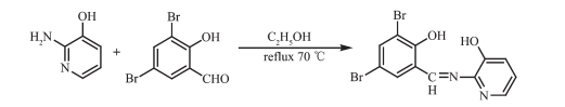
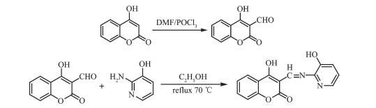
HL1 and HL2 were synthesized according to the following synthetic routes shown in Scheme 1 and 2.
H2L1: 3-Formyl-4-hydroxyl-comnarin was synthes-ized according to the analogous method[47]. POCl3 (10 mL, 2.00 mmol) was dropped in the anhydrous DMF solution (10 mL) of 4-hydroxyl coumarin (3.8 g, 20.00 mmol) with the reaction temperature sustained lower than 5 ℃. The reaction mixture was kept at room temperature for 2 h, then it was heated in the steam bath for 1 h at 70 ℃. After the mixed solution come to room temperature, it was mixed with ice water and neutralized with sodium carbonate, and the resulting yellow solid was collected and recrystallized using ethyl alcohol. Yield: 78.6%; m.p. 115~116 ℃. Anal. Calcd. for C10H6O4(%): C, 63.16; H, 3.18. Found(%): C, 63.68; H, 3.21.
A solution of 3-formyl-4-hydroxyl comnarin (190.03 mg, 1.00 mmol) in ethanol (2 mL) was added to a solution of 2-amino-3-hydroxy pyridine (110.05 mg, 1.00 mmol)) in ethanol (2 mL) and the mixture was subjected to heating at 70 ℃ for 12 h. After the mixed solution come to room temperature, the resulting white solid was collected. After cooling to room temperature the light yellow precipitate was filtered and washed successively with ethanol/petroleum ether (1: 4, V/V). The product was dried under reduced pressure to obtain 222.00 mg H2L1. Yield: 74.5%. m.p. 214~215 ℃. Anal. Calcd. for C15H10N2O4(%): C, 63.83; H, 3.57; N, 9.93. Found(%): C, 63.52; H, 4.22; N, 9.80.
H2L2: The ligand H2L2 was synthesized by a method similar to that of H2L1 except substituting 3-aldehyde-4-hydroxy coumarin with 3, 5-dibrominesa-licylaldehyde. Yield: 351.33 mg, 76.8 %. m.p. 204~205 ℃. Anal. Calcd. for C12H8Br2N2O2(%): C, 38.74; H, 2.17; N, 7.53. Found(%): C, 38.81; H, 2.16; N, 7.49.
Complex 1: A solution of Ni(Ⅱ) acetate monohy-drate (0.7 mg, 0.003 mmol) in ethanol (2 mL) was added dropwise to a solution of H2L1 (0.95 mg, 0.005 mmol) in acetone (4 mL). The color of the mixture turned to brown immediately, and then 2 drops of DMF were added in it, stirred for 1 h at room temperature. The mixture was filtered, and the filtrate was allowed to stand at room temperature for about a week. The solvent was partially evaporated, and single crystals suitable for X-ray crystallographic analysis were obtained. Yield: 25.6%. Anal. Calcd. for C18H15N3NiO5(%): C, 52.47; H, 3.67; N, 10.20. Found(%): C 53.47, H, 3.67; N, 10.87.
Complex 2: The complex 2 was synthesized with the similar method for complex 1. Yield: 28.6%. Anal. Calcd. for C15H15Br2N3O4Zn(%): C, 34.22; H, 2.87; N, 7.98. Found(%): C, 34.78; H, 2.87; N, 8.38.
The single crystals of the complexeswith approxi-mate dimensions of 0.40 mm× 0.11 mm× 0.08 mm (1) and 0.30 mm× 0.27 mm× 0.15 mm (2) were placed on a Bruker Smart 1000 CCD area detector. The reflec-tions were collected using graphite-monochromatized Mo Kα radiation (λ=0.071 073 nm). The Lp correc-tions were applied to the SAINT program[48] and semi-empirical correction were applied to the SADABS program[49]. The crystal structures were solved by the direct methods (SHELXS-2014)[50]. The hydrogen atoms of water molecules in the complex 2 were located from difference Fourier maps, and the other hydrogen atoms were generated geometrically. Details of the crystal parameters, data collection and refinements for complexes 1 and 2 are summarized in Table 1.
 下载:
导出CSV
下载:
导出CSV
| Empirical formula | C18H15N3NiO5 | C12H8Br2N2O3Zn·C3H7NO |
| Formula weight | 412.04 | 526.49 |
| Temperature/K | 297.16(10) | 293(2) |
| Crystal system | Monoclinic | Monoclinic |
| Space group | P21/c | P21/c |
| a/nm | 0.991 21(4) | 1.159 66(6) |
| b/nm | 1.411 92(6) | 1.394 83(4) |
| c/nm | 1.240 16(5) | 1.135 06(6) |
| β/(°) | 102.395(5) | 105.547(5) |
| Volume/nm3 | 1.695(13) | 1.768(14) |
| Z | 4 | 4 |
| Dc/(Mg·m-3) | 1.615 | 1.977 |
| μ/mm-1 | 1.18 | 5.93 |
| F(000) | 848 | 1 032 |
| θ range/(°) | 3.566 0~28.422 0 | 3.679 0~23.951 0 |
| Limiting indices | -16 ≤ h ≤ 17, -12 ≤ k ≤ 13, -15 ≤ l ≤ 14 | -14≤ h ≤ 7, -17 ≤ k ≤ 16, -13≤ l ≤ 14 |
| Reflection collected, unique | 6 323, 2 737 (Rint=0.026) | 7 039, 2 306 (Rint=0.047) |
| Completeness to θ=26.32°/% | 99.67 | 99.74 |
| Data, restraint, parameter | 3 335, 0, 246 | 3 478, 0, 229 |
| GOF on F2 | 1.047 | 0.912 |
| R1, wR2 [I > 2σ(I)] | 0.036 1, 0.081 5 | 0.053 3, 0.133 0 |
| Largest diff. peak and hole/(e·nm-3) | 520 and -322 | 836 and -766 |
CCDC: 1855900, 1; 1855901, 2.
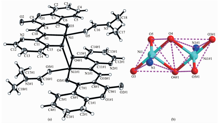
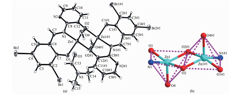
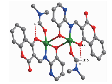
The single crystal structures and the coordination pattern diagram of complexes 1 and 2 are shown in Fig. 1 and 2. The selected bond lengths and angles of complexes 1 and 2 are listed in Table 2. X-ray crystallographic analysis reveals that both complexes 1 and 2 crystallize in the monoclinic system with space group P21/c. Complexes 1 and 2 can be described as binuclear M(Ⅱ) complexes (M=Ni or Zn), consist of two M(Ⅱ) ions, two (L2-) units and two coordinated solvent molecules (DMF for 1 and H2O for 2), in which the difference is that complex 2 contains two free DMF molecules.
Symmetry codes: #1:-x, 0.5+y, 0.5-z
Symmetry codes: #1:-x, 0.5+y, 0.5-z
下载:
导出CSV
| Complex 1 | |||||
| Ni1-O3 | 0.194 0(2) | Ni1-O5 | 0.197 8(2) | Ni1-O4#1 | 0.235 8(2) |
| Ni1-O4 | 0.194 4(2) | Ni1-N1 | 0.192 9(2) | ||
| O3-Ni1-O5 | 94.25(8) | O3-Ni1-O4 | 177.77(7) | O3-Ni1-O4#1 | 92.27(7) |
| O5-Ni1-O4#1 | 89.16(7) | O4-Ni1-O5 | 87.94(7) | O4-Ni1-O4#1 | 87.37(7) |
| N1-Ni1-O3 | 93.03(8) | N1-Ni1-O5 | 161.42(8) | N1-Ni1-O4#1 | 107.62(7) |
| N1-Ni1-O4 | 84.99(8) | Ni1-O4-Ni1#1 | 92.63(7) | C7-O3-Ni1 | 126.33(2) |
| Complex 2 | |||||
| Zn1-O1 | 0.196 5(5) | Zn1-O2#1 | 0.200 0(4) | Zn1-O2 | 0.209 8(4) |
| Zn1-O4 | 0.201 7(5) | Zn1-N1 | 0.206 1(5) | O2-Zn1#1 | 0.200 0(4) |
| O1-Zn1-O2#1 | 100.8(2) | O1-Zn1-O2 | 161.30(2) | O1-Zn1-O4 | 101.39(2) |
| O1-Zn1-N1 | 90.62(2) | O2#1-Zn1-O2 | 78.33(2) | O2#1-Zn1-O4 | 103.22(2) |
| O2#1-Zn1-N1 | 140.7(2) | O4-Zn1-O2 | 96.95(2) | O4-Zn1-N1 | 111.22(2) |
| Symmetry codes: #1: -x, 0.5+y, 0.5-z for complexes 1 and 2. | |||||
As presented in Fig. 1 and Fig. 2, the M(Ⅱ) ion (Ni for 1 and Zn for 2) was coordinated by one oxime nitrogen (N1) atoms and two deprotonated hydroxyl oxygen (O3, O4 in 1 and O1, O2 in 2) atoms of (L)2- units (L=L1 or L2, as well as one oxygen (O5 in 1 and O4 in 2) atom of the coordinated solvent molecule (DMF for 1 and H2O for 2), which constitute the [M(L)(solvent)] moiety. And then the O4 and O4A in 1 (and O2 and O2A in 2) atoms bridge the two [M(L)(solvent)] moieties to form the binuclear structure [Ni(L1)(DMF)]2 (1) and [Zn(L2(H2O)]2 (2). Thus, the central M(Ⅱ) ions are penta-coordinated and their coordination sphere is best described as a distorted tetragonal pyramid. In order to get the geometry adopted by M(Ⅱ) ions, the τ value was estimated to be τ=0.222 8 for 1 and τ=0.133 3 for 2[51-52]. The difference between complexes 1 and 2 is that the two [M(L)] moieties in complex 2 are almost planar with the distance of 0.007 7 nm but those in 1 are paralleled with the distance of 0.251 0 nm. As well as in complex 1 the coordinated DMF molecule is almost planar with [Ni(L1)] moiety but the the coordinated H2O molecule in complex 2 is almost perpendicular to the [Zn(L2] moiety.
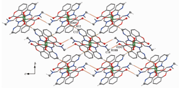
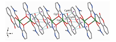
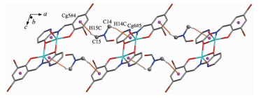
The intra- and intermolecular interactions data of complexes 1 and 2 are shown in Table 3 and 4. The crystal structure of complex 1 is stabilized by a pair of intramolecular non-classic hydrogen bonds C16-H16…O3 (Fig. 3, Table 3). Meanwhile, two pairs of intermolecular C18-H18B…O2 and C12-H12…O4 hydrogen bonds link neighboring molecules into 2D supramolecular network structure parallel to the bc planes (Fig. 4). Synchronously, complex 1 molecules are further linked by three pairs of intermolecular π…π stacking interactions(Cg1…Cg4, Cg2…Cg4 and Cg2…Cg3) between the benzene ring of adjacent complex 1 molecules to form the other 1D infinite chain along a axis (Fig. 5, Table 4). Thus, complex 1 molecules self-assemble via the intermolecular non-classic hydrogen-bonding and π…π stacking interac-tions to form the 3D supramolecular networks structure (Fig. 6). Consequently, the intermolecular non-classical hydrogen-bonding and π…π stacking interactions plays a very important role in the construction of supramolecular networks structure[53-59].
下载:
导出CSV
| D-H…A | d(D-H)/nm | d(H…A)/nm | d(D…A)/nm | ∠D-H…A/(°) | |
| Complex 1 | |||||
| C16-H16…O3 | 0.093 | 0.241 | 0.297 4(3) | 119 | |
| C12-H12…O4#1 | 0.093 | 0.246 | 0.337 6(3) | 169 | |
| C18-H18B…O2#2 | 0.096 | 0.252 | 0.346 2(4) | 166 | |
| Complex 2 | |||||
| C5-H5…O3#1 | 0.093 | 0.247 | 0.335 6(9) | 159 | |
| O4-H4A…O3#2 | 0.086 | 0.183 | 0.265 0(9) | 158 | |
| O4-H4B…N2#3 | 0.086 | 0.202 | 0.275 9(7) | 143 | |
| C15-H15C…Cg5#4 | 0.096 | 0.271 | 0.361 5(11) | 161 | |
| C14-H14C…Cg6#5 | 0.096 | 0.300 | 0.381 3(10) | 144 | |
| Cg5 and Cg6 are the centroids of benzene ring C1#4~C6#4 and the chelate ring C8#5-C12#5-O2#5-Zn1#5-N1#5 of complex 2, respectively; Symmetry codes: #1: x, 1/2-y, -1/2+z; #2: x, y, 1+z for 1; #1: x, 1/2-y, -1/2+z; #2: -x, -y, 2-z; #3: -x, -1/2+y, 3/2-z; #4: 1-x, -y, 2-z; #5: -x, -y, 2-z for 2. | |||||
下载:
导出CSV
| Ring (i) | Ring (j) | d(Cg…Cg)/nm | d(Cg(i)-perp)/nm | d(Cg(j)-perp)/nm |
| Cg1 | Cg4#3 | 0.360 38(15) | 0.332 92(9) | 0.327 87(11) |
| Cg2 | Cg4#3 | 0.349 87(14) | 0.330 91(8) | 0.331 93(11) |
| Cg2 | Cg3#3 | 0.367 44(13) | 0.328 74(9) | 0.330 08(10) |
| Cg1, Cg2, Cg3#3 and Cg4#3 are the centroids of ring C11-N1-Ni1-O4-C15, C7-O3-Ni1-N1-C10, C1#3-O1#3-C9#3 and C1#3~C6#3, respectively; Symmetry codes: #3: 1-x, -y, 1-z. | ||||
Hydrogen atoms, except those forming hydrogen bonds, are omitted for clarity
Symmetry codes: #1: x, 1/2-y, -1/2+z; #2: x, y, 1+z
Symmetry codes: #3: 1-x, -y, 1-z
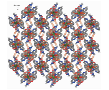
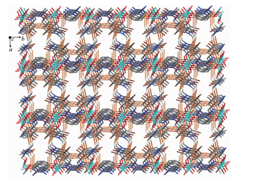
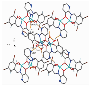
Complex 2 contains crystallizing DMF molecules and the oxygen atoms (O3) of the crystallizing DMF molecules have hydrogen-bond interactions with the coordinated water molecules and benzene rings of the L2- unit of one neighboring complex molecule, respe-ctively. Meanwhile, the coordinated water molecules are bonded to the neighboring complex molecule. Thus, the complex molecules and the crystallizing DMF molecules are linked by intermolecular hydrogen bonds to form a 2D-layer supramolecular structure parallel to the bc planes (Fig. 7, Table 3). Furthermore, this linkage is further stabilized by two pairs of intermolecular hydrogen bonds interactions (C15-H15C…Cg5 and C14-H14C…Cg6), which interlink the neighboring molecules into the other 1D infinite chain along the a axis (Fig. 8, Table 3). Thus, the crystal packing of complex 2 shows that a 3D supramolecular networks are formed through intermolecular O-H…O, O-H…N, C-H…O and C-H…π hydrogen bonding interactions[60-66] (Fig. 9).
Symmetry codes: #1: x, 1/2-y, -1/2+z; #2:-x, -y, 2-z; #3: -x, -1/2+y, 3/2-z
Symmetry codes: #4: 1-x, -y, 2-z; #5:-x, -y, 2-z
The FT-IR spectra of H2L1, H2L2 and their corresponding complexes 1 and 2 exhibit various bands in the 400~4 000 cm-1 region. The most impor-tant FT-IR bands for H2L1, H2L2 and its Ni(Ⅱ)and Zn(Ⅱ) complexes are listed in Table 5.
下载:
导出CSV
| cm-1 | |||||
| Compound | ν(O-H) | ν(C=N) | ν(Ar-O) | ν(M-N) | ν(M-O) |
| H2L1 | 3 447 | 1 619 | 1 200 | — | — |
| 1 | 3 442 | 1 605 | 1 193 | 478 | 438 |
| H2L2 | 3 460 | 1 607 | 1 203 | — | — |
| 2 | 3 437 | 1 600 | 1 196 | 576 | 506 |
The characteristic C=N stretching band of the free ligand H2L1 and H2L2 appeared at 1 619 and 1 607 cm-1, respectively, while those of complexes 1 and 2 were observed in the 1 605 and 1 600 cm-1, respe-ctively[67-75]. The C=N stretching frequencies were all shifted to lower frequencies by ca. 14 and 7 cm-1 upon complexation respectively, indicating a decrease in the C=N bond order due to the coordinated bond of the metal atom with the imino nitrogen lone pair[76-78]. The Ar-O stretching band of the ligands H2L1 and H2L2 occured at 1 200 and 1 203 cm-1, respectively, and those at 1 193 and 1 196 cm-1 for complexes 1 and 2. The lower frequency of the Ar-O absorption shift indicates that M-O bond is formed between the metal ions and the oxygen atoms of the phenolic groups[59]. In addition, the broad O-H group stretching band at 3 447 and 3 460 cm-1 in the free ligands H2L1 and H2L2 disappeared in complexes 1 and 2, indicating the oxygen atoms in the phenolic hydroxyl groups have been completely deprotonated and coordinated to the metal ions. Whereas, the stretching band at 3 437 cm-1 in the complex 2 is attributed to the stretching vibrations of the O-H group of the coordinated water. The FT-IR spectrum of complex 1 showed ν(M-N) and ν(M-O) vibration absorption frequencies at 478 and 438 cm-1 (or 576 and 506 cm-1 for complex 2), respe-ctively. These assignments are consistent with the literature frequency values.
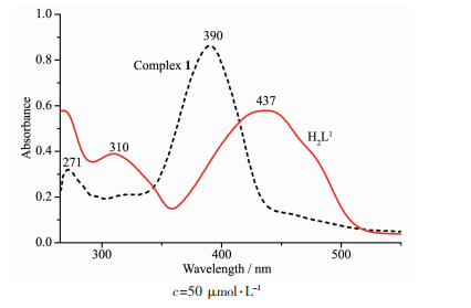
The absorption spectra of ligands H2L1, H2L2 and theirs corresponding Ni(Ⅱ) and Zn(Ⅱ) complexes 1 and 2 were determined in diluted DMF solution as shown in Fig. 10 and 11. The electronic absorption spectrum of free ligand H2L1 exhibited two absorption peaks at approximately 271 and 390 nm (Fig. 10). The former absorption peaks at 271 nm can be assigned to the π-π* transition of the benzene rings and the latter at 390 nm can be attributed to the intraligand π-π* transition of the C=N group[79]. Upon coordination of the ligand, the relatively intense absorption at 390 nm disappeared from UV-Vis spectra of complex 1, indicating that the amino nitrogen is involved in coordination with Ni(Ⅱ) ion[80]. The intraligand π-π* transition of the benzene ring is bathochromically shifted to 310 nm in complex 1, indicating the coordination of Ni(Ⅱ) ion with deprotonated L- unit. The new peak at 437 nm of complex 1 is assigned to L→M charge-transfer transition.
The absorption of complex 2 was obviously different from that of H2L2 owing to complexation (Fig. 11). For the free ligand there were two intense peaks centered at around 275 and 399 nm, assigned to π-π* transitions of the benzene rings of the benzaldehyde and C=N groups, respectively. Compared with the absorption peak of the free ligand, the absorption at 275 nm was slightly shifted hypsochromically to 272 nm of complex 2, indicating the coordination of Zn(Ⅱ)ion with deprotonated L unit. Meanwhile, the absorption peak at 399 nm disappeared from the UV-Vis spectrum of complex 2, which indicates that the amino nitrogen atom is involved in coordination to the metal atom. In addition, a new absorption peak was observed at 460 nm in complex 2, which is assigned to the L→M charge-transfer transition. This is chara-cteristic of a transition metal complex with Schiff base ligand[80].

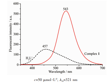
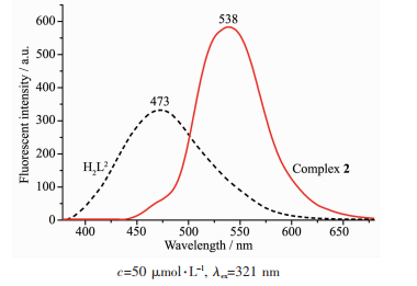
The emission spectra of ligands H2L1 and H2L2, complexes 1 and 2 were determined in diluted DMF solution at room temperature as shown in Fig. 12 and 13. The ligands H2L1 and H2L2 exhibited the relatively weak emission at 457 and 473 nm upon excitation at 321 and 351 nm, respectively. The blue emission should be assigned to intraligand π-π* transition[81-82]. Compared with the free ligands H2L1 and H2L2, an intense green emission at 543 and 538 nm for complexes 1 and 2 were observed upon excitation at 321 and 351 nm, respectively, which indicates that the addition of metal ions Ni(Ⅱ) and Zn(Ⅱ) induces the change of the fluorescence characteristics of the ligand, which may be due to the destruction of the intramolecular hydrogen bonding of the ligand and resulting in the enhancement of the planarity of the conjugated system[83-85]. The Stokes shift between the maximum wavelength of the fluorescence emission and the fluorescence excitation for H2L1, complex 1, H2L2 and complex 2 is 136, 222, 122 and 182 nm, respe-ctively, which indicates that the introduction of coumarin group is beneficial to the luminescence of ligand and its metal complex. And the red shifts in emission wavelength of complexes 1 and 2 compared with H2L1 and H2L2 might be related to the coor-dination of the metal ions to the ligands and increases of the rigidity of ligands, which can diminish the loss of energy via vibrational motions and increase the emission efficiency.
The syntheses, structural characterizations, and fluorescence properties of the Schiff base ligands H2L1, H2L2 and their corresponding binuclear Ni(Ⅱ) and Zn(Ⅱ) complexes 1 and 2 were discussed. Crystal structure analyses of complexes 1 and 2 showed that the Ni(Ⅱ) and Zn(Ⅱ) atoms all have penta-coordinate environ-ments and adopt distorted square pyramidal geome-tries. Complexes 1 and 2 are self-assembled into the 3D supramolecular networks through intermolecular hydrogen bonding, C-H…π and π…π stacking interactions. Furthermore, the optical properties of complexes 1 and 2 indicated that the coordination of metal ions Ni(Ⅱ) and Zn(Ⅱ) leads to the fluorescence enhancement of H2L1 and H2L2. Moreover, the Stokes shifts of H2L1 and complex 1 is larger than those of H2L2 and complex 2, which indicates that the intro-duction of coumarin group is beneficial to the luminescence of ligand and its metal complex.
Liu Y A, Wang C Y, Zhang M, et al. Polyhedron, 2017, 127:278-286 doi: 10.1016/j.poly.2017.02.007
Dong W K, Ma J C, Dong Y J, et al. Polyhedron, 2016, 115:228-235 doi: 10.1016/j.poly.2016.05.017
孙银霞, 董文魁, 王莉, 等.无机化学学报, 2009, 25:1478-1482 doi: 10.3321/j.issn:1001-4861.2009.08.028SUN Yin-Xia, DONG Wen-Kui, WANG Li, et al. Chinese J. Inorg. Chem., 2009, 25:1478-1482 doi: 10.3321/j.issn:1001-4861.2009.08.028
Sun Y X, Zhang S T, Ren Z L, et al. Synth. React. Inorg. Met.-Org. Nano-Met. Chem., 2013, 43:995-1000 doi: 10.1080/15533174.2012.753614
杨玉华, 郝静, 董银娟, 等.无机化学学报, 2017, 33:1280-1292 doi: 10.11862/CJIC.2017.150YANG Yu-Hua, HAO Jing, DONG Yin-Juan, et al. Chinese J. Inorg. Chem., 2017, 33:1280-1292 doi: 10.11862/CJIC.2017.150
Wu H L, Pan G L, Bai Y C, et al. J. Chem. Res., 2014, 38:211-217 doi: 10.3184/174751914X13933417974082
Yang H Q, Zhang L, Zhong L, et al. Angew. Chem. Int. Ed., 2007, 46:6861-6865 doi: 10.1002/(ISSN)1521-3773
Li X Y, Kang Q P, Liu L Z, et al. Crystals, 2018, 8:43 doi: 10.3390/cryst8010043
Wu H L, Pan G L, Bai Y C, et al. Res. Chem. Intermed., 2015, 41:3375-3388 doi: 10.1007/s11164-013-1440-5
Chen C Y, Zhang J W, Zhang Y H, et al. J. Coord. Chem., 2015, 68:1054-1071 doi: 10.1080/00958972.2015.1007965
Wu H L, Bai Y H, Zhang Y H, et al. Z. Anorg. Allg. Chem., 2014, 640:2062-2071 doi: 10.1002/zaac.v640.10
Hao J, Li L L, Zhang J T, et al. Polyhedron, 2017, 134:1-10 doi: 10.1016/j.poly.2017.05.060
Wu H L, Wang C P, Wang F, et al. J. Chin. Chem. Soc., 2015, 62:1028-1034 doi: 10.1002/jccs.v62.11
Song X Q, Liu P P, Xiao Z R, et al. Inorg. Chim. Acta, 2015, 438:232-244 doi: 10.1016/j.ica.2015.09.022
Dong W K, Li X L, Wang L, et al. Sens. Actuators B:Chem., 2016, 229:370-378 doi: 10.1016/j.snb.2016.01.139
Liu P P, Sheng L, Song X Q, et al. Inorg. Chim. Acta, 2015, 434:252-257 doi: 10.1016/j.ica.2015.05.026
Dong W K, Ma J C, Zhu L C, et al. New J. Chem., 2016, 40:6998-7010 doi: 10.1039/C6NJ00855K
Zhang H, Dong W K, Zhang Y, et al. Polyhedron, 2017, 133:279-293 doi: 10.1016/j.poly.2017.05.051
Dong X Y, Akogun S F, Zhou W M, et al. J. Chin. Chem. Soc., 2017, 64:412-419 doi: 10.1002/jccs.2017.64.issue-4
Tao C H, Ma J C, Zhu L C, et al. Polyhedron, 2017, 128:38-45 doi: 10.1016/j.poly.2017.02.040
Dong Y J, Dong X Y, Dong W K, et al. Polyhedron, 2017, 123:305-315 doi: 10.1016/j.poly.2016.12.010
Li G, Hao J, Liu L Z, et al. Crystals, 2017, 7:217 doi: 10.3390/cryst7070217
Dong W K, Sun Y X, Zhang, Y P, et al. Inorg. Chim. Acta, 2009, 362:117-124 doi: 10.1016/j.ica.2008.03.128
Dong W K, Zhang J, Zhang Y, et al. Inorg. Chem. Acta, 2016, 444:95-102 doi: 10.1016/j.ica.2016.01.034
Chai L Q, Tang L J, Chen L C, et al. Polyhedron, 2017, 122:228-240 doi: 10.1016/j.poly.2016.11.032
Chai L Q, Zhang K Y, Tang L J, et al. Polyhedron, 2017, 130:100-107 doi: 10.1016/j.poly.2017.04.010
Chen L, Dong W K, Zhang H, et al. Cryst. Growth Des., 2017, 17:3636-3648 doi: 10.1021/acs.cgd.6b01860
陆瑞娥, 李新然, 赵亚元, 等.无机化学学报, 2015, 31:1055-1062 http://www.wjhxxb.cn/wjhxxbcn/ch/reader/view_abstract.aspx?flag=1&file_no=20150527&journal_id=wjhxxbcnLU Rui-E, LI Xin-Ran, ZHAO Ya-Yuan, et al. Chinese J. Inog. Chem., 2015, 31:1055-1062 http://www.wjhxxb.cn/wjhxxbcn/ch/reader/view_abstract.aspx?flag=1&file_no=20150527&journal_id=wjhxxbcn
Wang P, Zhao L. Synth. React. Inorg. Met.-Org. Nano-Met. Chem., 2016, 46:1095-1101 doi: 10.1080/15533174.2015.1004416
Zhao L, Dang X T, Chen Q, et al. Synth. React. Inorg. Met.-Org. Nano-Met. Chem., 2013, 43:1241-1246 doi: 10.1080/15533174.2012.757236
Sun Y X, Wang L, Dong X Y, et al. Synth. React. Inorg. Met.-Org. Nano-Met. Chem., 2013, 43:599-603 doi: 10.1080/15533174.2012.751424
Dong W K, Ma J C, Zhu L C, et al. Cryst. Growth Des., 2016, 16:6903-6915 doi: 10.1021/acs.cgd.6b01067
董文魁, 王莉, 孙银霞, 等.无机化学学报, 2011, 27(2):372-376 http://www.wjhxxb.cn/wjhxxbcn/ch/reader/view_abstract.aspx?flag=1&file_no=20110228&journal_id=wjhxxbcnDONG Wen-Kui, WANG Li, SUN Yin-Xia, et al. Chinese J. Inorg. Chem., 2011, 27(2):372-376 http://www.wjhxxb.cn/wjhxxbcn/ch/reader/view_abstract.aspx?flag=1&file_no=20110228&journal_id=wjhxxbcn
Zhang H, Wu H L, Chen C Y, et al. J. Coord. Chem., 2016, 69:1577-1586 doi: 10.1080/00958972.2016.1185518
Chai, L Q, Liu G, Zhang Y L, et al. J. Coord. Chem., 2013, 66:3926-3938 doi: 10.1080/00958972.2013.857016
Duan J G, Liu G L. Transition Met. Chem., 2007, 32:702-705 doi: 10.1007/s11243-007-0240-4
Dong W K, He X N, Yan H B, et al. Polyhedron, 2009, 28; 1419-1428 doi: 10.1016/j.poly.2009.03.017
Dong W K, Feng J H, Wang L, et al. Transtion Met. Chem., 2007, 32:1101-1105 doi: 10.1007/s11243-007-0292-5
Dong W K, Sun Y X, Zhao C Y, et al. Polyhedron, 2010, 29:2087-2097 doi: 10.1016/j.poly.2010.04.006
Li L H, Dong W K, Zhang Y, et al. Appl. Organomet. Chem., 2017, 31:e3818 doi: 10.1002/aoc.v31.12
Li X Y, Chen L, Gao L, et al. RSC Adv., 2017, 7:35905-35916 doi: 10.1039/C7RA06796H
Song X Q, Wang L, Zheng Q F, et al. Inorg. Chim. Acta, 2012, 391:171-178 doi: 10.1016/j.ica.2012.04.007
Hu J H, Sun Y, Qi J, et al. Spectrochim. Acta A, 2017, 175:125-133 doi: 10.1016/j.saa.2016.12.009
Dong X Y, Kang Q P, Jin B X, et al. Z. Naturforsch., 2017, 72:415-420 doi: 10.1515/znb-2016-0268
Wu H L, Bai Y C, Zhang Y H, et al. J. Coord. Chem., 2014, 67:3054-3066 doi: 10.1080/00958972.2014.959507
Wu H L, Pan G L, Bai Y C, et al. J. Coord. Chem., 2013, 66:2634-2646 doi: 10.1080/00958972.2013.812725
Jia H R, Li J, Sun Y X, et al. Z. Krist.:New Cryst. Struct, 2018, 233(1):45-47 http://www.sciencedirect.com/science/article/pii/S0020169315006271
SAINT-Plus, Ver.6.02, Bruker Analytical X-ray System, Madison, WI, 1999.
Sheldrick G M. SADABS, Program for Empirical Absorption Correction of Area Detector Data, University of Göttingen, Germany, 1996.
Sheldrick G M. SHELXS-97, Program for the Solution and the Refinement of Crystal Structures, University of Göttingen, Germany, 1997.
Sun Y X, Xu L, Zhao T H, et al. Synth. React. Inorg. Met.-Org. Nano-Met. Chem., 2013, 43:509-513 doi: 10.1080/15533174.2012.740756
Addison A W, Rao T N, Reedijk J, et al. J. Chem. Soc. Dalton Trans., 1984, 7:1349-1356
Dong W K, Zhang X Y, Sun Y X, et al. Synth. React. Inorg. Met.-Org. Nano-Met. Chem., 2015, 45:956-962 doi: 10.1080/15533174.2013.862814
Dong Y J, Li X L, Zhang Y, et al. Supramol. Chem., 2017, 29:518-527 doi: 10.1080/10610278.2017.1285031
Wang B J, Dong W K, Zhang Y, et al. Sens. Actuators B:Chem., 2017, 247:254-264 doi: 10.1016/j.snb.2017.02.154
Wang L, Hao J, Zhai L X, et al. Crystals, 2017, 7:277 doi: 10.3390/cryst7090277
Ma J C, Dong X Y, Dong W K, et al. J. Coord. Chem., 2016, 69:149-159 doi: 10.1080/00958972.2015.1108410
Dong W K, Zhu L C, Dong Y J, et al. Polyhedron, 2016, 117:148-154 doi: 10.1016/j.poly.2016.05.055
Xu L, Zhu L C, Ma J C, et al. Z. Anorg. Allg. Chem., 2015, 641:2520-2524 doi: 10.1002/zaac.201500619
Dong W K, Wang Z K, Li G, et al. Z. Anorg. Allg. Chem., 2013, 639:2263-2268 doi: 10.1002/zaac.v639.12/13
Chai L Q, Huang J J, Zhang J Y, et al. J. Coord. Chem., 2015, 68:1224-1237 doi: 10.1080/00958972.2015.1019875
Wang L, Ma J C, Dong W K, et al. Z. Anorg. Allg. Chem., 2016, 642:834-839 doi: 10.1002/zaac.v642.15
Wang P, Zhao L. Asian J. Chem., 2015, 4:1424-1426 http://www.researchgate.net/publication/272379464_Synthesis_and_Crystal_Structure_of_Supramolecular_Copper(II)_Complex_Based_on_N2O2_Coordination_Sphere
Dong Y J, Ma J C, Zhu L C, et al. J. Coord. Chem., 2017, 70:103-115 doi: 10.1080/00958972.2016.1262537
Chai L Q, Wang G, Sun Y X, et al. J. Coord. Chem., 2012, 65:1621-1631 doi: 10.1080/00958972.2012.677836
Wu H L, Bai Y, Yuan J K, et al. J. Coord. Chem., 2012, 65:2839-2851 doi: 10.1080/00958972.2012.707314
Wu H L, Pan G L, Bai Y C, et al. J. Photochem. Photobiol. B, 2014, 135:33-43 doi: 10.1016/j.jphotobiol.2014.04.005
Song X Q, Peng Y J, Chen G Q, et al. Inorg. Chim. Acta, 2015, 427:13-21 doi: 10.1016/j.ica.2014.12.008
Hu J H, Li J B, Qi J, et al. New J. Chem., 2015, 39:843-848 doi: 10.1039/C4NJ01147C
Liu P P, Wang C Y, Zhang M, et al. Polyhedron, 2017, 129:133-140 doi: 10.1016/j.poly.2017.03.019
Chai L Q, Zhang H S, Huang J J, et al. Spectrochim. Acta A, 2015, 137:661-669 doi: 10.1016/j.saa.2014.08.084
Dong W K, Zhang F, Li N. Z. Anorg. Allg. Chem., 2016, 642:532-538 doi: 10.1002/zaac.v642.7
Wang P, Zhao L. Spectrochim. Acta Part A, 2015, 135:342-350 doi: 10.1016/j.saa.2014.06.129
Gao L, Wang F, Zhao Q, et al. Polyhedron, 2018, 139:7-16 doi: 10.1016/j.poly.2017.10.004
Dong W K, Ma J C, Dong Y J, et al. J. Coord. Chem., 2016, 69:3231-3241 doi: 10.1080/00958972.2016.1231302
Wu H L, Huang X C, Yuan J K, et al. Z. Naturforsch., 2011, 66b:1049-1055
Wu H L, Li K, Sun T, et al. Transition Met. Chem., 2011, 36:21-28 doi: 10.1007/s11243-010-9429-z
Wu H L, Wang K. T, Kou F, et al. J. Coord. Chem., 2010, 64:2676-2687 doi: 10.1007/s11243-014-9880-3
Zhang Y G, Shi Z H, Yang L Z, et al. Inorg. Chem. Commun., 2014, 39:86-89 doi: 10.1016/j.inoche.2013.10.035
Wang F, Gao L, Zhao Q, et al. Spectrochim. Acta Part A, 2018, 190:111-115 doi: 10.1016/j.saa.2017.09.027
Dong W K, Akogun S F, Zhang Y, et al. Sens. Actuators B:Chem., 2017, 238:723-734 doi: 10.1016/j.snb.2016.07.047
Song X Q, Cheng G Q, Liu Y A. Inorg. Chim. Acta, 2016, 450:386-394 doi: 10.1016/j.ica.2016.06.028
Zheng S S, Dong W K, Zhang Y, et al. New J. Chem., 2017, 41:4966-4973 doi: 10.1039/C6NJ04090J
Song X Q, Liu P P, Liu Y A, et al. Dalton Trans., 2016, 45:8154-8163 doi: 10.1039/C6DT00212A
Wang L, Zhao Q, Li X Y, et al. Z. Naturforsch. B, 2017, 72(12):947-953 doi: 10.1515/znb-2017-0107

Figure 1 (a) Molecular structure of complex 1 showing 30% probability displacement ellipsoids; (b) Coordination geometry diagram for Ni(Ⅱ) ions of complex 1
Symmetry codes: #1:-x, 0.5+y, 0.5-z

Figure 2 Molecular structure of complex 2 showing 30% probability displacement ellipsoids; (b) Coordination geometry diagram for Zn(Ⅱ) ions of complex 2
Symmetry codes: #1:-x, 0.5+y, 0.5-z

Figure 3 Intramolecular hydrogen bonding of complex 1
Hydrogen atoms, except those forming hydrogen bonds, are omitted for clarity

Figure 4 Part of 2D supramolecular structure of complex 1 linked by intermolecular hydrogen bonds parallel to the bc planes
Symmetry codes: #1: x, 1/2-y, -1/2+z; #2: x, y, 1+z

Figure 5 Part of 1D supramolecular structure linked by π… π stacking interactions along the a axis of complex 1
Symmetry codes: #3: 1-x, -y, 1-z

Figure 7 Part of 2D supramolecular structure linked by intermolecular hydrogen bonds parallel to the bc planes of complex 2
Symmetry codes: #1: x, 1/2-y, -1/2+z; #2:-x, -y, 2-z; #3: -x, -1/2+y, 3/2-z

Figure 8 Part of 1D supramolecular structure linked by C- H…π stacking interactions along the a axis of complex 2
Symmetry codes: #4: 1-x, -y, 2-z; #5:-x, -y, 2-z

Figure 10 UV-Vis absorption spectra of H2L1 and complex 1 in diluted DMF solution at room temperature

Figure 11 UV-Vis absorption spectra of H2L2 and complex 2 in diluted DMF solution at room temperature
Table 1. Crystal data and structure refinement for complexes 1 and 2
| Empirical formula | C18H15N3NiO5 | C12H8Br2N2O3Zn·C3H7NO |
| Formula weight | 412.04 | 526.49 |
| Temperature/K | 297.16(10) | 293(2) |
| Crystal system | Monoclinic | Monoclinic |
| Space group | P21/c | P21/c |
| a/nm | 0.991 21(4) | 1.159 66(6) |
| b/nm | 1.411 92(6) | 1.394 83(4) |
| c/nm | 1.240 16(5) | 1.135 06(6) |
| β/(°) | 102.395(5) | 105.547(5) |
| Volume/nm3 | 1.695(13) | 1.768(14) |
| Z | 4 | 4 |
| Dc/(Mg·m-3) | 1.615 | 1.977 |
| μ/mm-1 | 1.18 | 5.93 |
| F(000) | 848 | 1 032 |
| θ range/(°) | 3.566 0~28.422 0 | 3.679 0~23.951 0 |
| Limiting indices | -16 ≤ h ≤ 17, -12 ≤ k ≤ 13, -15 ≤ l ≤ 14 | -14≤ h ≤ 7, -17 ≤ k ≤ 16, -13≤ l ≤ 14 |
| Reflection collected, unique | 6 323, 2 737 (Rint=0.026) | 7 039, 2 306 (Rint=0.047) |
| Completeness to θ=26.32°/% | 99.67 | 99.74 |
| Data, restraint, parameter | 3 335, 0, 246 | 3 478, 0, 229 |
| GOF on F2 | 1.047 | 0.912 |
| R1, wR2 [I > 2σ(I)] | 0.036 1, 0.081 5 | 0.053 3, 0.133 0 |
| Largest diff. peak and hole/(e·nm-3) | 520 and -322 | 836 and -766 |
 下载: 导出CSV
Table 2. Selected bond lengths (nm) and bond angles (°) for complexes 1 and 2
| Complex 1 | |||||
| Ni1-O3 | 0.194 0(2) | Ni1-O5 | 0.197 8(2) | Ni1-O4#1 | 0.235 8(2) |
| Ni1-O4 | 0.194 4(2) | Ni1-N1 | 0.192 9(2) | ||
| O3-Ni1-O5 | 94.25(8) | O3-Ni1-O4 | 177.77(7) | O3-Ni1-O4#1 | 92.27(7) |
| O5-Ni1-O4#1 | 89.16(7) | O4-Ni1-O5 | 87.94(7) | O4-Ni1-O4#1 | 87.37(7) |
| N1-Ni1-O3 | 93.03(8) | N1-Ni1-O5 | 161.42(8) | N1-Ni1-O4#1 | 107.62(7) |
| N1-Ni1-O4 | 84.99(8) | Ni1-O4-Ni1#1 | 92.63(7) | C7-O3-Ni1 | 126.33(2) |
| Complex 2 | |||||
| Zn1-O1 | 0.196 5(5) | Zn1-O2#1 | 0.200 0(4) | Zn1-O2 | 0.209 8(4) |
| Zn1-O4 | 0.201 7(5) | Zn1-N1 | 0.206 1(5) | O2-Zn1#1 | 0.200 0(4) |
| O1-Zn1-O2#1 | 100.8(2) | O1-Zn1-O2 | 161.30(2) | O1-Zn1-O4 | 101.39(2) |
| O1-Zn1-N1 | 90.62(2) | O2#1-Zn1-O2 | 78.33(2) | O2#1-Zn1-O4 | 103.22(2) |
| O2#1-Zn1-N1 | 140.7(2) | O4-Zn1-O2 | 96.95(2) | O4-Zn1-N1 | 111.22(2) |
| Symmetry codes: #1: -x, 0.5+y, 0.5-z for complexes 1 and 2. | |||||
下载: 导出CSV
Table 3. Hydrogen-bonding interactions for complexes 1 and 2
| D-H…A | d(D-H)/nm | d(H…A)/nm | d(D…A)/nm | ∠D-H…A/(°) | |
| Complex 1 | |||||
| C16-H16…O3 | 0.093 | 0.241 | 0.297 4(3) | 119 | |
| C12-H12…O4#1 | 0.093 | 0.246 | 0.337 6(3) | 169 | |
| C18-H18B…O2#2 | 0.096 | 0.252 | 0.346 2(4) | 166 | |
| Complex 2 | |||||
| C5-H5…O3#1 | 0.093 | 0.247 | 0.335 6(9) | 159 | |
| O4-H4A…O3#2 | 0.086 | 0.183 | 0.265 0(9) | 158 | |
| O4-H4B…N2#3 | 0.086 | 0.202 | 0.275 9(7) | 143 | |
| C15-H15C…Cg5#4 | 0.096 | 0.271 | 0.361 5(11) | 161 | |
| C14-H14C…Cg6#5 | 0.096 | 0.300 | 0.381 3(10) | 144 | |
| Cg5 and Cg6 are the centroids of benzene ring C1#4~C6#4 and the chelate ring C8#5-C12#5-O2#5-Zn1#5-N1#5 of complex 2, respectively; Symmetry codes: #1: x, 1/2-y, -1/2+z; #2: x, y, 1+z for 1; #1: x, 1/2-y, -1/2+z; #2: -x, -y, 2-z; #3: -x, -1/2+y, 3/2-z; #4: 1-x, -y, 2-z; #5: -x, -y, 2-z for 2. | |||||
下载: 导出CSV
Table 4. π…π stacking interactions for complex 1
| Ring (i) | Ring (j) | d(Cg…Cg)/nm | d(Cg(i)-perp)/nm | d(Cg(j)-perp)/nm |
| Cg1 | Cg4#3 | 0.360 38(15) | 0.332 92(9) | 0.327 87(11) |
| Cg2 | Cg4#3 | 0.349 87(14) | 0.330 91(8) | 0.331 93(11) |
| Cg2 | Cg3#3 | 0.367 44(13) | 0.328 74(9) | 0.330 08(10) |
| Cg1, Cg2, Cg3#3 and Cg4#3 are the centroids of ring C11-N1-Ni1-O4-C15, C7-O3-Ni1-N1-C10, C1#3-O1#3-C9#3 and C1#3~C6#3, respectively; Symmetry codes: #3: 1-x, -y, 1-z. | ||||
下载: 导出CSV
Table 5. Main bands in IR spectra of H2L1, H2L2 and their Ni(Ⅱ) and Zn(Ⅱ) complexes
| cm-1 | |||||
| Compound | ν(O-H) | ν(C=N) | ν(Ar-O) | ν(M-N) | ν(M-O) |
| H2L1 | 3 447 | 1 619 | 1 200 | — | — |
| 1 | 3 442 | 1 605 | 1 193 | 478 | 438 |
| H2L2 | 3 460 | 1 607 | 1 203 | — | — |
| 2 | 3 437 | 1 600 | 1 196 | 576 | 506 |
下载: 导出CSV

 扫一扫看文章

扫一扫关注我们