Table 1.
Crystal data and structure refinements for the complex
Citation:
Ren‐Shu WANG, Jing FENG, Yuan‐Lan WANG, Chen‐Feng OUYANG. A Complex of Silver(Ⅰ) with N⁃O Chelating Agent and Phosphine Ligand: Design, Structures, and Biological Activity[J]. Chinese Journal of Inorganic Chemistry,
2022, 38(8): 1609-1622.
doi:
10.11862/CJIC.2022.167

N⁃O螯合配体协同膦配体的银(Ⅰ)混配配合物的设计、结构和生物活性
摘要:
合成了一种Ag(Ⅰ)配合物[Ag(Qina)(Tpp)2]·1.5H2O(Qina=2‐喹啉羧酸根,Tpp=三苯基膦)。运用单晶X射线衍射、IR、NMR和粉末X射线衍射对配合物的结构进行了表征。配合物属于单斜晶系,C2/c空间群,晶胞参数:a=3.195 90(13) nm,b=1.210 96(4) nm,c=2.319 74(7) nm,β=102.166(4)°,V=8.776 0(5) nm3,Z=8。通过Hirshfeld表面分析,推导出配合物的超分子结构和分子间弱作用力。同时,通过DNA结合活性测试、抗菌活性和癌细胞体外毒活性测试,评价了配合物的生物活性。
-
关键词:
- 银(Ⅰ)配合物
- / Hirshfeld表面电荷分析
- / 晶体结构
- / 生物活性
English
A Complex of Silver(Ⅰ) with N⁃O Chelating Agent and Phosphine Ligand: Design, Structures, and Biological Activity
Abstract:
A new Ag(Ⅰ) complex, [Ag(Qina) (Tpp)2]·1.5H2O (Qina=2 ‐ quinolinecarboxylate, Tpp=triphenylphosphine), was synthesized and successfully obtained as a single crystal. The structure of the complex was characterized by single‐crystal X‐ray diffraction, IR, NMR, and powder X‐ray diffraction. The results show that the complex belongs to monoclinic crystal system, C2/c space group, and the unit cell parameters are a=3.195 90(13) nm, b= 1.210 96(4) nm, c=2.319 74(7) nm, β=102.166(4)°, V=8.776 0(5) nm3, and Z=8. The supramolecular structures and weak intermolecular force of the complex were deduced by Hirshfeld surface analysis. Meanwhile, the biological activities of the complex were tested by evaluating DNA ‐ binding efficacy, antibacterial activity, and in vitro toxic activity of cancer cells.
-
Key words:
- Ag(Ⅰ) complex
- / Hirshfeld surface analysis
- / crystal structures
- / biological activity
-
0. Introduction
Since ancient times, ancestors have used silverware for food preservation, silver nitrate for disinfection, and then silver compounds went on the market in the last century as an anti‐inflammatory drug for burns represented by silver sulfadiazine[1‐2]. Silver, as a metal with biological activity and medicinal value, has been flowing throughout the long history of mankind. Because microorganisms have almost no drug resistance to silver compounds, silver compounds can coexist with a variety of antibiotics and occupy a place[3]. Firstly, silver ions can penetrate the surface of the cell wall and strongly bind to sulfur‐containing enzymes, or they can remove electrons from cell components, destroy the integrity and permeability of the cell wall, and finally interfere with cell metabolism and kill cells[4‐6]. Therefore, silver can play a role in the pharma‐ceutical neighborhood. Secondly, due to the existence of a large number of chloride ions in the human body environment, after the activation of silver ions, these chloride ions will combine with them and then convert into AgCl without toxicity[7]. As a traditional safe metal, silver is undoubtedly favored by developers of nonplatinum cancer chemotherapeutic drugs. Platinum‐based chemotherapy drugs have been criticized in clinical applications because of their high toxicity and side effects[8].
Quinoline compounds have excellent biological activities. As a result, many compounds in the quinoline ring family, such as plasmocin, bulaquine, and tafenoquine, have been used as medicines because of their antimalarial properties[9]. From the perspective of structural chemistry, it is found that the 2 ‐quinolinecarboxylate (Qina) ligand molecule has an electron‐rich anionic COO- group and a neutral N atom that can provide a lone electron pair. It is a classical nitrogenoxygen chelating ligand, so it is often selected as a metal chelating agent for metal wet selection and analytical chemistry[10‐11]. More importantly, the Qina ligand also has a wide range of biochemical activity significance[12]. The Qina ligand in the quinoline ring family has been reported to be used as a nucleic acid matrix to assist in the DNA laser desorption process[13]. In addition, Qina is a metabolite that eventually exists in urine after humans receive tryptophan through the kynurenine metabolic pathway[14‐16]. Studies have pointed out that the tryptophan kynurenine metabolic pathway can produce Qina because it is closely related to the role of intestinal flora[17‐18]. At the same time, there is also research evidence that the stem extract of Ephedra Far East has a strong inhibitory effect on Clostridium difficile and Clostridium perfringens[19]. These two bacteria are considered to be related to intestinal diseases in animals[20‐22]. The reason why Ephedra stem extract expresses antibacterial activity is that the extract contains the key substance Qina, which plays a vital role in the antibacterial process[19]. More specifically, studies have shown that Qina can also affect the expression of oncogene p53 at the gene and protein molecule level, and then express its inhibitory effect on LS180 human colon adenocarcinoma cells[23]. More pragmatically, thiostrepton, an antibiotic originally used as a feed additive, also has Qina functional group in its side chains that enables its molecules to express anticancer and antibacterial activities[24‐25]. In recent years, the improved synthesis of this antibiotic has often been carried out around the Qina group contained in its side chain[26‐27]. Correspondingly, there are also reports on the anticancer activity of metal complexes containing Qina ligand[28‐29]. Besides, related complexes are also used in luminescence[30], catalysis, functional group protection[31], and so on.
In the synthesis of small molecular complexes with mixed ligands, synthesizers usually select Lewis base bidentate chelating agents with nitrogen‐containing heterocycles as the auxiliary ligand. However, choosing bidentate or multidentate ligands as the main ligand may cause excessive steric hindrance to the secondary coordination of the auxiliary ligand, resulting in difficulties in the synthesis of target compounds or ligand dissociation[32‐33]. Recent reports have shown that the spatial obstacles and restrictions between primary and secondary ligands could be avoided by the use of phosphorus‐containing monodentate auxiliary ligands in synthesis[34]. Triphenylphosphine (TPP) is often used as a reducing agent, and it participates in various organic reactions after being converted to triphenylphosphine oxide in the synthesis process[35]. TPP is also a good candidate for monodentate ligands with multiple‐aromatic groups, which have good lipophilicity and can effectively penetrate the mitochondrial membrane[36]. It has been reported that the use of TPP in the synthesis of silver complexes can contribute to the improvement of the stability and light resistance of the silver complex[37]. In addition, the fluorescence activity of Ag‐TPP type complexes has also been studied[38].
PI3K kinase is a key signal molecule for cancer cell growth and one of the most active signal pathways in cancer cells[39‐40]. In addition, PI3K is closely related to the Warburg effect of glycolysis in cancer cells, that is, cancer cells use Warburg metabolism to maintain the activity of the PI3K signaling pathway, to ensure continuous growth and division of cancer cells[41‐42].
We previously reported a case of Ag‐Qina type linear coordination polymer with good DNA binding efficiency and moderate cancer cytotoxic activity[43]. Based on the excellent biological characteristics of Ag, Qina, and TPP, we are interested in the preparation and activity evaluation of small molecule complex with these three components and hope to use the combination of the three components to make them synergistically express more biological activities in the complementary process.
1. Experimental
1.1 Material and instruments
All reagents and solvents were commercially purchased and used without any further treatment. The elemental composition (including C, H, and N) of these samples was determined by a model Elementar Vario Macro cube. The powder X‐ray diffraction (PXRD) experiment was completed by using a Shimadzu 6100 diffractometer, which employed Cu Kα (0.154 056 nm) radiation and was operated at 40 kV and 40 mA in a 2θ range of 5°‐80°. The 1H NMR spectra of the complex in DMSO ‐ d6 solutions were recorded on a Bruker AVANCE Ⅲ 600 spectrometer. The FT ‐ IR spectra were recorded on a Bruker Equinox 55 FT‐IR spectrophotometer. In addition, the emission spectra were obtained on a Perkin‐Elmer LS55 fluorescence Spectrofluorometer.
1.2 Synthesis of the complex
0.3 mmol of silver acetate and 0.6 mmol of TPP were mixed in a small volume of toluene. The reaction system was heated, and the reaction was stopped when the system solution suddenly became clear. Toluene was removed by vacuum drying with an oil pump, and white bulk Ag‐TPP intermediate was collected. The intermediate was stirred with the mixed solvent of ethanol and distilled water (1∶1, V/V) containing Qina at room temperature. After carefully adjusting the pH value to 5.0, the reaction continued for at least 3 h. A large amount of white precipitate that appeared during the reaction was collected by filtration and washed with ethanol. The dried precipitate was recrystallized with methanol and acetonitrile (1∶1, V/V). The product solution was kept in a ventilated darkroom for about a week, and the complex was precipitated out of the solution in the form of colorless massive crystals. The yield was ca. 65.5% based on Ag. 1H NMR (600 MHz, DMSO-d6): δ 8.51 (d, J=8.4 Hz, 1H), 8.34 (d, J=8.4 Hz, 1H), 8.00 (d, J=7.72 Hz, 1H), 7.63 (d, J=8.4 Hz, 1H), 7.55 (t, J=7.08 Hz, 1H), 7.45‐7.36 (m, 7H), 7.33‐7.14 (m, 24H) (Fig.S1a, Supporting information). 13C NMR (101 MHz, DMSO‐d6): δ 166.84, 155.82, 144.87, 138.18, 133.25 (d, J=16.9 Hz), 132.49 (d, J=23.3 Hz), 130.51, 130.05, 129.10 (d, J=9.4 Hz), 128.64, 128.48, 128.19, 127.63, 122.21 (Fig. S1b). Elemental analysis Calcd. for C46H39AgNO3.5P2(%): C 66.43; H 4.72; N 1.68; Found(%): C 65.68; H 4.50; N 1.67. Selected FT‐IR bands (KBr, cm-1): 3 051 ν(Ar—H), 1 606 ν(C=C), 1 585 ν(C=C), 1 502 ν(C=C), 1 477 ν(C=C), 1 606 ν(O—C—O)asym, 1 351 ν(O—C—O)sym, 1 428 ν(C=N), 781ν(phenyl rings), 722 ν(phenyl rings), 688 ν(phenyl rings), 490 ν(C—P) (Fig.S2).
1.3 Single⁃crystal X⁃ray crystallography
An Agilent Gemini E single‐crystal X ‐ray diffractometer (Mo Kα, λ =0.071 073 nm) was applied for polymorphism analysis. Data indexing, integration, and absorption correction were carried out using the CrysAlisPro software package. Besides, the structure solution and refinement were done with the support of full‐matrix least‐squares procedures using the SHELXL‐2014 software package through the OLEX2 suite[44‐46]. All non ‐ hydrogen atoms were located in successive difference Fourier syntheses and refined by full‐matrix least ‐ squares on F2. The locations of all hydrogen atoms are theoretically correct.
1.4 Hirshfeld surface calculations
The CIF file of the complex molecule analysis was studied by the Crystal Explorer software[47]. All the obtained Hirshfeld surfaces were output with high resolution. Moreover, the complex has been analyzed by the coordinate range of 2D fingerprint plots. However, the calculated coordinate range of complex molecules was 0.1 to 0.28 nm.
1.5 Fluorescence spectrum
As a well‐known fluorescent molecule, ethidium bromide (EtBr) can embed into the DNA base, which shows fluorescence enhancement after fixation. The effects of complex differ in its concentration because of the competitive binding with calf thymus DNA (ct‐DNA)‐EtBr (cDNA=5 μmol·L- 1, cEtBr =1 μmol·L-1). In this work, a buffer solution with a pH value of 7.4 was prepared, containing 10 nmol·L-1 NaCl and 50 mmol·L-1 Tris‐HCl. Before the fluorescence test, the samples were incubated in the buffer solution for 4 h at 20 ℃. The excitation wavelength was set as 602.5 nm, while the emission scale was set in a range of 550‐750 nm.
1.6 Antibacterial efficacy tests
The cultivation of the bacterial liquid and plate smearing inoculation were completed according to the conventional procedures. Then, sterilized filter paper with a diameter of about 1 cm was used and immersed in a sterile aqueous solution of complexes with concentration gradients of 0.5, 1.0, 1.5, and 2.0 mmol·L-1. After 20 min, the filter papers were placed on central media plates containing Escherichia coli (E. coli), Staphylococcus aureus (S. aureus), and Paratyphoid salmonella (P. salmonella), and the concentration of each triplicate set. Finally, all plates were cultured in biochemical incubators at 37 ℃ for 24 h to complete the record of bacterial inhibition rings. In addition, the inhibitory effects of the complex with different concentrations on bacterial growth could be compared by drawing the bacterial growth inhibition curves.
A bacterial suspension (1 mL) and 10 mL of liquid culture medium were added in parallel to five sterile test tubes. Then, each test tube was sequentially added with 1 mL of the acetonitrile solution of the complex with the concentration of 0, 0.5, 1.0, 1.5, and 2.0 mmol·L-1, and placed in a biochemical incubator at 37 ℃ for constant temperature incubation for 24 h. The culture medium was transferred to the centrifuge tube and centrifuged at 1 000 r·min-1 for 10 min. After centrifugation, the supernatant of the bacterial solution was collected. The absorbance of the supernatant at a wavelength of 600 nm was measured using an ultraviolet spectrophotometer. The penicillin sodium solution with the same concentration gradient as the tested complex was set as the control group to compare the antibacterial efficacy of the Ag complex more objectively.
1.7 Cytotoxicity assay
The growth inhibitory effect of the metal complex on the HeLa and MCF‐7 cells was evaluated by the MTT assay. Briefly, the cells firstly were cultured in RPMI1640 containing 10% fetal bovine serum in an incubator at 37 ℃ under a CO2 atmosphere (φ =5%). Then each well of the 96‐well culture plate was precisely inoculated with 100 μL suspension containing 3×10⁴ cells and then incubated for 24 h. The solution of the complex in each well was diluted by the addition of 150 μL RPMI 1640 solution. Besides, dimethyl sulfoxide (DMSO) with a final concentration of not higher than 0.1% was added with three replication settings. After incubation for 72 h, the old solution in each well was washed using 200 μL of saline. Then, 100 μL of MTT solution (0.5 mg·mL-1) diluted 10 times with RPIM1640 was introduced into each well and then the cells were cultivated for another 3 h. Then, the media containing MTT was removed. Finally, the obtained formazan crystals were dissolved in 150 μL of DMSO at room temperature for 0.5 h. The absorbance performance was evaluated at 492 and 630 nm, respectively. The detailed effects of the cisplatin and carboplatin on the cell growth were investigated as a control team in another experiment.
1.8 Molecular docking
The PI3K enzyme protein crystal file number 3csf was downloaded from the PDB database. The open‐source PyRx 0.8 nested AutoDock Vina program was utilized to simulate the molecular docking of the protein and the target compound[48]. The visual projection of the 2D effect of the docking result was computed and output by Discovery Studio. 3D results of the docking were displayed and output using the Pymol program.
2. Results and discussion
2.1 Crystal structure determination
Single‐crystal X‐ray analysis shows that the com‐ plex crystallizes successfully in the monoclinic system with the C2/c space group. The data of a colorless sin‐ gle crystal of the complex was collected in a θ range of 2.58°‐29.324° with a total of 20250 reflections. There might be a bug with CheckCIF because there is a hydrogen bond O2W—H2W…O3W between the water molecules O2W and O3W. To make the cif file no warning, the influence of water molecules was avoided in the process of subsequent crystal structure analysis and calculation. As shown in Fig.S3, the peak positions of the simulated and experimental PXRD patterns matched each other. As shown in the thermogravimetric analysis curve in Fig. S4, the complex exhibited three steps of weight loss. The first weight loss corresponding to the removal of lattice water was 3.25% (Calcd. 3.46%) from 30 to 120 ℃, while the second weight loss corresponding to the removal of ligands was 79.88% (Calcd. 83.79%) from 120 to 300 ℃. The final residue is the metal oxide (Calcd. 27.87%). The crystal data and structural refinement of the complex are summarized in Table 1. The selected bond lengths and bond angles are listed in Table 2.
Table 1

 下载:
导出CSV
下载:
导出CSV
Parameter [Ag(Qina)(Tpp)2]·1.5H2O Parameter [Ag(Qina)(Tpp)2]·1.5H2O Empirical formula C46H39AgNO3.5P2 Z 8 Formula weight 831.59 Dc / (g·cm-3) 1.218 Crystal system Monoclinic μ / mm-1 0.571 Space group C2/c F(000) 3 416 a / nm 3.195 90(13) Independent reflection 10 070 b / nm 1.210 96(4) Goodness‐of‐fit on F2 1.011 c / nm 2.319 74(7) R [I≥2σ(I)] 0.066 3 β/(°) 102.166(4) wR [I≥2σ(I)] 0.161 5 Volume / nm3 8.776 0(5) Table 2
Table 2. Selected bond lengths (nm) and angles (°) for the complex下载:
导出CSV
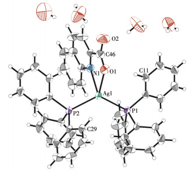
Ag1—P1 0.244 86(14) Ag1—P2 0.246 70(15) Ag1—O1 0.239 6(4) Ag1—N1 0.238 4(5) P1—Ag1—P2 126.90(5) O1—Ag1—P1 110.26(11) O1—Ag1—P2 114.86(12) N1—Ag1—P2 103.97(11) N1—Ag1—P1 117.18(11) N1—Ag1—O1 69.44(16) The complex presents a tetracoordinate mode. Each silver(Ⅰ) ion is coordinated by two phosphorus atoms from two TPP ligands and chelated by the oxygen and nitrogen atoms of the Qina ligand. The basic structure of the complex is exhibited in Fig. 1.
Figure 1
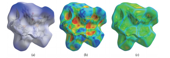
2.2 Hirshfeld surface analysis and structural details of the complex
From the dnorm analysis results in Fig. 2a, it can be inferred that the red dot region indicates the existence of some π interactions and hydrogen bond interaction regions between the complex molecules. Fig. 2b shows that the results of the shape index can be mutually confirmed with dnorm analysis. In the area where red dots appear in dnorm, the shape index will produce surface depressions at the corresponding positions, and it will still be red[49]. The shape index also shows that there are strong O…H and π interactions between molecules. Fig. 2c shows the smooth curvedness surface of molecules, which indicates that the intermolecular interactions tightly fit together.
Figure 2
 Figure 2. Hirshfeld surface of the complex: dnorm (a), shape index (b), and curvedness (c)
π‐stacking is widely present in biological macromolecular systems and often plays a key role in the stable stacking of DNA and RNA bases, the maintenance of polysaccharides and protein‐specific structures, and the recognition of protein‐ligand interactions[50‐51]. In addition, supramolecular interactions such as the π‐stacking network and hydrogen bond network between complex molecules often become potential action points for drug molecules to exert force on biological targets[52].
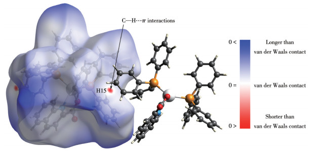
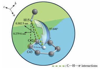
The dissection of dnorm (Fig. 3) shows that there is an edge ‐ to ‐ face type weak C—H… π interaction between the complex molecules along the crystal axis c. The H15 subordinate to C15 on the aromatic ring of the TPP ligand interacts with the aromatic ring Cg (C40‐C45) of another adjacent complex TPP ligand at a distance (H…Cg) of 0.321 3 nm. The closest distance between the H15 in the C—H… π interaction and the Csp2 atom of the aromatic ring is 0.279 6 nm, the secondary distance to the Csp2 atom of the aromatic ring is 0.302 5 nm, and the dihedral angle between the plane C42 ‐ H15 ‐ C43 and the plane C42 ‐ centroid ‐ C43 is 97.646°. The details of the geometric parameters of the weak interaction are shown in Fig. 4. This C—H… π interaction can meet the parameter range defined and required in published papers[53‐54].
Figure 3
Figure 4
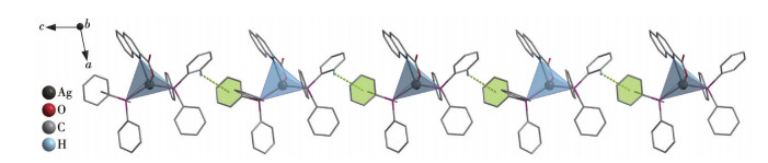
It is through the alternation of intermolecular C— H…π interactions that the 1D supramolecular configu‐ ration of the complex can be formed. The green dotted line in Fig. 5 shows this C —H…π interaction, and the 1D supramolecular chain structure of the complex is also shown in Fig. 5.
Figure 5
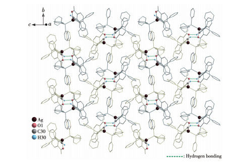
Based on the 1D chain of supramolecular structure, there is a pair of C—H…O type hydrogen bonds between the O1 of the carboxyl functional group of the Qina ligand and the CH group of the side chain. Each supramolecular chain can extend the 2D supramolecular surfaces on the bc plane through this effect. The bond parameters of the hydrogen bond pair are the same, the D…A distance is 0.261 3 nm, and ∠DHA= 139.078°, which conforms to the range of hydrogen bond parameters of this type[55]. The green dotted line in Fig. 6 shows the paired hydrogen bonding between the supramolecular chains and the 2D supramolecular surface structure of the complex.
Figure 6
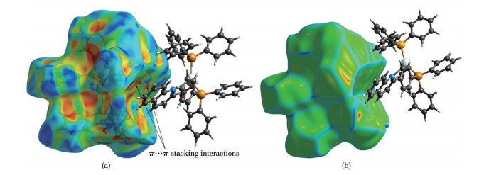
The shape index analysis in Fig. 7 shows that a large red concave surface appears above the quinoline fused ring of the Qina ligand, indicating that the complex molecules will have π … π stacking interactions here. The curvedness analysis also shows that the molecule surface above the quinoline fused ring is smooth and fitted to the adjacent molecule surface, which further illustrates that the π … π stacking interactions inferred from the results of the shape index analysis are established[56].
Figure 7
 Figure 7. π…π stacking interactions between two Qina molecules: shape index (a) and curvedness (b)
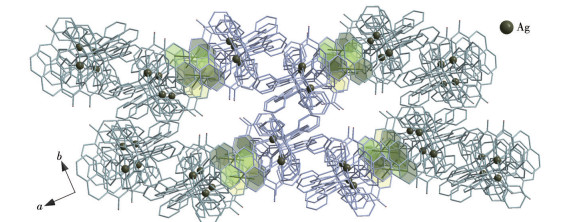
Along the crystal axis a, the 2D supramolecular planes depend on the equidistant double π …π interactions to maintain each other. These π‐action pairs occur between the fused rings of quinoline in the Qina ligandxs structure, and finally construct a complete crystal packing structure of the complex molecular system (Fig. 8). This can be confirmed by the conclusion of the Hirshfeld surface analysis. The intermolecular π ‐interaction surfaces match each other well, and the center‐to‐center distance is 0.377 6 nm, which is within the regular numerical range[57].
Figure 8
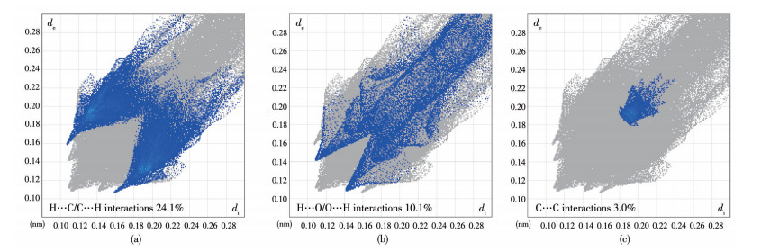
Two‐dimensional fingerprint plots can show in detail the quantitative relationship between the atomic charges between molecules[58]. As shown in Fig. 9, the interaction ratio of H…C/C…H is as high as 24.1%. The projected shape is a symmetrical sharp flying wing with a bright area in the center, which confirms the existence of C—H…π interactions between molecules. In addition, the H…O/O…H interactions account for 10.1%. The two wings of the projection map are symmetrical and very sharp, indicating that there is a strong O…H hydrogen bond between the molecules. The C…C interactions in the central area of the shadow map a regular and symmetrical pattern with a light spot in the center, indicating that there are π…π interactions between the molecules.
Figure 9
 Figure 9. Two‐dimensional fingerprint plots of the complex: (a) H…C/C…H interactions, (b) H…O/O…H interactions, (c) C…C interactions
The proportion of charge interaction between all atoms is illustrated in Table 3. The sum of the proportion of charge interaction between all atoms is 100%, indicating that the Hirshfeld surface analysis of the crystal is complete and strict. The analysis of the 2D fingerprint plots can be consistent with the analysis result of the crystal structure.
Table 3
Table 3. Hirshfeld surface calculations for the complex下载:
导出CSV
Interaction C…H H…O C…C H…H H…Ag C…N H…N Proportion / % 24.1 10.1 3.0 62.0 0.2 0.3 0.3 2.3 Fluorescence properties
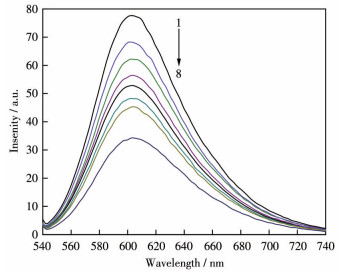
According to the fluorescence spectra of the complex‐EB‐DNA system (Fig. 10), in the EB‐DNA system, the sensitivity reached its maximum when the fluorescence wavelength was 602.5 nm. After the introduction of the complex, the fluorescence of the EB‐DNA system was quenched. With the gradual increase of the complex concentration, the fluorescence quenching became more obvious. The intensity was quenched from 77.63 to 34.23, with a difference of 43.40.
Figure 10
 Figure 10. Fluorescence spectra for the binding of the complex to EB‐DNA
1: cDNA=5.0 μmol·L-1, cEB=1.0 μmol·L-1; 2 ‐ 8: ccomplex=0.5, 1.0, 1.5, 2.0, 2.5, 3.0, 3.5 μmol·L-1+EB‐DNA
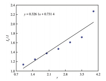
The Stern‐Volmer equation[59] I0/I=1+rKs can be used to quantitatively evaluate the competitive binding ability of the complex with the EB‐ DNA system, where I0 and I represent the fluorescence intensities with and without the complex, respectively; Ks refers to the quenching constants and r represents the ratio of the gradient concentration of the complex to that of DNA (ccomplex/cDNA). In addition, regarding I0/I and r scatter plots, the slope of the linear fitting can be obtained by the Ks value. Additionally, the linear fitting diagram is also reflected in Fig. 11. The Ks value of the complex was 0.326. In this case, the complex has displaced the EtBr molecules in the EtBr ‐DNA system to a certain extent. In addition, fluorescence studies show that the main mode of the complex interacting with DNA is intercalation[60].
Figure 11
2.4 Antibacterial activity
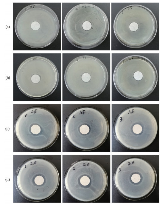
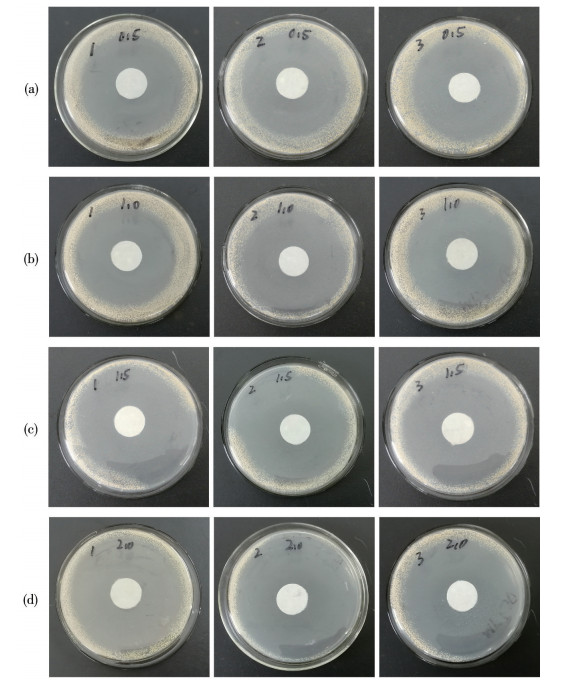
The complex showed certain inhibitory activity against S. aureus, and the area of the inhibitory ring increased with the increase of solution concentration. The comparative test shows that the antibacterial effect of the complex was not as good as that of penicillin sodium, as shown in Table 4. Fig. 12 and 13 show the bacteriostatic zones of the complex and penicillin sodium against S. aureus under different concentration gradients, respectively. In addition, the ligands and complex cannot inhibit P. salmonella and E. coli.
Table 4
Table 4. Diameter of the antibacterial ring of tested compounds下载:
导出CSV
c / (mmol·L-1) [Ag(Qina)(Tpp)2]·1.5H2 O Penicillin sodium Diameter / nm Average diameter / nm Diameter / nm Average diameter / nm 0.5 2.10 2.12 2.18 2.13 6.31 6.32 6.43 6.35 1 2.29 2.30 2.21 2.27 6.51 6.72 6.71 6.65 1.5 2.41 2.58 2.56 2.52 6.62 6.81 7.12 6.85 2 2.71 2.93 2.81 2.82 7.23 7.91 7.24 7.46 Figure 12
Figure 13
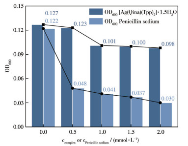
The inhibitory effects of the complex with different concentrations on bacterial growth can be compared by bacterial growth inhibition curves. According to the bacterial liquid system, the antibacterial action line of the test substance obtained by measuring the OD600 (the optical density at 600 nm) values is shown in Fig. 14. When the concentration of the complex reached 1.0 mmol·L-1, the inhibitory effect on S. aureus was obvious at first, and then gradually leveled off with the increase of the concentration of the complex. Consistent with the results of the bacteriostatic ring experiment, the complex showed a mild inhibitory effect on S. aureus, but its activity was inferior to penicillin sodium.
Figure 14
2.5 Cytotoxicity in vitro study
The complex was examined regarding the in vitro cytotoxic activity using an MTT assay. According to Table 5, it can be found that IC50 values were evaluated for two human cancer cells, and the lines of HepG2 and SKOV ‐ 3 are listed. The results clearly show that the complex exhibited a high level of anti‐tumor activity as well as a level of toxicity. In particular, the cytotoxicity of the complex was much better than that of cisplatin and carboplatin.
Table 5
Table 5. Cytotoxicity of tested compounds against tumor cells after 72 h incubation下载:
导出CSV
Compound IC50 / (μmol·L-1) HepG2 SKOV‐3 Qina > 100 > 100 Tpp > 100 > 100 [Ag(Qina)(Tpp)2]·1.5H2O 1.365 1.503 Cisplatin 15.297 15.797 Carboplatin 6.949 7.919 2.6 Molecular docking studies
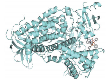
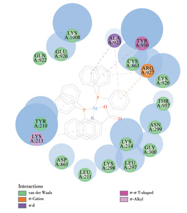
To further investigate the biological activity of the complex, we simulated the possible binding modes of the complex with PI3K kinase. Fig. 15 exhibits the possible combination of thermodynamically stable complexes with PI3K, where the complex to PI3K with computed binding affinity was -40.184 kJ·mol-1, respectively. The interaction model between the complex with PI3K can be displayed in 2D mapping pictures (Fig. 16). The aromatic ring groups of the auxiliary ligand can contribute to the multiple π‐binding sites for the complex and the enzyme.
Figure 15
Figure 16
3. Conclusions
A complex [Ag(Qina) (Tpp)2]·1.5H2O was synthesized and characterized mainly by X‐ray single‐crystal diffraction. The properties of the complex in DNA binding were tested via fluorescence spectra, and the results indicated that the complex behaves with fairly strong binding affinities in DNA interaction. In addition, the complex can be predicted to have potential enzymatic activity by molecular docking. Through in vitro cytotoxicity experiment, it was testified that the complex possessed a varied level of toxicity to inhibit HepG2, SKOV‐3, and cell lines. In particular, the complex expressed higher cytotoxicity than cisplatin and carboplatin. Finally, the complex also showed certain antibacterial activity. It is suggested that the silver complex could be further exploited as a potential anticancer pharmaceutical product.
Supporting information is available at http://www.wjhxxb.cn
Acknowledgments: This work is financially supported by the Youth Talent Growth Project of the Department of Education of Guizhou Province (Grant No.KY[2020]123) and Guizhou Key Laboratory of Coal Clean Utilization (Qiankehe Platform Talents [2020]2001). -
-
[1]
Medici S, Peana M, Crisponi G, Nurchi V M, Lachowicz J I, Remelli M, Zoroddu M A. Silver Coordination Compounds: A New Horizon in Medicine[J]. Coord. Chem. Rev., 2016, 327-328: 349-359. doi: 10.1016/j.ccr.2016.05.015
-
[2]
Fox C L Jr. Silver Sulfadiazine—A New Topical Therapy for Pseudomonas in Burns: Therapy of Pseudomonas Infection in Burns[J]. Arch. Surg., 1968, 96(2): 184-188. doi: 10.1001/archsurg.1968.01330200022004
-
[3]
Kuchar J, Rust J, Lehmann C W, Mohr F. Silver(Ⅰ) Complexes with Camphorsulfonato and Phosphine Ligands: Structural Diversity and Antibacterial Activity[J]. Inorg. Chem., 2020, 59(15): 10557-10568. doi: 10.1021/acs.inorgchem.0c00982
-
[4]
Kascatan‐Nebioglu A, Panzner M J, Tessier C A, Cannon C L, Youngs W J. N‐Heterocyclic Carbene‐Silver Complexes: A New Class of Antibiotics[J]. Coord. Chem. Rev., 2007, 251(5): 884-895.
-
[5]
Wakshlak R B K, Pedahzur R, Avnir D. Antibacterial Activity of Silver ‐ Killed Bacteria: The"Zombies"Effect[J]. Sci. Rep., 2015, 5(1): 9555. doi: 10.1038/srep09555
-
[6]
Mariam J, Sivakami S, Kothari D C, Dongre P M. Bioactivity of Albumins Bound to Silver Nanoparticles[J]. Protein J., 2014, 33(3): 258-266. doi: 10.1007/s10930-014-9553-2
-
[7]
Zhang S K, Du C, Wang Z Z, Han X G, Zhang K, Liu L H. Reduced Cytotoxicity of Silver Ions to Mammalian Cells at High Concentration Due to the Formation of Silver Chloride[J]. Toxicol. In Vitro, 2013, 27(2): 739-744. doi: 10.1016/j.tiv.2012.12.003
-
[8]
Mihorianu M, Franz M H, Jones P G, Freytag M, Kelter G, Fiebig H H, Tamm M, Neda I. N‐Heterocyclic Carbenes Derived from Imidazo‐[1, 5‐a]pyridines Related to Natural Products: Synthesis, Structure and Potential Biological Activity of Some Corresponding Gold and Silver Complexes[J]. Appl. Organomet. Chem., 2016, 30(7): 581-589.
-
[9]
Kavitha R, Nirmala S, Nithyabalaji R, Sribalan R. Biological Evaluation, Molecular Docking and DFT Studies of Charge Transfer Complexes of Quinaldic Acid with Heterocyclic Carboxylic Acid[J]. J. Mol. Struct., 2020, 1204: 127508. doi: 10.1016/j.molstruc.2019.127508
-
[10]
Högberg A G S, Madan K, Moberg C, Sjöberg B, Weber M, Muhammed M. Selective Reagents for Solvent Extraction of Metals—Ⅰ. Quinaldic Acids[J]. Polyhedron, 1985, 4(6): 971-977. doi: 10.1016/S0277-5387(00)84066-8
-
[11]
Shaw M J, Hill S J, Jones P. Chelation Ion Chromatography of Metal Ions Using High Performance Substrates Dynamically Modified with Heterocyclic Carboxylic Acids[J]. Anal. Chim. Acta, 1999, 401(1/2): 65-71.
-
[12]
Li M H, Dong X H, Zhang N, Jérôme F, Gu Y L. Eco‐Efficient Synthesis of 2‐Quinaldic Acids from Furfural[J]. Green Chem., 2019, 21(17): 4650-4655. doi: 10.1039/C9GC02206F
-
[13]
Song F H. Quinaldic Acid as a New Matrix for Matrix‐Assisted Laser Desorption/Ionization of Nucleic Acids. Rapid Commun[J]. Mass Spectrom., 2003, 17(15): 1802-1807.
-
[14]
Badawy A B B. Kynurenine Pathway of Tryptophan Metabolism: Regulatory and Functional Aspects[J]. Int. J. Tryptophan Res., 2017, 10: 1178646917691938.
-
[15]
Foster A C, Vezzani A, French E D, Schwarcz R. Kynurenic Acid Blocks Neurotoxicity and Seizures Induced in Rats by the Related Brain Metabolite Quinolinic Acid[J]. Neurosci. Lett., 1984, 48(3): 273-278. doi: 10.1016/0304-3940(84)90050-8
-
[16]
Williams M, Zhang Z, Nance E, Drewes J L, Lesniak W G, Singh S, Chugani D C, Rangaramanujam K, Graham D R, Kannan S. Maternal Inflammation Results in Altered Tryptophan Metabolism in Rabbit Placenta and Fetal Brain[J]. Dev. Neurosci., 2017, 39(5): 399-412. doi: 10.1159/000471509
-
[17]
Fila M, Chojnacki J, Pawlowska E, Szczepanska J, Chojnacki C, Blasiak J. Kynurenine Pathway of Tryptophan Metabolism in Migraine and Functional Gastrointestinal Disorders[J]. Int. J. Mol. Sci., 2021, 22(18): 10134. doi: 10.3390/ijms221810134
-
[18]
Fu Q Y, Tan Z, Shi L G, Xun W J. Resveratrol Attenuates Diquat‐Induced Oxidative Stress by Regulating Gut Microbiota and Metabolome Characteristics in Piglets[J]. Front. Microbiol., 2021, 12: 695155. doi: 10.3389/fmicb.2021.695155
-
[19]
Lee C H, Lee H S. Growth Inhibiting Activity of Quinaldic Acid Isolated from Ephedra Pachyclada against Intestinal Bacteria[J]. J. Korean Soc. Appl. Biol. Chem., 2009, 52(4): 331-335. doi: 10.3839/jksabc.2009.059
-
[20]
Sun X M, Savidge T, Feng H P. The Enterotoxicity of Clostridium Difficile Toxins[J]. Toxins, 2010, 2(7): 1848-1880. doi: 10.3390/toxins2071848
-
[21]
Silva R O S, Dorella F A, Figueiredo H C P, Costa É A, Pelicia V, Ribeiro B L D, Ribeiro M G, Paes A C, Megid J, Lobato F C F. Clostridium Perfringens and C. Difficile in Parvovirus‐Positive Dogs[J]. Anaerobe, 2017, 48: 66-69. doi: 10.1016/j.anaerobe.2017.07.001
-
[22]
Frederik S S, Mads F B, Endre S, Anders M B. Prevalence of Salmonella Species, Clostridium Perfringens, and Clostridium Difficile in the Feces of Healthy Elephants (Loxodonta Species and Elephas Maximus) In Europe[J]. J. Zoo Wildl. Med., 2021, 51(4): 752-760.
-
[23]
Langner E, Jeleniewicz W, Turski W A, Plech T. Quinaldic Acid Induces Changes in the Expression of P53 Tumor Suppressor Both on Protein and Gene Level in Colon Cancer LS180 Cells[J]. Pharmacol. Rep., 2019, 71(2): 189-193. doi: 10.1016/j.pharep.2018.10.016
-
[24]
Kelly W L, Pan L, Li C X. Thiostrepton Biosynthesis: Prototype for a New Family of Bacteriocins[J]. J. Am. Chem. Soc., 2009, 131(12): 4327-4334. doi: 10.1021/ja807890a
-
[25]
Nicolaou K C, Safina B S, Funke C, Zak M, Zécri F J. Stereocontrolled Synthesis of the Quinaldic Acid Macrocyclic System of Thiostrepton[J]. Angew. Chem. Int. Ed., 2002, 41(11): 1937-1940. doi: 10.1002/1521-3773(20020603)41:11<1937::AID-ANIE1937>3.0.CO;2-Y
-
[26]
Duan P P, Zheng Q F, Lin Z, Wang S F, Chen D D, Liu W. Molecular Engineering of Thiostrepton via Single"Base"‐Based Mutagenesis to Generate Side Ring‐Derived Variants[J]. Org. Chem. Front., 2016, 3(10): 1254-1258. doi: 10.1039/C6QO00320F
-
[27]
Zheng Q F, Wang S F, Duan P P, Liao R J, Chen D D, Liu W. An α/β‐Hydrolase Fold Protein in the Biosynthesis of Thiostrepton Exhibits a Dual Activity for Endopeptidyl Hydrolysis and Epoxide Ring Opening/Macrocyclization[J]. Proc. Natl. Acad. Sci. U. S. A., 2016, 113(50): 14318-14323. doi: 10.1073/pnas.1612607113
-
[28]
El‐bendary M M, Etaiw S E H. Structure and Applications of Organotin Complex Based on Trimethyltin Cation and Quinaldic Acid[J]. Appl. Organomet. Chem., 2018, 32(3): e4152. doi: 10.1002/aoc.4152
-
[29]
Silva E N, Silva P A B, Graminha A E, Oliveira P F, Damasceno J L, Tavares D C, Batista A A, Von Poelhsitz G. Synthesis, Characterization, Cytotoxic Activity, and Interactions with CT ‐DNA and BSA of Cationic Ruthenium(Ⅱ) Complexes Containing Dppm and Quinoline Carboxylates[J]. Bioinorg. Chem. Appl., 2017, 2017: 2562780.
-
[30]
Im Y H, Kang E, Kim I H, Kim D E, Shin H K, Lee B J. Synthesis and OLED Properties of Zinc Complexes Based on Quinaldic Acid[J]. Mol. Cryst. Liq. Cryst., 2014, 599(1): 105-111. doi: 10.1080/15421406.2014.935971
-
[31]
Tanaka S, Suzuki Y, Saburi H, Kitamura M. Soft Ruthenium and Hard Brønsted Acid Combined Catalyst for Efficient Cleavage of Allyloxy Bonds[J]. Application to Protecting Group Chemistry. Tetrahedron, 2015, 71(37): 6559-6568.
-
[32]
Shankar K, Kirillov A M, Baruah J B. Bottom Up Synthesis for Homo‐and Heterometallic 2, 3‐Pyridinedicarboxylate Coordination Compounds[J]. Polyhedron, 2015, 102: 521-529. doi: 10.1016/j.poly.2015.10.031
-
[33]
İlkimen H, Yenikaya C, Gülbandılar A, Sarı M. Synthesis and Characterization of a Novel Proton Salt of 2‐Amino‐6‐nitrobenzothiazole with 2, 6‐Pyridinedicarboxylic Acid and Its Metal Complexes and Their Antimicrobial and Antifungal Activity Studies[J]. J. Mol. Struct., 2016, 1120: 25-33. doi: 10.1016/j.molstruc.2016.04.068
-
[34]
Banti C N, Papatriantafyllopoulou C, Manoli M, Tasiopoulos A J, Hadjikakou S K. Nimesulide Silver Metallodrugs, Containing the Mitochondriotropic, Triaryl Derivatives of Pnictogen; Anticancer Activity against Human Breast Cancer Cells[J]. Inorg. Chem., 2016, 55(17): 8681-8696. doi: 10.1021/acs.inorgchem.6b01241
-
[35]
Trost B M, Machacek M R, Tsui H C. Development of Aliphatic Alcohols as Nucleophiles for Palladium‐Catalyzed DYKAT Reactions: Total Synthesis of (+)‐Hippospongic Acid A[J]. J. Am. Chem. Soc., 2005, 127(19): 7014-7024. doi: 10.1021/ja050340q
-
[36]
Banti C N, Hatzidimitriou A G, Kourkoumelis N, Hadjikakou S K. Diclofenac Conjugates with Biocides through Silver(Ⅰ) Ions (CoMeD′s); Development of a Reliable Model for the Prediction of Anti‐proliferation of NSAID′s‐Silver Formulations[J]. J. Inorg. Biochem., 2019, 194: 7-18. doi: 10.1016/j.jinorgbio.2019.01.020
-
[37]
Richards V N, Rath N P, Buhro W E. Pathway from a Molecular Precursor to Silver Nanoparticles: The Prominent Role of Aggregative Growth[J]. Chem. Mater., 2010, 22(11): 3556-3567. doi: 10.1021/cm100871g
-
[38]
李中峰, 张振伟, 崔洋哲, 刘敏, 杨玉平, 金琼花. 两种基于硫醇配体的银(Ⅰ)配合物的合成、表征和晶体结构[J]. 无机化学学报, 2016,32,(1): 139-144. LI Z F, ZHANG Z W, CUI Y Z, LIU M, YANG Y P, JIN Q H. Syntheses, Characterizations and Crystal Structures of Two Kinds of Silver(Ⅰ) Complexes Derived from Thiol Ligand[J]. Chinese J. Inorg. Chem., 2016, 32(1): 139-144.
-
[39]
Umadevi M, Muthuraj V, Vanajothi R. Structural, Cytotoxicity and Molecular Docking Studies of Some Quinoline Schiff Bases and Their Pd(Ⅱ), Mn(Ⅱ) and Ru(Ⅱ) Complexes[J]. J. Mol. Struct., 2020, 1221: 128778. doi: 10.1016/j.molstruc.2020.128778
-
[40]
Xie P, Williams D S, Atilla‐Gokcumen G E, Milk L, Xiao M, Smalley K S M, Herlyn M, Meggers E, Marmorstein R. Structure‐Based Design of an Organoruthenium Phosphatidyl‐Inositol‐3‐Kinase Inhibitor Reveals a Switch Governing Lipid Kinase Potency and Selectivity[J]. ACS Chem. Biol., 2008, 3(5): 305-316. doi: 10.1021/cb800039y
-
[41]
Liu M, Kuo F S, Capistrano K J, Kang D, Nixon B G, Shi W, Chou C, Do M H, Stamatiades E G, Gao S Y, Li S, Chen Y B, Hsieh J J, Hakimi A A, Taniuchi I, Chan T A, Li M O. TGF ‐ β Suppresses Type 2 Immunity to Cancer[J]. Nature, 2020, 587(7832): 115-120. doi: 10.1038/s41586-020-2836-1
-
[42]
Li S, Liu M, Do M H, Chou C, Stamatiades E G, Nixon B G, Shi W, Zhang X, Li P, Gao S Y, Capistrano K J, Xu H, Cheung N K V, Li M O. Cancer Immunotherapy via Targeted TGF ‐β Signalling Blockade in TH Cells[J]. Nature, 2020, 587(7832): 121-125. doi: 10.1038/s41586-020-2850-3
-
[43]
Wang R S, Feng J, Lei Y Z, Chen D M, Lian M L. New 3D Supramolecular Ag(Ⅰ) Coordination Polymer Crystal Containing 2‐Quinaldic Acid Radical Biological Ligand: Crystal Structure and Anticancer Activity[J]. Cryst. Res. Technol., 2018, 53(8): 1800065. doi: 10.1002/crat.201800065
-
[44]
Dolomanov O V, Bourhis L J, Gildea R J, Howard J A K, Puschmann H. OLEX2: A Complete Structure Solution, Refinement and Analysis Program[J]. J. Appl. Crystallogr., 2009, 42(2): 339-341. doi: 10.1107/S0021889808042726
-
[45]
Sheldrick G M. Crystal Structure Refinement with SHELXL[J]. Acta Crystallogr. Sect. C, 2015, C71: 3-8.
-
[46]
Sheldrick G M. SHELXT—Integrated Space‐Group and Crystal‐Structure Determination[J]. Acta Crystallogr. Sect. A, 2015, A71: 3-8.
-
[47]
Spackman M A, Jayatilaka D. Hirshfeld Surface Analysis[J]. CrystEngComm, 2009, 11(1): 19-32. doi: 10.1039/B818330A
-
[48]
Dallakyan S, Olson A J. Small‐Molecule Library Screening by Docking with PyRx//Hempel J E, Williams C H, Hong C C. Chemical Biology. Methods in Molecular Biology: Vol. 1263. New York: Humana Press, 2014: 243‐250
-
[49]
王彦斌, 于盟, 张雨, 苏琼, 董文魁. 基于非对称salamo型N2O4配体的四核镍(Ⅱ)和锌(Ⅱ)配合物的合成、晶体结构、Hirshfeld表面分析与荧光性质[J]. 无机化学学报, 2020,36,(10): 1967-1976. doi: 10.11862/CJIC.2020.215WANG Y B, YU M, ZHANG Y, SU Q, DONG W K. Syntheses, Crystal Structures, Hirshfeld Surfaces Analyses and Fluorescence Properties of Two Tetranuclear Nickel(Ⅱ) and Zinc(Ⅱ) Complexes Based on an Unsymmetrical Salamo‐like N2O4‐Donor Ligand[J]. Chinese J. Inorg. Chem., 2020, 36(10): 1967-1976. doi: 10.11862/CJIC.2020.215
-
[50]
Ma Y, Zhu Y J, Wang C, Pan D L, Liu S, Yang M Y, Zhang X P, Yang X T, Zhao W T, Zhou X Y, Li Y D, Pan Y F, Sun J, Wang S H, Guan Z, Zhang L H, Yang Z J. Annealing Novel Nucleobase ‐Lipids with Oligonucleotides or Plasmid DNA Based on H‐Bonding or π‐π Interaction: Assemblies and Transfections[J]. Biomaterials, 2018, 178: 147-157. doi: 10.1016/j.biomaterials.2018.06.012
-
[51]
Zhuang W R, Wang Y, Cui P F, Xing L, Lee J, Kim D, Jiang H L, Oh Y K. Applications of π‐π Stacking Interactions in the Design of Drug‐Delivery Systems[J]. J. Controlled Release, 2019, 294: 311-326. doi: 10.1016/j.jconrel.2018.12.014
-
[52]
Bhattacharyya M K, Dutta D, Nashre‐ul‐Islam S M, Frontera A, Sharma P, Verma A K, Das A. Energetically Significant Antiparallel π‐Stacking Contacts in Co(Ⅱ), Ni(Ⅱ) and Cu(Ⅱ) Coordination Compounds of Pyridine‐2, 6‐Dicarboxylates: Antiproliferative Evaluation and Theoretical Studies[J]. Inorg. Chim. Acta, 2020, 501: 119233. doi: 10.1016/j.ica.2019.119233
-
[53]
Suezawa H, Yoshida T, Umezawa Y, Tsuboyama S, Nishio M. CH/π Interactions Implicated in the Crystal Structure of Transition Metal Compounds—A Database Study[J]. Eur. J. Inorg. Chem., 2002, (12): 3148-3155.
-
[54]
Li S, Li G L, Wang W, Liu Y, Cao Z M, Cao X L, Huang Y G. A 2D Metal‐Organic Framework Interpenetrated by a 2D Supramolecular Framework Assembled by CH/π Interactions[J]. Inorg. Chem. Commun., 2021, 130: 108705. doi: 10.1016/j.inoche.2021.108705
-
[55]
Basu Baul T S, Chaurasiya A, Vasquez‐Ríos M G, Höpfl H. Zinc(Ⅱ) Complexes Constructed from an Adamantane‐Functionalized Pyridine Schiff Base—Influence of the Counterion on the Supramolecu‐ lar Organization by Means of C—H…O, C—H…N, C—H… π and π…π Interactions[J]. J. Mol. Struct., 2022, 1247: 131241. doi: 10.1016/j.molstruc.2021.131241
-
[56]
Muddassir M. A New 1D Cu(Ⅱ)‐W(Cn)8 Based Coordination Polymer: Crystallographic Structural Architecture, Hirshfeld Surface, DFT and Luminescent Analyses[J]. J. Organomet. Chem., 2020, 926: 121499. doi: 10.1016/j.jorganchem.2020.121499
-
[57]
Zahra R, Masoud M, Mohammad C, Joel T M. Accurate DFT Studies on Crystalline Network Formation of a New Co(Ⅱ) Complex Bearing 8‐Aminoquinoline[J]. Inorg. Chim. Acta, 2018, 473: 152-159. doi: 10.1016/j.ica.2017.12.033
-
[58]
Burkhanova T M, Babashkina M G, Taskin‐Tok T, Sharov A V, Safin D A. Naphthalene‐Based Bis‐N ‐Salicylidene Aniline Dyes: Crystal Structures, Hirshfeld Surface Analysis, Computational Study and Molecular Docking with the SARS ‐ CoV ‐ 2 Proteins[J]. J. Iran. Chem. Soc., 2022, 19(5): 1979-1991. doi: 10.1007/s13738-021-02438-y
-
[59]
Boaz H, Rollefson G K. The Quenching of Fluorescence[J]. Deviations from the Stern‐Volmer Law. J. Am. Chem. Soc., 1950, 72(8): 3435-3443.
-
[60]
Almaqwashi A A, Zhou W, Naufer M N, Riddell I A, Yilmaz Ö H, Lippard S J, Williams M C. DNA Intercalation Facilitates Efficient DNA‐Targeted Covalent Binding of Phenanthriplatin[J]. J. Am. Chem. Soc., 2019, 141(4): 1537-1545. doi: 10.1021/jacs.8b10252
-
[1]
-
Figure 2 Hirshfeld surface of the complex: dnorm (a), shape index (b), and curvedness (c)
Figure 7 π…π stacking interactions between two Qina molecules: shape index (a) and curvedness (b)
Figure 9 Two‐dimensional fingerprint plots of the complex: (a) H…C/C…H interactions, (b) H…O/O…H interactions, (c) C…C interactions
Figure 10 Fluorescence spectra for the binding of the complex to EB‐DNA
1: cDNA=5.0 μmol·L-1, cEB=1.0 μmol·L-1; 2 ‐ 8: ccomplex=0.5, 1.0, 1.5, 2.0, 2.5, 3.0, 3.5 μmol·L-1+EB‐DNA
Table 1. Crystal data and structure refinements for the complex
Parameter [Ag(Qina)(Tpp)2]·1.5H2O Parameter [Ag(Qina)(Tpp)2]·1.5H2O Empirical formula C46H39AgNO3.5P2 Z 8 Formula weight 831.59 Dc / (g·cm-3) 1.218 Crystal system Monoclinic μ / mm-1 0.571 Space group C2/c F(000) 3 416 a / nm 3.195 90(13) Independent reflection 10 070 b / nm 1.210 96(4) Goodness‐of‐fit on F2 1.011 c / nm 2.319 74(7) R [I≥2σ(I)] 0.066 3 β/(°) 102.166(4) wR [I≥2σ(I)] 0.161 5 Volume / nm3 8.776 0(5)  下载: 导出CSV
Table 2. Selected bond lengths (nm) and angles (°) for the complex
Ag1—P1 0.244 86(14) Ag1—P2 0.246 70(15) Ag1—O1 0.239 6(4) Ag1—N1 0.238 4(5) P1—Ag1—P2 126.90(5) O1—Ag1—P1 110.26(11) O1—Ag1—P2 114.86(12) N1—Ag1—P2 103.97(11) N1—Ag1—P1 117.18(11) N1—Ag1—O1 69.44(16)
下载: 导出CSV
Table 3. Hirshfeld surface calculations for the complex
Interaction C…H H…O C…C H…H H…Ag C…N H…N Proportion / % 24.1 10.1 3.0 62.0 0.2 0.3 0.3
下载: 导出CSV
Table 4. Diameter of the antibacterial ring of tested compounds
c / (mmol·L-1) [Ag(Qina)(Tpp)2]·1.5H2 O Penicillin sodium Diameter / nm Average diameter / nm Diameter / nm Average diameter / nm 0.5 2.10 2.12 2.18 2.13 6.31 6.32 6.43 6.35 1 2.29 2.30 2.21 2.27 6.51 6.72 6.71 6.65 1.5 2.41 2.58 2.56 2.52 6.62 6.81 7.12 6.85 2 2.71 2.93 2.81 2.82 7.23 7.91 7.24 7.46
下载: 导出CSV
Table 5. Cytotoxicity of tested compounds against tumor cells after 72 h incubation
Compound IC50 / (μmol·L-1) HepG2 SKOV‐3 Qina > 100 > 100 Tpp > 100 > 100 [Ag(Qina)(Tpp)2]·1.5H2O 1.365 1.503 Cisplatin 15.297 15.797 Carboplatin 6.949 7.919
下载: 导出CSV
-


 扫一扫看文章
计量
- PDF下载量: 6
- 文章访问数: 1491
- HTML全文浏览量: 244
