Figure 1.
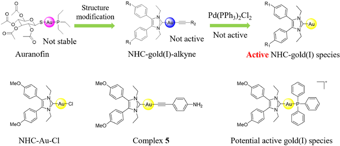
The design of NHC-gold(I)-alkyne complexes by bioorthogonal chemistry.
NHC-gold(I)-alkyne complexes induced hepatocellular carcinoma cell death through bioorthogonal activation by palladium complex in living system
Yunlong Lu , Yuanhao Liu , Zhenlin Liang , Xiaoyan Ma , Lijuan Liu , Zhenfan Wen , Iogann Tolbatov , Alessandro Marrone , Wukun Liu

Hepatocellular carcinoma (HCC) is a global health challenge and remains a serious problem worldwide [1]. It is estimated that above 1 million people will be affected by HCC annually by 2025 [2]. The occurrence and mortality of HCC are increasing, while successful therapeutic approaches remain elusive for most patients [3]. The most successful treatment for early-stage HCC is surgery such as transarterial chemoembolizationor (TACE) or liver transplantation. Even though the great progress has been made in HCC treatment, the late-stage HCC requires more reasonable therapies and improved prognosis [4,5]. Most of the HCC were developed on the basis of liver inflammation which was usually caused by hepatitis B virus (HBV) or hepatitis C virus (HCV) infections, and inflammation is the most direct reflection of immunity [6]. Recent studies showed that increased PD-1/PD-L1 expressions have been observed in HCC patients and immunotherapy have shown promise against HCC [7,8]. However, overcoming tumor-induced immune tolerance in HCC is still an extremely challenging task [9].
Bioorthogonal chemistry allows organic synthesis that can generally perform in living organisms and cells [10-12]. They are intended to covalently modify biomolecules with non-native functional groups under biological conditions [13]. Transition metal-mediated bioorthogonal activation is a powerful tool to selectively convert inactive prodrugs into their active forms [14]. However, the applications of bioorthogonal reactions, especially for transformations in living cells or animals, are less occurred and far from the limit. To the best of our knowledge, organometallic transmetallation is known to be an essential step in cross coupling reactions by transferring aryl, vinyl, alkynyl groups, etc., to metal catalysts (e.g., Pd) [15].
Thioredoxin reductase (TrxR) is a key reductase and plays a significant role in maintaining intracellular redox homeostasis in living system [16]. In recent years, TrxR has been considered as a promising cancer target based on clinical characteristics since TrxR is overexpressed in malignant cancer cells compared to noncancerous tissues. Recent study showed that TrxR inhibition can enhance cancer immunity through inducing immunogenic cell death (ICD), which is an advantage to achieve therapeutic purpose [17]. Gold complexes (e.g., auranofin) exhibited strong anticancer bioactivities toward a variety of cancers by inhibiting TrxR on the active site of Sec498 [18,19]. Usually, the organogold(I) complexes are less stable and can easily be metabolized by thiol-containing biomolecules, and the coordinated ligands are mostly lost before reaching the targets [20,21]. However, recently we found that the N-heterocyclic carbene (NHC)-gold(I)-alkyne complexes are more stable than we usually thought. This organogold(I) complexes display a reasonably high stability under physiological conditions and they can potentially undergo transmetallation reaction [22]. We therefore hypothesize that organic gold(I) alkyne complexes can be activated by other metallic catalysts in living system to exhibit anticancer bioactivities. Herein, we report the Pd(PPh3)2Cl2 catalyst, which is very lipophilic that can easily penetrate cell membrane, and can activate NHC-gold(I)-alkyne complexes to release active gold species to exhibit antiproliferative activities (Fig. 1).
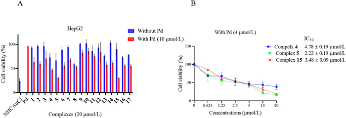
We prepared gold(I) alkyne complex analogues by employing the privileged scaffold 4, 5-diarylimidazole NHC that we have developed previously [23,24]. The preparation of final complexes underwent single step synthesis by employing substituted (1, 3-diethyl-4, 5-diphenyl-1H-imidazol-2-yl) gold(I) bromide and substituted alkynes to generate the final complexes (Scheme S1 in Supporting information). All of the complexes had been tested in HepG2 cells at 20 µmol/L in the presence and absence of Pd(PPh3)2Cl2 (10 µmol/L). Complexes 4 and 15 demonstrated weak antiproliferative activity with survival rates of 73.14% ± 6.56% and 90.03% ± 9.05% without Pd catalyst, while they exhibited significant antiproliferative activity in the presence of Pd catalyst, with the inhibition rates increased to 47.31% ± 2.51% and 30.09% ± 1.13% (Fig. 2). After initial screen of these complexes at 20 µmol/L on HepG2 cells, we focused on the best three complexes 4, 5 and 15 and tested IC50 values of them. The results showed that the IC50 values of complexes 4, 5 and 15 on HepG2 cells were 4.78 ± 0.19 µmol/L, 2.22 ± 0.19 µmol/L and 3.48 ± 0.09 µmol/L in presence of 4 µmol/L Pd catalyst. Complex 5 has the best antiproliferative activity against HepG2 cells. We therefore selected complex 5 for further study and analysis (Fig. 2).
Generally, gold(I) complexes can easily interact with thiol-containing proteins or biomolecules. Initially, the stability of complex 5 were tested in phosphate buffer solution (PBS) (pH 7.4). The results showed that there is no significant change of complex 5 in PBS solution after 24 h of incubation by UV-vis spectrophotometry. We then investigated the stability of complex 5 in PBS buffer containing GSH. After incubation of complex 5 with GSH, the UV-vis spectrophotometry did not show significant changes, which displayed that NHC-gold(I)-alkyne complex 5 was stable in PBS GSH solution (Fig. S1 in Supporting information).
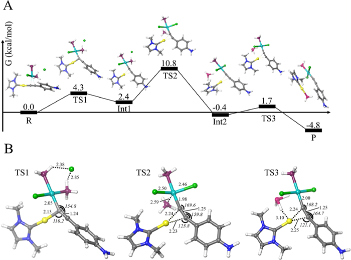
To explain the possible mechanism of transmetallation process, density functional theory (DFT) calculations were performed to elucidate mechanism of reaction. In order to decrease the computational burden, we have simplified Pd(PPh3)2Cl2 to Pd(PH3)2Cl2, the MeO-phenyls in the NHC-gold(I) complex were substituted by hydrogens, ethyls were substituted by methyls. Our calculations have shown that the chloride detachment from Pd(PH3)2Cl2 has the reaction Gibbs free energy of -38.7 kcal/mol, thus exhibiting strong exergonicity. Furthermore, such a strong exergodicity determines the almost complete dissociation of complex Pd(PH3)2Cl2, and thus the Pd(PH3)2Cl+ is the actual species reacting with the gold complex, whereas chloride ions are released in the bulk. Hence, the chloride ions are available to non-covalently interact with the reacting PdCl2(PH3)+-NHC-Au-(4-amino-phenyl-acetylene) adduct, thus forming a neutral reactant (Fig. 3). We then used mass spectrometry to determine the products of the reaction which confirmed the calculative results (Fig. S2 in Supporting information).
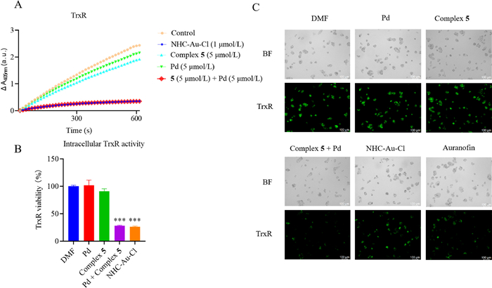
Since Pd-mediated transmetallation generates labile NHC-gold(I) species that is potentially active for TrxR bioactivity. We then test complex 5 for enzyme and cell based bioactivity. Complex 5 exhibited almost no activity against TrxR purified enzyme and it indicated that NHC-gold(I)-alkyne complexes are very stable. The gold(I) species cannot be exposed to selenol-containing TrxR. Similar phenomenon was observed in Pd catalyst group and Pd catalyst cannot inhibit TrxR at 5 µmol/L. However, it can significantly inhibit TrxR enzyme when Pd catalyst was added to the solution of complex 5, similar to positive control NHC-Au-Cl at 1 µmol/L (Figs. 4A and B). Next, the intracellular TrxR activity was also tested by the immunofluorescence. The TrxR green probe was freshly prepared, and incubated for 6 h to detect the intracellular TrxR activity of each different groups (Fig. 4C). The results showed that Pd catalyst can activate and transfer the gold(I) in gold complex 5 to its active forms to exhibit TrxR inhibition bioactivity.
Complex 5 with Pd catalyst can inhibit TrxR bioactivity and therefore disturb the intracellular redox states. As shown in Fig. S3 (Supporting information), the complex 5 with Pd catalyst treatment group generated strong fluorescent signal compared to control group, complex 5 group and Pd catalyst group. Indeed, the inhibition of TrxR activity by gold complexes has been demonstrated to trigger mitochondrial dysfunction and mitochondrial membrane potential (MMP) damage [25]. We then investigated the MMP levels by the JC-10 kit and observed an increase in green (monomer) fluorescence in the complex 5 group in the presence of Pd catalyst compared to other groups. These results indicated that bioorthogonal reaction of complex 5 can induce MMP damage in cells (Fig. S4 in Supporting information). The accumulation of ROS may trigger multiple cell death pathways which will lead to cell death. The flow cytometry analysis showed that complex 5 and Pd catalyst can induce HepG2 cells apoptosis. After 24 h treatment of complex 5 (2 µmol/L), Pd catalyst (4 µmol/L), complex 5 and Pd catalyst (2 µmol/L and 4 µmol/L), auranofin (2 µmol/L), NHC-Au-Cl (2 µmol/L). The apoptosis rate increased in complex 5 and Pd catalyst co-treatment group better than control and auranofin group (Fig. S5 in Supporting information). The cell cycle arrest experiment was also conducted and the results were documented in Fig. S6 (Supporting information). This study showed that complex 5 and Pd catalyst can arrest HepG2 cells in G0/G1 phase which is more significant than positive control group. This is strong evidence to show that complex 5 and Pd catalyst can effectively inhibit cancer cell growth.
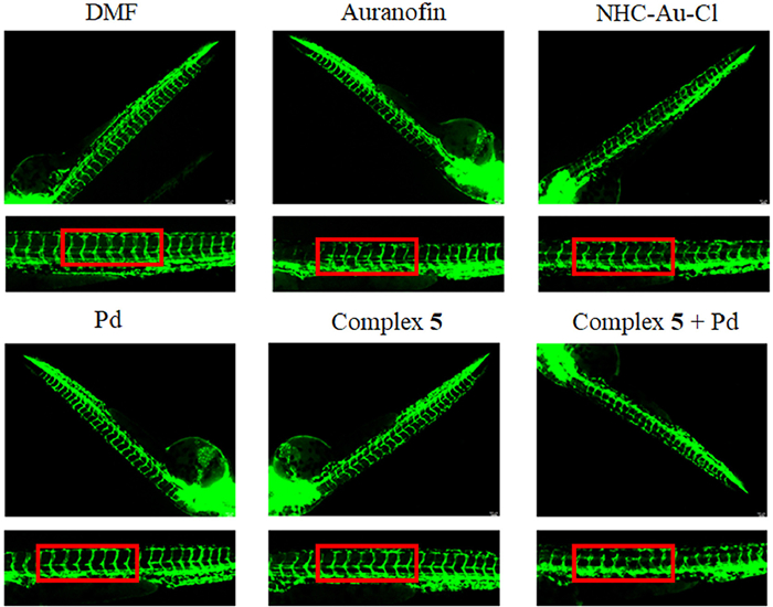
It has been reported that TrxR contributes to tumor growth and development not only by the inhibition of apoptotic events but also by the stimulation of angiogenesis. Therefore, inhibition of TrxR can suppress angiogenesis by NHC-gold(I)-alkyne complexes [26]. Tumor cells can produce high levels of angiogenic factors which contribute to the formation of abnormal vascular network. We then testified whether the bioorthogonal reaction of complex 5 and Pd catalyst can inhibit angiogenesis in zebrafish embryos. As shown in Fig. 5, after treatment of complex 5 and Pd catalyst along with other groups for 48 h, incomplete vascular morphology was observed in complex 5 and Pd catalyst group, which confirmed that the active gold species were released and could inhibit tumor angiogenesis. This is in vivo evidence to show that complex 5 can inhibit cancer cell migration and metastasis in the presence of Pd catalyst.
In summary, we have developed a novel bioorthogonal activation approach that can transform stable organogold(I) complexes into active gold(I) species in living systems in a controllable manner. This method is based on a combination of organic Pd catalyst Pd(PPh3)2Cl2 with an NHC-gold(I)-alkyne complexes to perform transmetallation reaction and gold(I) activation. The NHC-gold(I)-alkyne complex remained in an "off" state before being activated by Pd catalyst. In the in vitro environment, the in situ activation produced high level active gold(I) species that exhibited antiproliferative bioactivity by inhibiting TrxR, increasing ROS, MMP and inducing cell cycle arrest and apoptosis. The bioorthogonal activation of NHC-gold(I)-alkyne complex can also induce angiogenesis inhibition, which further enhance the anticancer effects. The above results showed that our approach represented a promising strategy for HCC treatment.
The authors declare that they have no known competing financial interests or personal relationships that could have appeared to influence the work reported in this paper.
We thank the financial supports of the National Natural Science Foundation of China (Nos. 82173684 and 82204181), Nanjing University of Chinese Medicine National Natural Science Foundation of China Counterpart Funding (No. XPT82204181), General Project of Basic Science in Colleges and Universities of Jiangsu Province (No. 21KJB350007), Jiangsu Provincial Health Commission (No. Z2021057), the Open Project of Chinese Materia Medica First Class Discipline of Nanjing University of Chinese Medicine (No. 2020YLXK010), the Priority Academic Program Development of Jiangsu Higher Education Institutions (Integration of Chinese and Western Medicine). We also thank for the experimental support from the Experiment Center for Science and Technology, Nanjing University of Chinese Medicine. The research is also financially supported by the "Innovative and Entrepreneurial Team" Program of Jiangsu Province (2020). And our team is leaded by Prof. Ming Zhao, Nanjing University of Chinese Medicine.
Supplementary material associated with this article can be found, in the online version, at doi:
J.M. Llovet, J. Zucman-Rossi, E. Pikarsky, et al., Nat. Rev. Dis. Primers 2 (2016) 16018.
J.M. Llovet, R.K. Kelley, A. Villanueva, et al., Nat. Rev. Dis. Primers 7 (2021) 6.
R.L. Siegel, K.D. Miller, A. Jemal, CA Cancer J. Clin. 70 (2020) 7-30. doi: 10.3322/caac.21590
R.M. Feng, Y.N. Zong, S.M. Cao, R.H. Xu, Cancer Commun. 39 (2019) 22. doi: 10.1186/s40880-019-0368-6
S. Mancarella, S. Krol, A. Crovace, et al., Cancers (Basel) 11 (2019) 1510. doi: 10.3390/cancers11101510
M. Bian, R. Fan, Z. Yang, et al., J. Med. Chem. 65 (2022) 1848-1866. doi: 10.1021/acs.jmedchem.1c01248
C. Kole, N. Charalampakis, S. Tsakatikas, et al., Cancers (Basel) 12 (2020) 2859. doi: 10.3390/cancers12102859
D. Liu, K.F. Staveley-O'Carroll, G. Li, J. Clin. Cell. Immunol. 6 (2015) 376.
E. Breous, R. Thimme, J. Hepatol. 54 (2011) 830-834.
S. Jia, S. Yang, H. Ji, et al., Chin. Chem. Lett. 31 (2020) 1104-1108.
G. Zou, K. Zhang, W. Yang, et al., Chin. Chem. Lett. 32 (2021) 3252-3256.
Z. Shao, C. Zhang, X. Zhu, et al., Chin. Chem. Lett. 30 (2019) 2169-2172.
R.E. Bird, S.A. Lemmel, X. Yu, Q.A. Zhou, Bioconjugate Chem. 32 (2021) 2457-2479. doi: 10.1021/acs.bioconjchem.1c00461
Y. Long, B. Cao, X. Xiong, et al., Angew. Chem. Int. Ed. 60 (2021) 4133-4141. doi: 10.1002/anie.202013366
J.J. Hirner, Y. Shi, S.A. Blum, Acc. Chem. Res. 44 (2011) 603-613. doi: 10.1021/ar200055y
M. Bian, R. Fan, S. Zhao, W. Liu, J. Med. Chem. 62 (2019) 7309-7321. doi: 10.1021/acs.jmedchem.8b01595
Z. Xu, J. Xu, S. Sun, et al., Redox Biol. 54 (2022) 102351.
M. Bian, R. Fan, G. Jiang, et al., J. Med. Chem. 63 (2020) 9197-9211. doi: 10.1021/acs.jmedchem.0c00257
Y. Lu, X. Ma, X. Chang, et al., Chem. Soc. Rev. 51 (2022) 5518-5556. doi: 10.1039/d1cs00933h
T. Zou, C.T. Lum, C.N. Lok, et al., Chem. Soc. Rev. 44 (2015) 8786-8801.
B. Bertrand, A. Casini, Dalton Trans. 43 (2014) 4209-4219.
A.S.K. Hashmi, C. Lothschütz, R. Döpp, et al., Angew. Chem. Int. Ed. 48 (2009) 8243-8246. doi: 10.1002/anie.200902942
W. Liu, K. Bensdorf, M. Proetto, et al., J. Med. Chem. 54 (2011) 8605-8615. doi: 10.1021/jm201156x
W. Liu, K. Bensdorf, M. Proetto, et al., J. Med. Chem. 55 (2012) 3713-3724. doi: 10.1021/jm3000196
M. Bian, Y. Sun, Y. Liu, et al., Chem. Eur. J. 26 (2020) 7092-7108. doi: 10.1002/chem.202000045
I. Ott, X. Qian, Y. Xu, et al., J. Med. Chem. 52 (2009) 763-770. doi: 10.1021/jm8012135

Figure 2 The antiproliferation bioactivity of NHC-gold(I)-alkyne complexes. (A) The antiproliferative bioactivity of complexes 1-17 at 20 µmol/L with or without Pd catalyst (Pd(PPh3)2Cl2). (B) The IC50 values of complexes 4, 5 and 15 on HepG2 cells.

Figure 3 (A) Reaction pathway. All values represent Gibbs free energies in kcal/mol. R, TS, Int, and P stand for reactants, transition state, intermediate, and products, respectively. Color scheme employed in this figure and in other figures below: Au (yellow), Pd (cyan), Cl (green), P (burgundy), N (blue), C (grey), H (white). (B) Transition states. All bonds in angstroms, all angles in degrees.

Figure 4 (A) The purified TrxR enzyme activity by NHC-Au-Cl (1 µmol/L), complex 5 (5 µmol/L), Pd catalyst (5 µmol/L), and complex 5 with Pd catalyst (5 µmol/L, respectively). (B) Cellular TrxR inhibition assay after incubation for 24 h with Pd catalyst (4 µmol/L), complex 5 (2 µmol/L), Pd catalyst (4 µmol/L) and complex 5 (2 µmol/L), NHC-Au-Cl (2 µmol/L). n = 3, ***P < 0.001. (C) Immunofluorescence images of TrxR probe assay to measure the binding affinity of TrxR with complex 5 in the presence of Pd catalyst, Pd catalyst (4 µmol/L), complex 5 (2 µmol/L), Pd catalyst (4 µmol/L) and complex 5 (2 µmol/L), NHC-Au-Cl (2 µmol/L), auranofin (2 µmol/L) (scale bar = 100 µm).

 扫一扫看文章

扫一扫关注我们
 DownLoad:

 下载:
下载:
