Citation:
Huihui Hu, Zhen Zhang, Yifen Fang, Lei Chen, Jun Wu. Therapeutic poly(amino acid)s as drug carriers for cancer therapy[J]. Chinese Chemical Letters,
2023, 34(6): 107953.
doi:
10.1016/j.cclet.2022.107953
Therapeutic poly(amino acid)s as drug carriers for cancer therapy
English
Therapeutic poly(amino acid)s as drug carriers for cancer therapy
junwuhkust@ust.hk (J. Wu). 1 These authors contributed equally to this work.
Received Date:
04 August 2022 Accepted Date:
23 October 2022 Revised Date:
20 October 2022 Available Online:
15 June 2023
Abstract:
As one of the top global health problems, the effective treatment of cancer is one of the most urgent clinical challenges. Currently, the main treatments for cancer include surgery, chemotherapy, radiotherapy, and gene therapy etc. Chemotherapy is one of the most commonly used treatments, however it has limitations such as highly toxic side effects and low drug utilization rate that limit its application. Gene therapy, as an emerging cancer treatment, has limitations such as drug instability, off-target effects and low internalization efficiency. Poly(amino acid)s carriers with good biocompatibility, degradability and multifunctionality as drug carriers have received much attention, as they can reduce the toxic side effects of chemotherapy, improve drug utilization, and enhance the internalization efficiency and utilization of gene drugs. However, little attention has been paid to the nature of the carriers themselves. This paper reviews the immunomodulatory, anti-inflammatory, antioxidant, internalization-promoting and apoptosis-promoting functions of poly(amino acid)s drug carriers in tumor therapy to provide a theoretical basis for different carrier-drug-adapted synergistic therapies.
With continuous economic development, industrial production has damaged the environmental homeostasis of human existence. Factors such as pollution in the living environment, aging population, poor lifestyles and the competitive pressure for social development have led to the incidence and mortality of cancer is increasing. Cancer has become one of the three major killers threatening human health and one of the top health problems worldwide [1]. In accordance with the latest global cancer data reported in 2020, 19.3 million new cancer cases and 9.9 million deaths were covered worldwide in 2020, while 4.57 million new cases and 3 million deaths were covered in China, ranking first in the world [2]. The increasing incidence of cancer, the trend of the cancer happening in young individuals and the difficulty of treatment not only impose heavy economic burdens on society but also bring huge mental and economic burdens on patients. Therefore cancer prevention and treatment research still has a long way to go. Currently, cancer treatment mainly includes radiotherapy, surgical therapy and pharmacotherapy, which includes traditional chemotherapy and emerging genetic drug therapies [3]. Surgical therapy is not applicable to metastatic tumors, advanced tumors, or tumors occurring in important and sensitive areas, and the surgical site may become infected and cause secondary damage. Radiotherapy is not applicable to metastatic tumors and may induce lesion formation [1,4,5]. Chemotherapeutic drugs can combat tumors through a variety of mechanisms, including interfering nucleic acid biosynthesis, affecting DNA structure and function, interfering transcriptional processes and preventing RNA synthesis, interfering protein synthesis and function, and affecting hormone homeostasis. Recently, emerging vascular blocking agents that cut off tumor nutrients and oxygen supply by blocking tumor neovascularization have emerged as a new option for chemotherapy. Compared to radiation and surgery, which are local treatments, chemotherapy is a systemic treatment. After administration, chemotherapeutic drugs are randomly distributed to most organs of the body through the circulatory system, and have a certain inhibitory effect on diffuse tumors and advanced tumors that have metastasized. However, due to the lack of specificity of small-molecule chemotherapeutic drugs, which have the same killing effect on tumor cells, normal cells and immune cells, chemotherapy often causes strong toxic side effects and affects the function of the immune system [1,5]. Gene therapy, a potentially novel therapeutic approach, allows the delivery of DNA or siRNA into patient-specific cells to promote or inhibit the expression of target proteins. However, free nucleic acids have low cellular uptake and are unstable in circulation owing to it can be degraded by nucleases, which limits the further application of gene therapy [6]. To reduce the toxic side effects of chemical drugs, increase the targeting and utilization of drug and enhance the internalization efficiency and stability of gene drugs, synthetic polymers and composites can be used as drug carriers to load anticancer drugs to promote effective cancer treatment. Poly(amino acid)s are widely used to load anticancer drugs owing to their unique advantages and multifunctionality.
Amino acids are one of the basic substances in the composition of the human body and the basis of the metabolism of life, and they contain at least one amino group and one carboxyl group [7]. There are 20 amino acids in the adult body, which can be divided into essential and non-essential amino acids. All amino acids are amino acids that are essential to the body, among which essential amino acid cannot be synthesized in the body or are synthesized at a rate that is far from adequate for the body's needs and must be supplemented from food, including methionine, valine, lysine etc., while non-essential amino acids can be obtained through organism synthesis or conversion from other amino acids, including glycine, alanine, and proline etc. Amino acids are essential components of proteins. Meanwhile certain amino acids like leucine play essential roles in processes such as cell signaling and gene expression [8], and certain amino acids like methionine have antioxidant capacity [9]. Amino acid also maintains the nitrogen balance in the body and produces carbon monoxide in the process of decomposition, all of which are irreplaceable as the basic material of life metabolism by other substances [7]. In addition, certain amino acids are drugs in their own right and have therapeutic effects on some diseases, such as tryptophan, tyrosine [10], L-arginine [11-13], leucine and isoleucine [14,15]. They also produce vital secondary metabolic molecules that can maintain normal physiological conditions [10]. At present, amino acids have been used in various fields such as food, medicine, agriculture, and feed, creating high value and improving the quality of life with good prospects for development. At the same time, because amino acids are highly biosafe, have a variety of functional groups, and some amino acids are positively charged in the physiological environment, they are used as drug carriers.
Poly(amino acid)s are a class of polymeric materials formed from natural amino acids and their derivatives through main chain peptide bonds [16,17]. Peptides are low molecular polymers formed by the dehydration and condensation of amino acids [18]. Both poly(amino acid)s and peptides are composed of various amino acid monomers linked by peptide bonds, and both can form regular secondary structures such as α-helicES or β-foldS [17,19]. However, poly(amino acid)s mostly refer to homogeneous peptides (which may also contain two or more homogeneous peptide chains), while peptides are designed short chains of less than or equal to forty amino acids in length [17,20]. Peptides of different lengths and amino acid sequences have different functions that can regulate a variety of human physiological functions at the cellular level, such as cell membrane and nuclear membrane transport [21,22], endosome escape [23], targeting of tumor cells [24], pro-apoptosis [25]. Different lengths and types of poly(amino acid)s have different properties, such as water solubility, chargeability, and therapeutic properties, so they may have different functions, which we summarize and review in this paper. They also have the advantages of good biocompatibility, biodegradability, stability, and ease of self-assembly, showing great potential for applications in the field of targeted drug delivery [18]. While both poly(amino acid)s and peptides can be used for drug transport, poly(amino acid)s offer better designability and self-assembly capabilities. And compared with other delivery carriers, poly(amino acid) carriers have many advantages, such as easy surface modification, good biocompatibility and biodegradability [16,17,26], and some poly(amino acid) carriers also have certain therapeutic properties.
Wu et al. previously proposed therapeutic slow and controlled-release materials, including amino acid biopolymers, which contain hydrophobic poly(amino acid)s [27,28], pharmaceutical amino acids [29], exosome carriers [30], liposome systems [31,32], and therapeutic polyhydroxy acid biomaterials, which contain erulic acid [33], ursolic acid [34] and salicylic acid [35]. These materials with therapeutic functions can achieve the slow and controlled release of anti-cancer drugs, and the carriers themselves can also enhance the efficacy of cancer treatment, which is important for the effective treatment of cancer. In this paper, we review therapeutic poly(amino acid)s carriers with many functions, such as immunomodulation, anti-inflammatory, antioxidant, internalization-promoting, and cancer cell apoptosis-promoting, so that we can take advantage of them to improve immunotherapy efficacy and promote internalization of drugs for effective cancer treatment.
2.
Synthesis of poly(amino acid)s
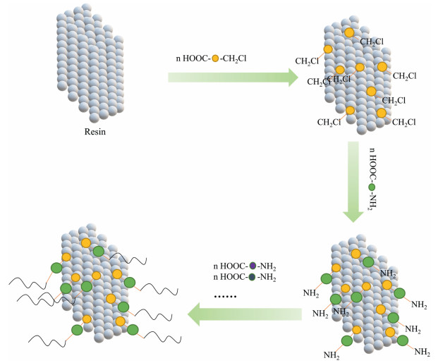
Poly(amino acid)s can be synthesized from amino acids using conventional solid-phase synthesis (Fig. 1). Solid-phase synthesis includes two methods, Fmoc and tBoc, Since Fmoc has more advantages than tBoc, the Fmoc method is used in most cases. However, conventional solid-phase synthesis can only be used to synthesize short peptides and oligomers. For poly(amino acid)s with polymerization degree greater than 100, a large number of unreacted monomers and by-products appear, which affect the yield and purity of the products. For this reason, scientists have developed various methods for the preparation of amino acids after continuous experiments, among which the ring-opening technique (ROP) of α-amino acids-N-carboxylic anhydrides (NCAs) has been widely used for its low cost, high degree of polymerization, and high yield and has been improved to synthesis poly(amino acid)s with controllable molecular weights, low PDIs, and sophisticated polymeric architectures due to the development of catalysts, optimization of reaction conditions, exploration of the initiating systems [36-38].
Figure 1
Figure 1.
Solid phase synthesis. Firstly, the chloromethyl is introduced on the solid phase carrier. It is subsequently reacted with the amino acid protected by the amino group, and the first amino acid is solidly loaded onto the resin. Afterwards, multiple amino acids are introduced in the same manner to form poly(amino acid)s.
Poly(amino acid)s have various advantages: (1) They have many reactive groups (e.g., carboxyl, sulfhydryl, phenyl and amino groups) as Fig. S1 (Supporting information) shows, which can be used to prepare bioactive materials with various functions and control the loading and release of the loaded substances for targeted drug delivery when used as carriers. Some poly(amino acid) carriers can also act directly as biomedical drugs [39], such as cationic amino acids polylysine can carry electronegative DNA or RNA segments as gene vectors [39], the anionic carboxyl groups from poly(glutamic acid)s and poly(aspartic acid)s can induce biomineralization [40]. (2) The hydrophilicity of poly(amino acid)s can be controlled by adjusting the type, ratio, sequence and content of the constituent amino acid monomers to facilitate the preparation of systems containing drugs with different properties by different methods [17]. (3) Functionalized amino acids can be introduced to make poly(amino acid)s biologically active, such as histidine containing imidazole ring to induce nanocarriers to escape from endosomes [41], and arginine containing guanidine to enable nanocarriers to enter cells directly through cell membranes [42]. (4) The unique secondary structure (α-helix, β-folding) helps to drive the assembly of its main chain into specific structures, bringing some specific biofunctionability [43,44]. (5) Poly(amino acid)s have good biocompatibility, degradability, and are easy to be functionalized. All of these has led to a high priority for poly(amino acid)s in the field of drug delivery [17].
4.
Poly(amino acid)s carriers with therapeutic function
4.1
Poly(amino acid)s carriers with immunomodulatory function
A tumor is not only a collection of tumor cells, but also includes tumor cells, stromal cells, immune cells (lymphocytes, dendritic cells, monocytes/macrophages, granulocytes, mast cells, etc.) and various cytokines [45]. Stromal cells, immune cells and various cytokines constitute the tumor microenvironment and influence tumor growth and the therapeutic effects of drugs [46]. Various cytokines in the tumor microenvironment, such as interleukin-10 (IL-10), indoleamine-2,3-dioxygenase (IDO) [47], transforming factor-β (TGF-β) and chemokines, are immunosuppressive factors that inhibit the activation, migration and differentiation of immune cells [48]. Tumor cells produce large amounts of lactic acid during metabolism, and high concentrations of lactic acid affect macrophage polarization and immune function in tumor tissues. On the one hand, large amounts of lactic acid increase the expression level of the M2-type macrophage marker molecule type I arginase, reduce the M1-type macrophage marker molecule nitric oxide synthase and decrease M1-type cytokine secretion. High concentrations of lactate reduce the expression of MHC I and MHC II in tumor-associated macrophages, weakening the antigen-presenting ability of macrophages, thus helping tumor cells escape from immunological effects [49]. Moreover, the high presence of lactate in tumor tissues inhibits the proliferation of T cells, reduces the secretion of cytokine type II interferon (IFN-γ), suppresses the cytotoxic effects of CD8+ T cells [50] and NK cells [51], and weakens the immune function of the body. In addition, tumor cells inhibit the killing effect of cytotoxic T lymphocytes (CTL) by upregulating anti-apoptotic molecules, such as programmed death ligand-1 (PD-L1) and B lymphocytoma-2. PD-L1, which is expressed on the surface of dendritic cells and macrophages binds to the programmed death receptor (PD-1) on the surface of T cells, preventing the overactivation of T cells and diminishing the immune function of the body. Many tumor cells and tumor-associated cells also highly express PD-L1, inhibiting the immune response by the PD-1/PD-L1 pathway [47]. Stromal cells in tumor tissues can mediate T cell exocytosis, such as CXC chemokine ligand 12 produced by tumor-associated fibroblasts, which can lead to exocytosis of activated T cells from tumor tissues and reduce the number of tumor-infiltrating T cells [52,53]. In addition, tumor cells can express suppressive cytokines that recruit immunosuppressive cells including myeloid suppressor cells (MDSCs) and regulatory T cells (Tregs), which suppress the immune process of tumor cells [47].
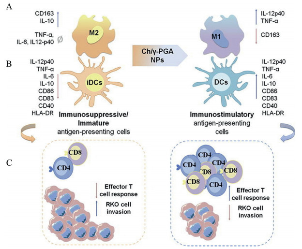
All these factors create an immunosuppressive microenvironment in the tumor tissue, which leads to the continued growth of tumor cells escape the policing of the immune system. By contrast, certain poly(amino acid)s can regulate the immunosuppressive microenvironment by enhancing the immune response or counteracting the negative immunoregulatory effects of tumor cells. For example, R-methyltryptophan (D-1MT) can inhibit the recruitment of Treg and MDSC and increase the proportion of CD8+ T cells. L-Phenylalanine can restore T cell number and function by suppressing MDSCs. Poly(γ-glutamic acid) combined with chitosan can inhibit the polarization of macrophages to M2-type macrophages and promote T cell proliferation. At the same time, some positively charged poly(amino acid)s can mediate the antigen delivery by macrophages through electrostatic adsorption, which can lead to positive immune response and IL-12 release from macrophages. So use these poly(amino acid)s delivery system to regulate the immunosuppressive microenvironment to promote effective treatment of tumors. Wang et al. [54] synthesized four different configurations of block copolymers mPEG-PA by mPEG-NH2-initiated ring-opening polymerization of alanine NCA with different chirality and investigated their sol-gel phase transition behavior. It was shown that poly-D-alanine induced T cell proliferation and differentiation and activated T cells by increasing the expression of TNF-α, IL-β and IL-6, which in turn generated a positive immune response. When it encapsulates tumor immunotherapeutic drugs to the tumor, it can improve immunotherapeutic efficacy by promoting immunomodulation. Yu et al. [9] used D-1MT, which can be oxidized by reactive oxygen species (ROS) as the raw material and synthesized a poly(amino acid)-based tri-block polymer (P(Me-D-1MT)-PEG-P(Me-D-1MT)) by ring-opening polymerization using PEG-NH2 as the initiator. In the tumor microenvironment, the P(Me-D-1MT)-PEG-P(Me-D-1MT) hydrogel can release D-1MT, inhibiting the production of IDO, which lead to Treg and MDSC recruitment was inhibited and the proportion of CD8+ T cells was saliently increased. The expression of aPD-1 was also promoted by the D-1MT released to improve the patient's response to aPD-1 and prolong aPD-1 antitumor time, leading to the effect of aPD-1 tumor immunotherapy was enhanced. All of these factors overcome the immunosuppressive state of the tumor. Lim et al. [55] synthesized amphiphilic poly(L-lysine)-g-poly(L-phenylalanine) (PLL-g-Phe) by grafting poly(L-phenylalanine) (Phe) onto poly(L-lysine) (PLL) and prepared cationic polymeric NPs containing squalene (CASq) using nanoemulsion method. NPs exhibit a high drug-loading rate, good stability and good biocompatibility. In addition, NPs are positively charged in the tumor microenvironment. But cell membrane is negatively charged. So the NPs can mediate the antigen delivery by macrophages through electrostatic adsorption. Also, NPs can promote a positive immune response and IL-12 release from macrophages, which in turn can stimulate Th1-mediated immune response. Wu et al. [27] developed an L-phenylalanine polymer, which is called metabolic reprogramming immunosurveillance activation nanomedicine (MRIAN). MRIAN can degrade to L-phenylalanine, which can suppress PKM2 activity, inhibit the metabolism of glucose and reduce the level of ROS in MDSCs, significantly deregulating immunosuppressive functions, and inducing differentiation of MDSCs to immune cells that have functions, such as NK cells, macrophages, and DCs. Since MDSCs suppress T cell proliferation during tumor progression, MRIAN significantly restored T cell numbers and function by suppressing MDSCs to re-establish immunosurveillance in T-cell acute lymphoblastic leukemia(T-ALL) mice. Li et al. [56] developed a tumor microenvironments (TMEs)-adapted poly-peptide composite based on thermosensitive hydrogel and semi-deprotected poly(L-lysine) to sequentially deliver regorafenib and transform growth factor-β inhibitor. It is shown that poly(L-lysine) nanogel can reduce the recruitment of tumor-associated macrophages (TAMs) and MDSCs, increase tumor infiltration of CD8+ T cells, and promote the polarization of macrophages from M2 to M1 types, effectively inhibiting tumor growth. Castro et al. [57] synthesized chitosan/poly(γ-glutamic acid) nanoparticles (Ch/γ-PGA NP) from chitosan (Ch) and poly(γ-inhibited the polarization of macrophages to M2-type macrophages (Fig. 2A) and induced an immunostimulatory dendritic cell phenotype (Fig. 2B), promoting T cell proliferation and suppressing colorectal cancer cell invasion (Fig. 2C) [58].
Figure 2
Figure 2.
Ch/γ-PGA NPs mechanism of action. (A) Ch/γ-PGA NPs promote the conversion of M2-type macrophages to M1-type macrophages, promote positive immune response, reduce CD163 expression, and promote IL-12p40 and TNF-a secretion. (B) Ch/γ-PGA NPs, which are synthesized by the co-acervation method, induce an immunostimulatory dendritic cells phenotype and improve the endocrinology of the proinflammatory cytokines TNF-a, IL-12p40, IL-6 and the expression of the synergistic stimulation molecules CD86, CD40 and HLA-DR. (C) These transitions promote the activation and proliferation of CD4+ and CD8+ T cells, while also suppressing the invasive capacity of colorectal cancer cells. Copied with permission [57]. Copyright 2017, Elsevier Publishing Group.
4.2
Poly(amino acid)s carriers with anti-inflammatory function
Tumor growth depends not only on individual genetic mutations, but also on the inflammatory cells, immune cells, and blood vessels in the microenvironment. Uncontrolled inflammation is an important factor leading to changes in the tumor microenvironment, which is significantly correlated with tumor progression, invasion, and metastasis [59-61]. Tumors are called "incurable wounds" and pro-tumorigenic inflammation is one important mark of cancer [62]. Inflammation can be classified into two categories: acute and chronic. While acute inflammation is easily cured, chronic inflammation is not and can persist for a long time. A small percentage of chronic inflammation may turn into cancer, which is a vital cause of cancer development and has different functions along with tumor development [63]. For example, in viral hepatitis, the patient's infection is mostly acute at the beginning, when it can be cured with early treatment. However, if it is delayed or improperly treated or if the patient has an immune deficiency, it can lead to chronic inflammation. These chronic inflammatory diseases can cause cellular and tissue changes, and if not effectively cleared over time, they can develop into cirrhosis or even liver cancer. At the same time, viruses also stimulate infected cells and their surrounding cell to secrete inflammatory factors that alter the cellular microenvironment and promote cancer cell proliferation, infiltration, and metastasis. Certain inflammatory cells, like TAM, assist tumor cells proliferate by secreting growth factors, promote the development of new blood vessels by secreting vascular endothelial growth factor (VGEF) [64], accelerate tumor incursion of normal tissues and metastasis to other organs by secreting matrix metalloproteinases (MMPs) [65], and inhabit intrinsic and acquired immunity by secreting a variety of immunosuppressive factors such as interleukin (IL)-10 [66]. In addition, studies have demonstrated that certain inflammatory cells also render tumor cells resistant chemotherapy [67]. Löffler et al. [68] and Li [69] demonstrated that the pro-inflammatory cytokine IL-6 induced microRNA-21 expression in a STAT3 signaling pathway-dependent manner, facilitating tumor cell proliferation and suppressing apoptosis. Tili et al. [70] found that inflammatory stimulation upregulated microRNA-155 expression by transfecting in vitro cell cultures, which lead to the promotion of mammary cells proliferation and mutation. Both microRNA-155 overexpression and the inflammatory environment can downregulate WEE1 expression [71] and facilitate tumor cell proliferation.
Therefore, inhibiting inflammation at the tumor site is beneficial for effective cancer treatment. M1 and M2-type macrophages are important bridges between inflammation and cancer development and have a "double-edged sword" effect on tumors. M1-type macrophages can inhibit tumor growth while M2-type macrophages can promote tumor growth [72]. Among them, tumor-associated macrophages belong to M2-type macrophages, which are the main components of the tumor microenvironment, are important for tumor metastasis and can be potential targets for oncology therapy [73]. Therefore, certain poly(amino acid)s like can suppress the inflammatory response by inhibiting the inflammatory signaling pathway of macrophages, thus promoting the preferential treatment of cancer. Studies have shown that glycosylated poly(amino acid)s can be used for anti-inflammatory [74]. When the side chains of amino acid residues are attached to glycosyl groups to form nonionic water-soluble glycosylated poly(amino acid)s, the multivalent nature of their side chain glycosyl groups can enhance the affinity of poly(amino acid)s materials for protein receptors, which in turn can participate in vital activities such as molecular recognition and can also be used for inhibiting inflammation [75]. So we can construct drug delivery system with anti-inflammatory function based on glycosylated poly(amino acid)s. Bertozzi et al. [75] used a nickel metal catalyst with a lipid structure to initiate the ring-opening polymerization of glycosylated serine and alanine NCA, and introduced sialic acid and its derivatives to the side chain through an enzyme-catalyzed reaction, thus preparing a glycosylated poly(amino acid) with a lipid C-terminus. The glycosylated poly(amino acid) (pS9L) exhibited a high binding capacity to Siglec-9 and a high specificity for binding to the same cell surface receptor in "cis", which inhibit LPS-induced inflammatory signaling pathway in macrophages and inhibit the endocytosis of macrophage by regulating mitogen-activated protein kinases (MAPK) signal as Fig. S2 (Supporting information) shows. Although glycol-poly(amino acid)s are not currently used in the transport of anticancer drugs and effective treatment of cancer, it provides ideas for the treatment of cancer at the anti-inflammatory level and preventing tumor recurrence. Furthermore Wu et al. synthesized poly(ferulic acid) and poly(ursolic acid) with anti-inflammatory function ferulic acid and ursolic acid as hydroxy acid monomers, respectively, and studies have shown that poly(ferulic acid) [33] and poly(ursolic acid) [34] still have anti-inflammatory function. Therefore, the polymers basing on amino acid monomers with anti-inflammatory functions such as glutamine, tryptophan and their derivatives may also have anti-inflammatory functions, which can be used as anti-cancer drug carriers to exert drug delivery and anti-inflammatory functions. It also provides ideas for synthesis of poly(amino acid) carriers with anti-inflammatory functions.
4.3
Poly(amino acid)s carriers with antioxidant function
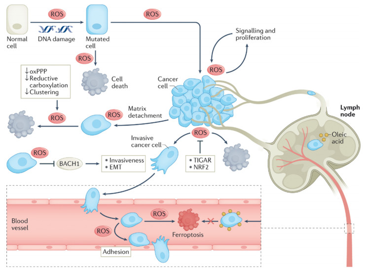
ROS are atoms, groups of atoms or molecules with unpaired electrons generated during cell metabolism, which are highly unstable and short-lived, such as superoxide anions (O2−) and hydrogen peroxide (H2O2) [76,77]. The abnormally active metabolism of tumor cells consumes a large amount of oxygen, making the tumor usually in a hypoxic environment. Hypoxia leads to more ROS production in mitochondria [78], which causes mutations in base pairs and DNA mainly by changing the structure of single or double strands DNA, leading to abnormal activation of gene mutations in tumor cells [79] and inactivation of tumor suppressor genes [77,80]. ROS can cause abnormal mitosis in cells and induce the transformation of normal cells to tumor cells [81]. ROS can regulate extracellular-regulated protein kinases (EKP) by shifting the inhibition of MAPK phosphatase through oxidative stress. Then the P13/Akt and ERK signaling pathways are activated, increasing the endogenous level of ROS production and the expression levels of antioxidants by repressing various transcription factors of FOXOs. Antioxidants such as nuclear factor erythroid 2-related factor 2 (NRF2) and TIGAR inhibit ROS-mediated cell death and promote tumor development (Fig. 3). In addition, ROS oxidize the tumor tissue matrix and help tumor cells metastasize [82], and the presence of large amounts of ROS inhibits T cell activation, creating an immunosuppressive tumor microenvironment that allows tumor cells to escape immune killing [83]. Meanwhile, continuous production of ROS can stimulate neovascularization to help tumor cells obtain more nutrients and facilitate tumor cell metastasis [84,85]. However, tumor cell death occurs when ROS levels are too high [86]. Therefore, tumor cells are sensitive to loss of antioxidant capacity due to reduced NADPH production, such as reduced oxidative pentose phosphate pathway (oxPPP) activity, reduced carboxylation and increased mitochondrial ROS (mtROS) production due to the inhibition of tumor cell aggregation (Fig. 3). In some cases specific anti-tumor immunity is activated due to ROS-induced tumor cell death [87]. In conclusion, although ROS can cause damage and reduce cell viability to some extent in almost all cell types, including tumor cells [88]. It is worth noting that the high tolerance of tumor cells will gain more benefits in a state of oxidative stress, and several studies have shown that high ROS expression often occurs in malignant tumor lesions in vivo, suggesting a close "tumor-ROS" tandem effect, promoting tumor growth and metastasis [89], so antioxidant therapy may bring more benefits for tumor treatment.
Figure 3
Figure 3.
Interconnection between ROS and tumor progression. ROS can promote the tumorigenesis, proliferation, metastasis by causing DNA damage, acting as signaling molecules, increasing the expression levels of antioxidant and so on. In addition, the stronger ROS effect causes apoptosis of normal cells as well as tumor cells. Copied with permission [77]. Copyright 2022, Springer Nature Publishing Group.
The main purpose of antioxidants is to prevent the accumulation of excess ROS and to remove excess ROS. Certain poly(amino acid)s can consume ROS through chemical reactions to achieve antioxidant effects and promote effective cancer treatment, such as poly(amino acid)s based on L-methionine, which are easily oxidized by ROS to methionine sulfoxide because of the thioether bond contained in methionine, which reduces ROS, so most poly(amino acid)s with antioxidant functions are based on methionine. Yu et al. [9] used L-methionine and D-1MT, which can be oxidized by ROS, as raw materials and synthesized poly(amino acid)-based tri-block polymer (P(Me-D-1MT)-PEG-P(Me-D-1MT)) hydrogels via ring-opening polymerization with macromolecular PEG-NH2 as the initiator. The P(Me-D-1MT)-PEG-P(Me-D-1MT) hydrogel can be injected in-situ in tumors and has a biostimulatory response to drug release. It shows that the hydrogel can effectively reduce ROS levels in the cellular microenvironment. Quan et al. [90] synthesized an ROS-responsive poly(ethylene glycol)-poly(methionine) (PEG-P(Met)), which can deliver the pro-oxidant drug piperlongumine (PL) to cancer cells safely and effectively. It can reduce ROS through the oxidation of the thioether bond contained in methionine. Zhao [91] prepared methionine as an active monomer that could be polymerized in situ, and then triggered the polymerization of the active monomer with a four-armed poly(ethylene glycol)-amino group as initiator to obtain block polypeptides, which could form stable hydrogels in aqueous solution, and the hydrophobic interactions between their blocks formed reversible physically cross-linked hydrophobic micro-regions within the hydrogels, making them available to encapsulate the hydrophobic anticancer drug and as a drug carrier to achieve slow release of the drug to promote effective cancer treatment. It was shown that in the tumor microenvironment with large amounts of ROS, methionine on the poly(methionine) side chain could be oxidized, resulting in a reduction of ROS in the tumor microenvironment. Xu [92] synthesized Met-based PEA and then conjugated PEG to the ends of the Met-PEA chains to synthesize amphiphilic polymers that can be autonomously loaded as nanoparticles. The nanoparticles include a hydrophobic Met-PEA core that can accommodate hydrophobic anticancer drugs, which have a hydrophilic PEG hull that reduces protein absorption from the blood on the outside of the nanoparticles and prolongs the blood circulation time. When the sulfhydryl group in cysteine is attached to the glycoside, thioether bonds are present in the polymer, which can also clear ROS via an oxidation. For example, Deming et al. [93] synthesize side-chain glycosylated poly(cysteine) by NCA ring-opening polymerization, which has thioether that can react with ROS, so it can be used to deliver anti-cancer drugs and exert its antioxidant ability to promote effective treatment of cancer.
4.4
Poly(amino acid)s carriers with apoptosis-promoting function
The proton sponge effect refers to the ability of the lysosome to trap a large number of protons when the pH in the lysosome drops causing an inward flow of Cl− and water molecules, resulting in osmotic swelling of the lysosome. The particles with cations bind to the cell membrane and enter the cell through endocytosis and then form endosomes, which fuse with the lysosomes (Fig. S3A in Supporting information). The unsaturated amino groups on the particles chelate the protons provided by the proton pump, which is continuously open, leading to a continuous proton influx. To prevent further acidification and balance the charge within the lysosome, each proton leads to the retention of one chloride ion and one water molecule in the lysosome (Fig. S3B in Supporting information), triggering lysosomal swelling and rupture (Fig. S3C in Supporting information) [94,95]. However, the rupture of lysosomes allows the entry of the encapsulated drug into the cytoplasm, while the spillover of lysosomal contents and release of intracellular Ca2+ lead to cytotoxicity [96]. The spillover of lysosomal enzymes can contribute to the activation of certain proteins, which in turn can damage mitochondria. Triggering of Ca2+-regulated permeability transition pores in mitochondria also leads to reduced ATP production and cell apoptosis [94].
Certain amino acids such as lysine, arginine, leucine, and histidine are positively charged. Therefore, drug carriers containing these amino acids can promote apoptosis of tumor cells through the proton sponge effect and facilitate effective cancer therapy. Chang et al. [23] synthesized a pH- and ROS-responsive drug delivery system. Paclitaxel and glucose were coupled to a paclitaxel polymer precursor (DEX-TK PTX) via a bond that could be cleaved by ROS. Subsequently, poly(L-histidine) (PHis) and Lapa were packaged in micelles formed by DEX-TK PTX to construct the drug delivery system PLP NPs. These PLP NPs have good biocompatibility, long cycle time and can accumulate in tumor tissues because of the enhanced permeability and retention (EPR) effects. PLP NPs can enter cancer cells via the endocytic pathway and are later delivered into the lysosome, where PHis are protonated in the acidic environment of the lysosome, leaving the proton pump continuously open, leading to the passive entry of chloride ion and water molecules into the lysosome, causing the lysosome to swell and rupture. Various enzymes within lysosomes are released into the cytoplasm, promoting mitochondrial damage and apoptosis in tumor cells. Yi et al. [25] synthesized an intelligent multi-stage tumor-targeting drug carrier based on n-butylamine-poly(L-lysine)-b-poly(L-cysteine) (PLL-PLC), and then functionalized it with folic acid (FA) and 1,2-dicarboxylic acid cyclohexene anhydride (DCA). Combining FA with poly(L-lysine) fragment enhances the tumor targeting ability. Grafting DCA on poly(L-lysine) fragment can make nanoparticles maintain negative charge under normal physiological conditions but rapidly convert to positive charge under the tumor microenvironment, the charge reversibility of nanoparticles can not only improve their blood circulation stability, but also provide excellent "proton sponge" effect, which promotes the absorption of drugs in tumors and the escape of endosomes, promoting the delivery of tumor-targeted drugs and apoptosis of tumor cells.
5.
Poly(amino acid)s carriers with internalization-promoting functions
Although this type of poly(amino acid) carriers do not have therapeutic function itself, they can improve the internalization efficiency of conventional chemotherapeutic drugs and transfection efficiency of gene drugs, enhancing the therapeutic effect of the drugs and promoting effective cancer treatment. So we also provide an overview of this type of poly(amino acid) carriers here. Key factors affecting the internalization pathway include the size [97] and charge of the complex [98], with large particles internalized via giant cell drinking action, whereas smaller complexes (less than 200 nm) generally follow the lattice-protein pathway. Polymeric nanoparticles modified with peptides of the same size can be internalized by lattice-protein and cytoplasmic membrane microcapsules. Positively charged or neutral complexes are transported via electrostatic interaction or fluid phase, whereas anionic complexes are mediated by endocytosis through the cytoplasmic membrane microcapsule. Anionic complexes cannot bind to the cell membrane by electrostatic interaction and therefore must be delivered by other means, such as the introduction of targeting peptides or molecular coatings [99,100]. Gene therapy, an emerging and effective technique for cancer treatment, involves the delivery of DNA or siRNA into patient-specific cells to promote or inhibit the expression of target proteins [101,102]. The main problem limiting the application of gene therapy is the difficulty in transporting large, easily disintegrated and negatively charged nucleic acid molecules into the cell nucleus [103].
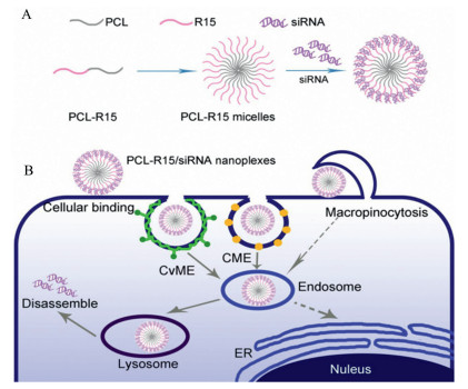
Since cell membranes are negatively charged and nucleic acid molecules are also negatively charged, it is difficult for nucleic acid molecules to enter the cell in the absence of carriers, While some poly(amino acid)s like poly(L-lysine) and poly(L-histidine) are positively charged in the tumor microenvironment, and the positively charged drug-carrying system has strong electrostatic interactions with negatively charged membranes, the poly(amino acid)s with positive charge can enhance cellular uptake [17,104,105]. In addition to the cationic nature of polyarginine, which provides the positive charge required for membrane penetration, the head of arginine also contains a guanidine group, which gives it better cell-penetration ability [42,106]. Mou et al. used a cationic polymer, poly(L-lysine) (PLL), for end-group functionalization and introduced hydrophobic poly(leucine) in the backbone to enable the carrier to self-assemble into size-stabilized nanoparticles by pro-hydrophobic interaction and reduce cytotoxicity. The end-group was then modified using poly(L-lysine) to increase the charge density. Arginine was then added to improve the membrane penetration ability of the carrier and facilitate the endocytosis of the drug delivery system. The endocytosis of the drug-loaded system has been shown to improve the internalization of nucleic acid molecules. Parelkar et al. [107] constructed a series of comb-shaped poly(cyclooctene-grafted-oligo-lysine) polymers containing oligo-lysine of different lengths that were immobilized on a hydrophobic polymer backbone. They greatly improve the internalization efficiency of DNA. Yamanouchi et al. [108] prepared a new series of poly(ester amide)s (PEA)s that can easily soluble in water and exhibit good biodegradability. They consist of three components: diols, diacids and amino acids. For example, they designed poly(ester amide)s based on L-Arg. It was confirmed that Arg-PEAs, acting as gene carriers can promote the internalization of cells. This is because the guanidine group in polyarginine can promote Arg-PEAs enter cancers cells. Sun et al. [109] synthesized a novel H6R6 peptide that can increase the internalization efficiency by incorporating histidine residues into 6-polyarginine. They then prepared an H6R6-CS copolymer that can deliver siRNA by introducing the H6R6 peptide into CS. It was shown that the H6R6-CS copolymer has a higher cellular internalization efficiency than ungrouped CS. Wu et al. [21] developed a new series of poly(ethylene ether ester amides) (Arg PEEAs) based on L-arginine to deliver genetic drugs. It was shown that this vector improved the efficiency of gene endocytosis and transfection. They also used poly(lactic-co-glycolic acid) (PLGA) and polycation (PC) based on L-arginine to design a polymeric NP platform with a positively charged hull, which can improve the efficiency of drug endocytosis [110]. Zhang et al. [111] synthesized a diblock polymer, PCL-R15, based on polyarginine and then constructed PCL-R15/siRNA nanoplexes. Polyarginine molecules confer favorable properties to PCL-R15/siRNA nanoplexes (Fig. 4A), such as the ability to bind to cells without relying on heparan sulfate proteoglycans, diverse internalization pathways and easy intracellular release of drugs, which greatly improves the efficiency of gene internalization. In addition, these poly(amino acid)s carriers allow nucleic acid-based drugs to enter the cytoplasm through endosomal escape (Fig. 4B). In addition, certain short peptides like NLS have nuclear localization ability, and their introduction into poly(amino acid)s carriers is more conducive to the nuclear delivery of gene drugs [22,112].
Figure 4
Figure 4.
Synthesis and internalization mechanism of PCL-R15/siRNA nanoplexes. (A) Assembly process of PCL-R15/siRNA nanoplexes. PCL-R15 amphiphilic block polymer can be formed into micelles by self-assembly. Then the negatively charged siRNA was electrostatically adsorbed onto the surface of the positively charged micelles. (B) Mechanism of action of PCL-R15/siRNA nanoplexes. PCL-R15/siRNA nanoplexes enter cells through lectin, caveolae mediated endocytosis and macropinocytosis. The free siRNA is then released into the cytoplasm by the action of lysosomes to function. Copied with permission [111]. Copyright 2020, American Chemical Society Publishing Group.
Poly(amino acid)s carriers have various functions such as immunomodulation, anti-inflammation, anti-oxidation, cancer cell apoptosis-promoting, and internalization-promoting, which can be used for drug delivery and effective cancer treatment. Thus, poly(amino acid)s have present good development prospects in drug delivery. For example, poly(amino acid)s with immunomodulatory functions can be used in combination with immunotherapeutic drugs to enhance the immune response and counteract the negative immunomodulatory effects of tumor cells, thus enhancing the efficacy of immunotherapy. Anti-inflammatory poly(amino acid)s can be used in combination with traditional chemical drugs to reduce the large inflammatory response caused by massive tumor cell death and reduce the risk of cancer recurrence. Poly(amino acid)s with antioxidant properties can be used for the delivery of gene therapy drugs or tradition chemical drugs, reducing drug resistance and risk of cancer recurrence. Poly(amino acid)s that promote internalization and apoptosis can be combined with gene therapy drugs to improve the stability, internalization efficiency and transfection of gene therapy drugs, thereby enhancing the efficacy of gene therapy. Those poly(amino acid)s carriers with immunomodulatory, anti-inflammatory, antioxidant and cancer cell apoptosis-promoting functions, which we also call therapeutic poly(amino acid)s carriers, inhibit the growth and metastasis of tumors and promote effective tumor treatment by regulating the tumor microenvironment and improving drug utilization. In conclusion, the selection of suitable poly(amino acid) carriers in combination with existing chemotherapeutic drugs or gene therapy drugs can achieve a "1 + 1 > 2" effect (Table S1 in Supporting information), providing a theoretical basis for the clinical application of poly(amino acid) carriers.
However, there are certain limitations of poly(amino acid)s as carriers for drug delivery, such as their drug-carrying capacity and drug encapsulation rate need to be further improved. Poly(amino acid) fragments are susceptible to the changes of physiological environment, resulting in degradation of poly(amino acid)s, causing the release of drug before reaching the target site, which decreases the utilization of the drug and increases the toxic side effects on normal tissues, etc. These factors limit the further application of poly(amino acid)s carriers. How to overcome these limitations may be one of the directions for the development of poly(amino acid)s carriers.
Declaration of competing interest
The authors declare that they have no known competing financial interests or personal relationships that could have appeared to influence the work reported in this paper.
Acknowledgments
This work was supported by the National Natural Science Foundation of China (Nos. 51973243 and 52173150), the Shenzhen Basic Research Project (No. JCYJ20190807155801657), the International Cooperation and Exchange of the National Natural Science Foundation of China (No. 51820105004).
[1]
E. Pérez-Herrero, A. Fernández-Medarde, Eur. J. Pharm. Biopharm. 93 (2015) 52–79. doi: 10.1016/j.ejpb.2015.03.018
[2]
H. Sung, J. Ferlay, R.L. Siegel, et al., CA Cancer J. Clin. 71 (2021) 209–249. doi: 10.3322/caac.21660
[3]
M.A. Zaimy, N. Saffarzadeh, A. Mohammadi, et al., Cancer Gene Ther. 24 (2017) 233–243. doi: 10.1038/cgt.2017.16
[4]
B.A. Chabner, T.G. Roberts Jr., Nat. Rev. Cancer 5 (2005) 65–72. doi: 10.1038/nrc1529
[5]
F. Qi, L. Zhao, A. Zhou, et al., Biosci. Trends 9 (2015) 16–34. doi: 10.5582/bst.2015.01019
[6]
T. Kurtanich, N. Roos, G. Wang, et al., SLAS Technol. 24 (2019) 151–160. doi: 10.1177/2472630318811108
W. Hu, M. Ying, S. Zhang, J. Wang, J. Biomed. Nanotechnol. 14 (2018) 1359–1374. doi: 10.1166/jbn.2018.2590
[18]
C. Zhang, W. Wu, R.Q. Li, et al., Adv. Funct. Mater. 28 (2018) 106385901.
[19]
Z. Song, Z. Han, S. Lv, et al., Chem. Soc. Rev. 46 (2017) 6570-6599. doi: 10.1039/C7CS00460E
[20]
D.M. Copolovici, A.I. Lupitu, Peptide-based systems for drug delivery, in: S. Koutsopoulos (Ed. ), Peptide Applications in Biomedicine, Biotechnology and Bioengineering, Woodhead Publishing, Sawston Cambridge, 2018, pp. 409-429.
[21]
J. Wu, D. Yamanouchi, B. Liu, C.C. Chu, J. Mater. Chem. 22 (2012) 18983–18991. doi: 10.1039/c2jm33753c
[22]
K.L. Jiang, L. Zhong, X.Q. Yang, et al., Oncol. Lett. 14 (2017) 7091–7098.
D.B. Bitoque, J. Morais, A.V. Oliveira, et al., Biosci. Rep. 41 (2021) BSR20201026. doi: 10.1042/BSR20201026
Figure 1
Solid phase synthesis. Firstly, the chloromethyl is introduced on the solid phase carrier. It is subsequently reacted with the amino acid protected by the amino group, and the first amino acid is solidly loaded onto the resin. Afterwards, multiple amino acids are introduced in the same manner to form poly(amino acid)s.
Figure 2
Ch/γ-PGA NPs mechanism of action. (A) Ch/γ-PGA NPs promote the conversion of M2-type macrophages to M1-type macrophages, promote positive immune response, reduce CD163 expression, and promote IL-12p40 and TNF-a secretion. (B) Ch/γ-PGA NPs, which are synthesized by the co-acervation method, induce an immunostimulatory dendritic cells phenotype and improve the endocrinology of the proinflammatory cytokines TNF-a, IL-12p40, IL-6 and the expression of the synergistic stimulation molecules CD86, CD40 and HLA-DR. (C) These transitions promote the activation and proliferation of CD4+ and CD8+ T cells, while also suppressing the invasive capacity of colorectal cancer cells. Copied with permission [57]. Copyright 2017, Elsevier Publishing Group.
Figure 3
Interconnection between ROS and tumor progression. ROS can promote the tumorigenesis, proliferation, metastasis by causing DNA damage, acting as signaling molecules, increasing the expression levels of antioxidant and so on. In addition, the stronger ROS effect causes apoptosis of normal cells as well as tumor cells. Copied with permission [77]. Copyright 2022, Springer Nature Publishing Group.
Figure 4
Synthesis and internalization mechanism of PCL-R15/siRNA nanoplexes. (A) Assembly process of PCL-R15/siRNA nanoplexes. PCL-R15 amphiphilic block polymer can be formed into micelles by self-assembly. Then the negatively charged siRNA was electrostatically adsorbed onto the surface of the positively charged micelles. (B) Mechanism of action of PCL-R15/siRNA nanoplexes. PCL-R15/siRNA nanoplexes enter cells through lectin, caveolae mediated endocytosis and macropinocytosis. The free siRNA is then released into the cytoplasm by the action of lysosomes to function. Copied with permission [111]. Copyright 2020, American Chemical Society Publishing Group.

 DownLoad:

 下载:
下载:


