bilan_wang@163.com (B. Wang). 1 These authors contributed equally to this work.
Received Date:
04 December 2022 Accepted Date:
13 February 2023 Revised Date:
06 February 2023 Available Online:
15 November 2023
Abstract:
Finding more effective and safe non-viral vectors to transfer genes into cancer cells has become the key of immune gene therapy for cancer. Herein a triblock compound MPEG2000–PDLLA4000–MPEG2000 modified by cationic liposome DOTAP was used as a non-viral vector DOTAP/MPEG2000–PDLLA4000–MPEG2000 (DMPM) to effectively transfer interleukin (IL)-12 plasmid (pIL-12) into tumor tissue. IL-12 produced by transfected tumor cells successfully inducing lymphocyte proliferation and promoting interferon-γ (IFN-γ) secretion, which resulted in tumor cells death. The ability of DMPM to transfer pIL-12 and the immune effect induced by IL-12 in cells had been explored. The anti-tumor effect, mechanism and safety of pIL-12/DMPM in mice cancer model were investigated in this study. Our results showed that the pIL-12 transferred by DMPM was highly expressed both in CT26 cells and B16-F10 cells. IL-12 expressed in the culture supernatant of transfected tumor cells stimulated lymphocyte proliferation and promoted IFN-γ secretion. The experimental result confirmed that pIL-12/DMPM therapy significantly reduced tumor growth in mice model. We designed the nanocomposite DMPM to deliver pIL-12 for cancer treatment and explored its therapeutic efficacy and the underlying anti-tumor mechanism. Our study suggested pIL-12 loaded by DMPM complex would be an effective strategy for cancer treatment.
According to the World Health Organization’s global cancer data in 2018, cancer has become the most life-threatening disease in the 21st century [1]. There are many reasons account for this phenomenon, such as difficulties in early diagnosis of cancer, easy metastasis of tumor, limited effect of radiotherapy and chemotherapy [2]. Therefore, to find a new therapy, which can overcome the shortcomings of the current treatment is urgent. Since the cancer associated with immunotherapy was recorded by William Coley in the 19th century, the application of immunotherapy in cancer has opened a new era [3–6]. Tumor immunotherapy is a treatment method to control and eliminate tumor by restarting and maintaining tumor immune cycle and restoring normal anti-tumor immune response [7]. Currently, immunotherapy has made it possible to provide more efficient and safe cancer treatment [8].
Cancer gene therapy is a kind of immunotherapy that strengthens anti-tumor immune responses through modifying or replacing defective genes or introduce foreign genes to express natural products [9]. Many cytokines are thought to affect the host immune system and play an important role in tumor microenvironment [10,11]. Interleukin-12 (IL-12) is a cytokine that plays an important role in immune system [12]. In the process of T cell activation, IL-12 induces the differentiation of effector cells and memory cells via directly regulating cell cycle, DNA synthesis and repair, protein translation and metabolism [13–16]. IL-12 is reported to promote the expansion of T-cells and natural killer (NK) cells and enhance their cytotoxic activity In the tumor immune microenvironment, IL-12 sponsors tumor rejection through lymphoid tissue carrying NK P46 cytotoxic receptor [17]. In brief, IL-12 built a significant bridge between innate and adaptive immunity, mediated by interferon-γ (IFN-γ) secretion, and has been widely studied both in basic and clinical research [18–25]. Additionally, IL-12 is capable of inducing the polarization of CD4+ T cells subset to Th1 cells during inflammation.
In order to weaken tumor immune resistance, IL-12 protein or IL-12 expressing gene are often designed in combination with other anti-tumor drugs [26,27]. Study showed that IL-12 combined with paclitaxel can enhance the anti-tumor immune response and regulate the tumor microenvironment [28]. However, the lack of selectivity and nonspecific immune response associated with the use of viral vectors remains a major problem when IL-12 is locally delivered and sustainably expressed through gene therapy [29,30]. Therefore, how to effectively transport IL-12 into tumor microenvironment to play an anti-tumor role has become the key of our study.
Another important aspect of cancer gene therapy is to find an effective gene carrier to transfer the target gene into cells. A growing number of gene delivery strategies are being employed for immunotherapy in applications ranging from disease prevention to cancer therapy [31,32]. Virus vectors were discovered and widely used in the early days, however, the use of viral vectors was limited because they can cause host immune response and the loss of infected cells, and the expression of foreign genes is transient [33]. New insight into gene delivery method has been focused on non-viral vectors. Given the advantages of readily available, structure controllable, low toxicity, low host immune response, non-viral vectors are considered to be the most promising gene delivery system [34,35]. Lipid-based DNA vectors and polymeric DNA vectors are widely used as a kind of non-viral vector to deliver gene into tumor cells [36]. Furthermore, the non-viral vector can improve its transfection efficiency through appropriate modification, making it the promising gene carrier for cancer therapy.
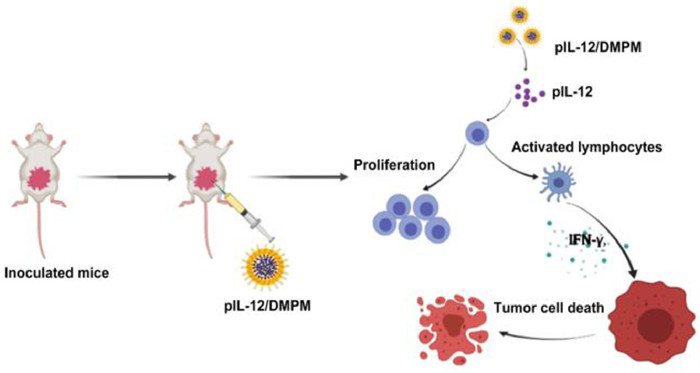
In summary, we aim to design a safe and efficient non-viral vector, which can effectively deliver IL-12 plasmid (pIL-12) to tumor tissue and trigger immune mechanism through high concentration effect in local tissue. In our study, successful delivery of immune stimulatory pIL-12 gene was achieved by self-assembled DMPM nanoparticles, which enhanced immune activation, showing remarkable anti-tumor effects (Scheme 1). We modified polyethylene glycol monomethyl ether (MPEG)2000–racemic polylactic acid (PDLLA)4000–polyethylene glycol monomethyl ether (MPEG)2000 copolymers with 1,2-dioleoyl-3-trimethylammonium-propane (DOTAP) as pIL-12 vector to achieve the goal of long cycle, high affinity and good biodegradability in vivo. After pIL-12 entrapped by the complex of DOTAP/MPEG2000–PDLLA4000–MPEG2000 (DMPM) in vitro, pIL-12 gene entered tumor tissue through local administration and successfully expressed IL-12 to play anti-tumor role through regulating the immune system.
Scheme 1
Scheme 1.
Pattern of immune response induced by pIL-12/DMPM in mice colon cancer model. The administration of pIL-12/DMPM successfully led to IL-12 expression in tumor tissue, which causes lymphocyte proliferation and stimulates lymphocyte to release IFN-γ, thus resulting in tumor cell apoptosis.
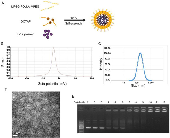
The pIL-12/DMPM complexes were prepared by self-assembly method. First, DOTAP and MPEG2000–PDLLA4000–MPEG2000 were assembled to form core-shell DMPM as gene vector. As shown in the Fig. 1A, on the inner surface of the shell structure, the hydrophilic head of DOTAP shows a positive charge, which attracts negatively charged pIL-12. Fig. 1B showed that the zeta potential of pIL-12/DMPM was 7.83 mV and according to particle size distribution spectrum, the average particle size of pIL-12/DMPM was 181.88 nm (Fig. 1C). Fig. 1D indicated the spherical shape of pIL-12/DMPM nanocomposite observed under transmission electron microscopy (TEM). Characterization of pIL-12 encapsulation capacity of DMPM was conducted by gel retardation method. As shown in Fig. 1E, lanes 1–3 were the brightest, indicating the band of naked pIL-12. When pIL-12 was completely entrapped by the DMPM complex, the band was dark, as shown in lanes 10–12, and the mass ratio of pIL-12 to DMPM is 1:50.
Figure 1
Figure 1.
Preparation and characterization of pIL-12/DMPM. Structure diagram of pIL-12/DMPM. (B) Zeta potential of pIL-12/DMPM. (C) Particle size distribution spectrum of pIL-12/DMPM. (D) TEM image of pIL-12/DMPM. Scale bar 10 nm. (E) Gel electrophoresis results. The charge ratio of pIL-12 to DMPM in lanes 4–6 is 1:12.5, lanes 7–9 is 1:25, lanes 10–12 is 1:50.
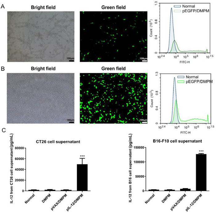
In the analysis of transfection efficiency, Fig. 2A demonstrated the expression of pEGFP in CT26 cells, and the flow cytometry result showed that the transfection efficiency of DMPM was 48.55%. Fig. 2B showed the expression of pEGFP in B16-F10 cells under fluorescence microscope, and the transfection efficiency was 54.29% by flow cytometry. The results demonstrated that DMPM can effectively transferred pEGFP into tumor cells. In order to investigate whether pIL-12 was successfully expressed in tumor cells in vitro, we used enzyme-linked immunosorbent assay (ELISA) method to detect the content of IL-12 in the supernatant of transfected cells. As shown in Fig. 2C, the expression level of IL-12 in pIL-12/DMPM group was significantly higher than that in control groups. The results showed that pIL-12 was highly expressed in the transfected cells.
Figure 2
Figure 2.
Transfection efficiency of DMPM. The transfection efficiency was detected by fluorescence microscope and flow cytometry assay after (A) CT26 cells and (B) B16-F10 cells transfected with pEGFP/DMPM for 48 h. (C) IL-12 expression in vitro. After CT26 cells and B16-F10 cells transfected with DMPM, pVAX/DMPM and pIL-12/DMPM for 48 h, the secretion of IL-12 in the supernatant was detected by ELISA kit. Scale bar: 100 µm. The data were expressed as mean ± standard error of mean (SEM), n = 3. ***P < 0.001 vs. normal group. These results represent three independent parallel experiments.
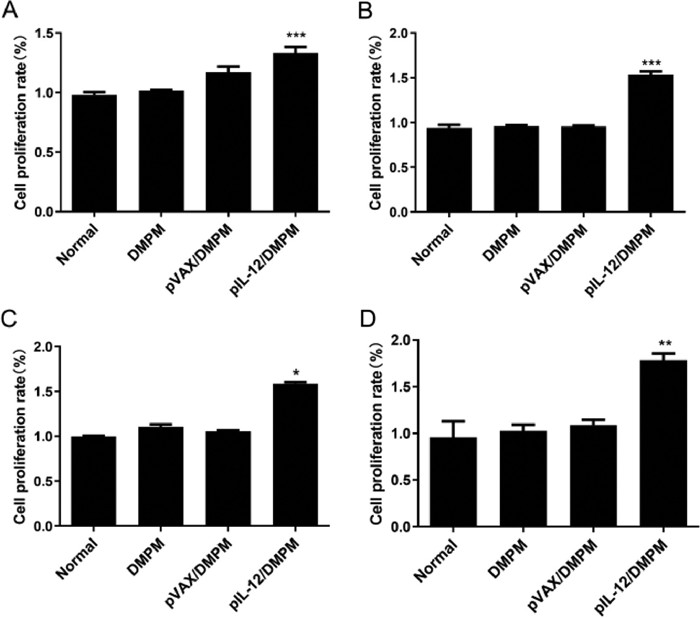
For further study, we examined the effect of expressed IL-12 on lymphocyte proliferation. CT26 cells or B16-F10 cells were transfected with DMPM, pVAX/DMPM and pIL-12/DMPM for 24 h, then, the supernatant from four groups were collected to stimulate lymphocytes for 48 h, respectively. The activity of lymphocytes was detected by 3-(4,5-dimethylthiazol-2-yl)−2,5-diphenyl tetrazolium bromide (MTT) assay and cell counting kit-8 (CCK-8) method. As shown in Fig. 3, MTT and CCK-8 results indicated that the supernatant of tumor cells transfected with pIL-12/DMPM could stimulate lymphocyte proliferation, and the cell proliferation rate of pIL-12/DMPM group was up-regulated compared with the control groups. Moreover, we detected tumor cells apoptosis by flow cytometry after co-culture of transfected tumor cells and lymphocytes, Fig. S1 (Supporting information) showed that the apoptosis rate of tumor cells transfected with pIL-12/DMPM was apparently higher than that of other control groups. The results showed that DMPM could effectively transfect pIL-12 into tumor cells to release IL-12, thus stimulating lymphocytes to kill tumor cells.
Figure 3
Figure 3.
Lymphocytes proliferation study. The supernatants of CT26 cells or B16-F10 cells transfected with DMPM, pVAX/DMPM and pIL-12/DMPM were used to stimulate lymphocytes from healthy BALB/c mouse spleen or C57 mouse spleen. The activity of lymphocytes was detected MTT assay and CCK-8 method. MTT assay: (A) CT26 cells and (B) B16-F10 cells. CCK-8 method: (C) CT26 cells and (D) B16-F10 cells after 48 h. The data were expressed as mean ± SEM, n = 3. P < 0.05, **P < 0.01, ***P < 0.001 vs. normal group. These results represent three independent parallel experiments.
On the other hand, we measured the expression of IFN-γ cytokines by activated lymphocytes. The supernatant of untreated CT26 cells and B16-F10 cells and the supernatant of tumor cells transfected with DMPM, pVAX/DMPM and pIL-12/DMPM were collected and used to stimulate lymphocytes. Next, the lymphocytes supernatant of each group was collected and measured by ELISA kit for IFN-γ secretion. The result was shown in Fig. S2 (Supporting information), the secretion of IFN-γ in pIL-12/DMPM group was obviously increased than that in other groups.
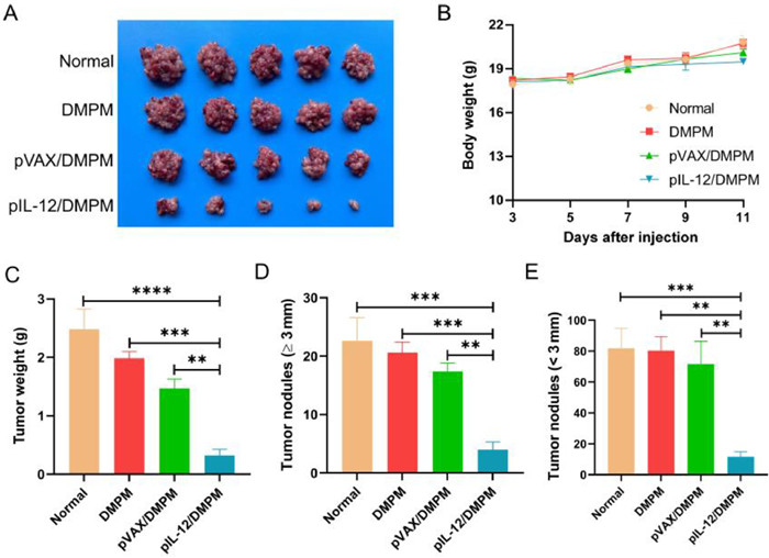
In addition to exploring the anti-tumor effect of pIL-12/DMPM in vitro, we established a colon cancer and melanoma models to further validate the anti-tumor effect of pIL-12/DMPM in mice. All animal experiments were approved by the Animal Experimental Ethics Committee of State Key Laboratory Biotherapy (SKLB), Sichuan University. As shown in Fig. 4 and Fig. S3 (Supporting information), with the increase of time, the weight of mice in the control groups (Normal, DMPM and pVAX/DMPM) and the experimental group (pIL-12/DMPM) increased gradually, and there was no distinct difference. Compared with the control groups, the weight of tumor in the experimental group decreased obviously, which means that pIL-12/DMPM inhibited the growth of tumor in mice. At the same time, the number of tumor nodules in the experimental group was significantly less than that in the control groups, which was consistent with the results of tumor weight.
Figure 4
Figure 4.
Therapeutic effect of pIL-12/DMPM in mice colon cancer model. (A) Tumor image of normal, DMPM, pVAX/DMPM and pIL-12/DMPM treatment groups. (B) Body weight of mice in different groups. (C) Tumor weight of different groups. (D) Tumor nodules (≥3 mm) number of different groups. (E) Tumor nodules (<3 mm) number of different groups. The data were expressed as mean ± SEM, n = 5. **P < 0.01, ***P < 0.001, ****P < 0.0001.
We carried out experiments to explore the apoptosis and the proliferation of tumor cells after treatment with pIL-12/DMPM. The tumor tissues were analyzed by proliferating cell nuclear antigen (PCNA) staining and terminal-deoxynucleoitidyl transferase mediated nick end labeling (TUNEL) staining. Cell proliferation was identified by calculating the proportion of PCNA positive cells. The experimental results in Fig. S4 (Supporting information) showed that compared with other groups, the number of PCNA positive cells in pIL-12/DMPM group was the least, indicating strong inhibitory effect on the proliferation of tumor cells. The apoptosis of tumor cells was identified by calculating the number of TUNEL staining positive cells. Fig. S5 (Supporting information) revealed that the TUNEL staining results showed that pIL-12/DMPM treatment could induce tumor cell apoptosis in vivo, and the apoptosis rate was much higher than that of the control groups. Therefore, the treatment of mouse colon cancer with pIL-12/DMPM can inhibit the proliferation of tumor cells and induce the apoptosis of tumor cells.
Angiogenesis is usually activated in some normal physiological processes, such as wound healing. On the contrary, if angiogenesis is activated and continues to provide nutrition and oxygen for tumor growth, it will make the tumor grow and metastasize rapidly. In order to explore the effect of pIL-12/DMPM on tumor angiogenesis, tumor tissues were stained with CD31 antibody. The results in Fig. S6 (Supporting information) showed active tumor neovascularization in normal, DMPM and pVAX/DMPM groups, while the number of CD31 positive vessels in pIL-12/DMPM group was significantly reduced compared with other groups, suggesting an inhibitory effect on tumor angiogenesis. Therefore, pIL-12/DMPM treatment can inhibit tumor angiogenesis, block the conditions of rapid growth and metastasis of tumor tissue, and inhibit tumor growth.
In order to evaluate the safety of pIL-12/DMPM treatment, mice blood serum was collected and detected by a blood biochemical analyzer (Roche, Switzerland). The serological biochemical results in Fig. S7 (Supporting information) showed that there was no significant difference between the experimental group and the control groups, indicating safety for application in vivo.
In summary, pIL-12 could be effectively transfected into CT26 cells and B16-F10 cells by DMPM, thus to stimulate lymphocyte proliferation, promote IFN-γ factor release, and promote tumor cells apoptosis in vitro. In vivo, pIL-12/DMPM remarkably inhibited tumor growth by inhibiting tumor cell proliferation, restrain tumor angiogenesis and promoting tumor cell apoptosis. Therefore, pIL-12/DMPM has good transfection efficiency, considerable anti-tumor effect and high safety, so it can be a potential anti-tumor drug for tumor gene therapy.
Declaration of competing interest
The authors declare that they have no known competing financial interests or personal relationships that could have appeared to influence the work reported in this paper.
Acknowledgments
This work was supported by the National Natural Science Foundation of China (No. 81972347) and the Key R&D Projects of the Science and Technology Department of Sichuan Province (No. 2022YFS0324).
Supplementary materials
Supplementary material associated with this article can be found, in the online version, at doi:10.1016/j.cclet.2023.108224.
[1]
F. Bray, J. Ferlay, I. Soerjomataram, et al., CA Cancer J. Clin. 68 (2018) 394–424. doi: 10.3322/caac.21492
[2]
K.D. Miller, L. Nogueira, A.B. Mariotto, et al., CA Cancer J. Clin. 69 (2019) 363–385. doi: 10.3322/caac.21565
[3]
R.S. Riley, C.H. June, R. Langer, M.J. Mitchell, Nat. Rev. Drug Disc. 18 (2019) 175–196. doi: 10.1038/s41573-018-0006-z
Scheme 1
Pattern of immune response induced by pIL-12/DMPM in mice colon cancer model. The administration of pIL-12/DMPM successfully led to IL-12 expression in tumor tissue, which causes lymphocyte proliferation and stimulates lymphocyte to release IFN-γ, thus resulting in tumor cell apoptosis.
Figure 1
Preparation and characterization of pIL-12/DMPM. Structure diagram of pIL-12/DMPM. (B) Zeta potential of pIL-12/DMPM. (C) Particle size distribution spectrum of pIL-12/DMPM. (D) TEM image of pIL-12/DMPM. Scale bar 10 nm. (E) Gel electrophoresis results. The charge ratio of pIL-12 to DMPM in lanes 4–6 is 1:12.5, lanes 7–9 is 1:25, lanes 10–12 is 1:50.
Figure 2
Transfection efficiency of DMPM. The transfection efficiency was detected by fluorescence microscope and flow cytometry assay after (A) CT26 cells and (B) B16-F10 cells transfected with pEGFP/DMPM for 48 h. (C) IL-12 expression in vitro. After CT26 cells and B16-F10 cells transfected with DMPM, pVAX/DMPM and pIL-12/DMPM for 48 h, the secretion of IL-12 in the supernatant was detected by ELISA kit. Scale bar: 100 µm. The data were expressed as mean ± standard error of mean (SEM), n = 3. ***P < 0.001 vs. normal group. These results represent three independent parallel experiments.
Figure 3
Lymphocytes proliferation study. The supernatants of CT26 cells or B16-F10 cells transfected with DMPM, pVAX/DMPM and pIL-12/DMPM were used to stimulate lymphocytes from healthy BALB/c mouse spleen or C57 mouse spleen. The activity of lymphocytes was detected MTT assay and CCK-8 method. MTT assay: (A) CT26 cells and (B) B16-F10 cells. CCK-8 method: (C) CT26 cells and (D) B16-F10 cells after 48 h. The data were expressed as mean ± SEM, n = 3. P < 0.05, **P < 0.01, ***P < 0.001 vs. normal group. These results represent three independent parallel experiments.
Figure 4
Therapeutic effect of pIL-12/DMPM in mice colon cancer model. (A) Tumor image of normal, DMPM, pVAX/DMPM and pIL-12/DMPM treatment groups. (B) Body weight of mice in different groups. (C) Tumor weight of different groups. (D) Tumor nodules (≥3 mm) number of different groups. (E) Tumor nodules (<3 mm) number of different groups. The data were expressed as mean ± SEM, n = 5. **P < 0.01, ***P < 0.001, ****P < 0.0001.

 DownLoad:

 下载:
下载:


