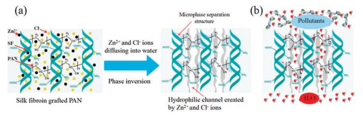
Figure 1.
The formation mechanism (a) and filtration process of SF-g-PAN membrane (b)
Biologically inspired silk fibroin grafted polyacrylonitrile filtration membrane prepared in ZnCl2 aqueous solution
Jingang Li , Shuwen Li , Xiaohui Wang , Fan Fan , Kongyin Zhao , Junfu Wei , Linhua Zhang , Dunwan Zhu

Membrane separation is one of the most promising technologies in water treatment, desalination and biomedical fields [1-3]. At present, the main method of preparing porous membranes is phase inversion. In the phase transformation process, the polymer solution is immersed in the non-solvent, and the phase separation occurs through the rapid diffusion exchange between the solvent and the non-solvent at the membrane interface. However, traditional membranes have many disadvantages such as membrane fouling, lack of response, and the difficulty to obtain narrow distributed pores [4].
Many methods have been used to prepare homoporous membranes to improve the separation efficiency [5-8]. Peinemann et al. [7] prepared homoporous membrane by non-solvent induced phase inversion of block copolymers. Wang et al. [8] prepared homoporous membranes from the blended amphiphilic block copolymers. But these methods are complex and cost much, and cannot produce large quantities of membranes.
In this case, the emergence of aquaporins (AQPs) provides inspiration for the design of high permeability and high selectivity artificial membrane. Due to the presence of AQPs, natural biological membranes often exhibit excellent transport performance [9]. AQPs has high permeability and can effectively intercept other ions and molecules. In order to reproduce the excellent transport performance of AQPs outside the cells, the scientists conducted a lot of research in recent years [10-12]. Many kinds of inorganic materials were utilized to construct efficient water channels within membranes [13, 14]. Bi et al. [15] prepared a novel thin film nanocomposite membrane by interfacial polymerization method with the incorporation of graphene quantum dots. Due to the formation of artificial water channel in polyamide layer, the pure water flux of membrane is greatly improved. The artificial water channel is of great significance to explore the structure and transport mechanism of water channel.
Polyacrylonitrile (PAN) was widely used in textiles and membranes [16]. In order to improve the moisture absorption performance of PAN fiber, the homogeneous grafting of PAN onto silk fibroin (SF) was investigated. The slow diffusion of zinc chloride in coagulation bath results in dense fibers [17]. SF is composed of three amino acids, glycine (45%), alanine (30%) and serine (12%). Amino acid residues endow SF with hydrophilicity and pH sensitivity [18]. However, no literature was reported about the use of SF-g-PAN as filtration membrane [19].
In this paper, inspired by the structure and transport mechanism of AQPs, ZnCl2 aqueous solution was used as solvent to obtain a casting solution by homogeneous grafting of PAN onto the SF without pore forming agent. Then SF-g-PAN filtration membrane was fabricated by putting the casting solution into a water coagulation bath. Phase inversion occurred when Zn2+ and Cl- ions gradually diffused into water, creating a well-connected ion channel network. The microphase separation of SF and PAN also forms a channel at the interface. Therefore, the obtained SF-g-PAN membrane has a narrow pore size distribution, high permeability and high selectivity, which can solve the contradiction between the membrane permeability and selectivity. The anti-fouling property, pore size distribution, dye rejection and pH-responsive property of the membrane were investigated.
Fig. 1 shows the formation and filtration mechanism of SF-gPAN membrane. Phase inversion occurred when Zn2+ and Cl- ions gradually diffused into water and many microporous were formed. The microphase inversion structure appeared between PAN and SF. Then SF-g-PAN filtration membrane with narrow pore size distribution was obtained. The mechanism of the microphase inversion can be investigated in detail through multi-scale computer simulation.
Fig. S1 (Supporting information) shows the preparation process of SF-g-PAN membrane. In a flask containing 52.2 mL 60 wt% ZnCl2 aqueous solution 3.5 g SF was dissolved at 55 ℃ to obtain a homogenous solution. When the temperature was down to 26 ℃, 10 mL AN and 0.8 mL 10 wt% (NH4)2S2O8 was added. After 3 min, 1 mL 10 wt% NaHSO3 was added and the reaction was performed for 30 min under stirring. The homogenous SF-g-PAN solution was degassed and 3–6 g of the solution was poured on a clean flat glass and unrolled by a glass rod winding copper wire at both ends (the diameter of the copper wire was 0.40 mm). The glass was immediately immersed in deionized water at room temperature for 2 h. The SF-g-PAN filtration membrane was obtained after washing rudimental ions with deionized water.
Fig. S2 (Supporting information) shows the schematic diagram and digital photo of the homemade cross-flow equipment. BSA aqueous solution (0.5 g/L) was used to investigate the anti-fouling property of the membrane. Amaranth and direct yellow 27 aqueous solutions (30 mg/L) were used to research the rejection property of the membrane. The concentrations of BSA or dye solution in feed and permeate solutions were measured by a UV spectrophotometer (UV-1100). The flux (J, L m-2 h-1) and the rejection (R, %) were calculated by following equations [20]:
|
|
|
|
where V is the permeate volume (L), A is the membrane area (m2), t is the time (h), Cp and Cf are the BSA or dye concentrations of permeate and feed solution, respectively.
The pure water flux (PWF) of SF-g-PAN filtration membrane was determined at 0.1 MPa and denoted as Jw1 (L m-2 h-1). Then, the feed solution was switched to BSA solution for 30 min, and the flux of BSA solution was denoted as JB1. The process was repeated for four times. The anti-fouling property of the membrane was determined by the flux recovery rate (FRR) [20]:
|
|
where Jwi is the PWF of i time.
Fig. 2 shows the morphologies of SF-g-PAN filtration membrane. The SF-g-PAN filtration membrane was white and opaque and the thickness was 0.342 mm. From Fig. 2b it is found that the wet membrane surface exhibited some well-distributed undulating peaks and valleys, which were caused by the different phaseinversion rates on different positions of the casting solution. The surface SEM image also approved the rough structure of the membrane surface. The cross-section SEM image of the membranes displays a dense structure without any fingerlike holes, which is different from most traditional polymeric membranes formed by a phase-inversion process [21].
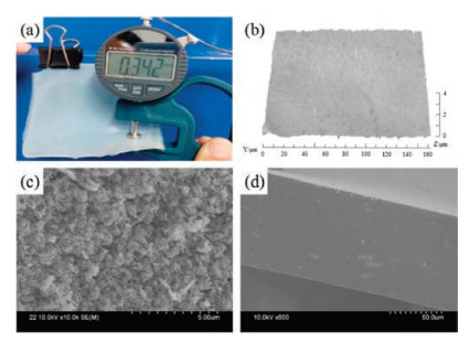
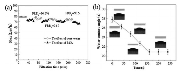
Fig. 3 shows the antifouling and hydrophilic properties of SF-g-PAN membrane. The SF-g-PAN membrane exhibits excellent protein anti-fouling property. The FRR1 was 96.6%. After three consecutive BSA filtrations, the FRR3 still reached 93.5% without any washing operation. When a water droplet (1 μL) contacted the membrane surface, it spread out quickly and the contact angle was 28°, and it changed to 22° after 150 s (Fig. 3b).
To determine the permeate size cut-off of the SF-g-PAN membranes prepared in this study, rigid dyes were used as probes [22]. Different molecular weight dyes vary in molecular size. The larger the molecular weight is, the larger the corresponding molecular size. Fig. 4a shows the flux and rejection rate of SF-g-PAN membrane using seven different molecular weight dyes as feed solutions at 0.1 MPa. The molecular weight and absorption peak wavelength of the seven dyes are showed in Table S1 (Supporting information). The stable flux of all these seven dyes is above 80 L m-2 h-1. The rejection rate for the dyes with a molecular weight greater than 660 was above 95%. However, the rejection for the dyes with a molecular weight below 600 was less than 30%. This indicates that SF-g-PAN filtration membrane pore size distribution is narrow, which can be finely separated. There is a high rejection rate for organic compounds with a molecular weight greater than 660, while the rejection rate for organic compounds with a molecular weight less than 600 is very low. The membrane can be applied to fine separation of drugs and biological samples [23].
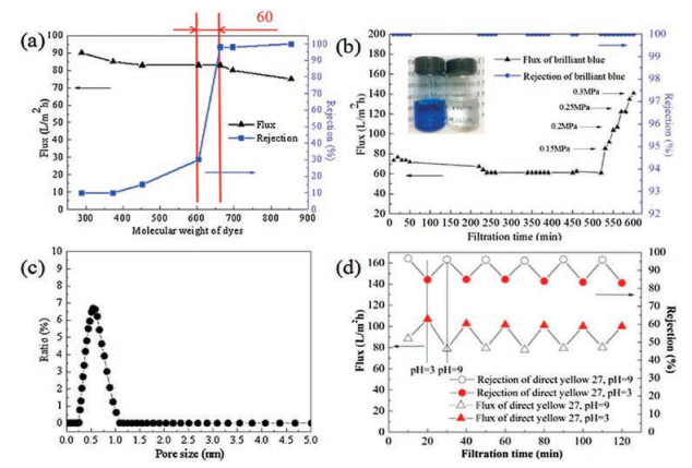
Fig. 4b shows the flux and rejection rate of brilliant blue solution under different operating pressure of SF-g-PAN membrane. The flux of the membrane is as high as 140 L m-2 h-1 at 0.3 MPa, but the rejection rate of brilliant blue is still close to 100%. The membrane has excellent performance both at lower operating pressures and higher operating pressures.
Fig. 4c shows the pores distribution of SF-g-PAN membrane. The membrane had a narrow pore size distribution (0.25–1.04 nm) and the average pore size was 0.55 nm. This value is consistent with the permeate size cut-off of the SF-g-PAN membranes. SF-g-PAN membrane is expected to finely separate the samples with very similar molecular weight, although the mechanism of forming the narrow-distributed pores needs further investigation. The capability of the SF-g-PAN filtration membrane to separate small molecules predominantly by size could open new applications in the biotechnology and food industries. From Fig. 4d, it is found that when the pH of direct yellow 27 solution alternately switched at 3.0 and 9.0, the flux repeatedly occurred at 101.5 ±1.62 and 78.9 ±1.17 L m-2 h-1, and the rejection occurred at 83.5% ± 0.74% and 95.8% ± 0.43%, respectively. SF-g-PAN membrane exhibited good pH sensitivity, which was caused by the ionization of amino acid residues in SF at different pH values [18].
Inspired by the structure and transport mechanism of AQPs, SF-g-PAN filtration membrane was prepared without using any organic solvent and pore-forming agent. SF-g-PAN membrane exhibited anti-fouling properties and pH sensitivity. The rejection rate for the dyes with a molecular weight greater than 660 was above 95%, while the rejection rate for the dyes with a molecular weight below 600 was less than 30%, indicating that the SF-g-PAN filtration membrane possesses a narrow pore size distribution. The pore size distribution of SF-g-PAN membrane is about 0.25– 1.04 nm. The pure water flux can reach 80 L m-2 h-1 at 0.1 MPa. The membrane is promising for practical applications in fine separation, dye desalination, waste water treatment and biomedical fields.
The research is supported by the National Natural Science Foundation of China (Nos. 51678409, 51708407, 21476172), Tianjin Science Technology Research Funds of China (Nos. 16JCZDJC37500, 15JCZDJC38300), Program for Innovative Research Team in University of Tianjin (No. TD13-5042), and Science Foundation for the Youth Teachers of Peking Union Medical College (No. 2014ZLGC0754).
Supplementary material related to this article can befound, in the online version, at doi:https://doi.org/10.1016/j.cclet.2018.07.016.
Q. Nie, F. Ran, C. He, et al., Chin. Chem. Lett. 22(2011) 370-373. doi: 10.1016/j.cclet.2010.10.017
J. Guo, K. Zhao, X. Zhang, et al., Mater. Lett. 157(2015) 112-115. doi: 10.1016/j.matlet.2015.05.080
J. Shan, W. Wei, D. Yonghong, et al., Chin. Chem. Lett. 29(2018) 390-394. doi: 10.1016/j.cclet.2018.01.006
B. Van der Bruggen, M. Mänttäri, M. Nyström, Sep. Purif. Technol. 63(2008) 251-263. https://www.sciencedirect.com/science/article/pii/S1385894713002829#!
W. Xu, M. Guo, J. Liu, et al., J. Biomed. Nanotechnol. 14(2018) 179-189. doi: 10.1166/jbn.2018.2465
V. Yangali-Quintanilla, S.K. Maeng, T. Fujioka, et al., J. Membr. Sci. 362(2010) 334-345. doi: 10.1016/j.memsci.2010.06.058
K.V. Peinemann, V. Abetz, P.F.W. Simon, Nat. Mater. 6(2007) 992-996. doi: 10.1038/nmat2038
M. Wei, W. Sun, X. Shi, et al., Macromolecules 49(2016) 215-223. doi: 10.1021/acs.macromol.5b02133
H. Sui, B.G. Han, J.K. Lee, et al., Nature 414(2001) 872-878. doi: 10.1038/414872a
W. Zhang, B. Jiang, P. Yang, Chin. Chem. Lett. 27(2016) 1339-1344. doi: 10.1016/j.cclet.2016.06.044
W. Li, J. Peng, L. Tan, et al., Biomaterials 106(2016) 119-133. doi: 10.1016/j.biomaterials.2016.08.016
Y. Wang, L. Chen, L. Tan, et al., Biomaterials 35(2014) 6972-6985. doi: 10.1016/j.biomaterials.2014.04.099
X. Cheng, Z. Wang, X. Jiang, et al., Prog. Mater. Sci. 92(2017) 258-283. doi: 10.1007/s12274-017-1635-y
C. Tang, Z. Wang, I. Petrinić, et al., Desalination 368(2015) 89-105. doi: 10.1016/j.desal.2015.04.026
R. Bi, Q. Zhang, R. Zhang, et al., J. Membr. Sci. 553(2018) 17-24. doi: 10.1016/j.memsci.2018.02.010
S. Liu, C. Lin, S. Lin, R. Fu, Y. Huang, et al., J. Biomed. Nanotechnol. 12(2016) 732-742. doi: 10.1166/jbn.2016.2201
Y. Sun, Z. Shao, J. Zhou, et al., J. Appl. Polym. Sci. 69(1998) 1089-1097. doi: 10.1002/(ISSN)1097-4628
M.A. Rahman, A.F. Ismail, A. Mustafa, Mater. Sci. Eng. A 448(2007) 275-280. doi: 10.1016/j.msea.2006.10.042
N. Padma, J. Macromol. Sci. Part C-Polym. Rev. 14(1976) 193-213. doi: 10.1080/15321797608065769
K. Zhao, X. Zhang, J. Wei, et al., J. Membr. Sci. 492(2015) 536-546. doi: 10.1016/j.memsci.2015.05.075
F. Liu, N.A. Hashim, Y. Liu, et al., J. Membr. Sci. 375(2011) 1-27. doi: 10.1016/j.memsci.2011.03.014
A. Asatekin, E.A. Olivetti, A.M. Mayes, J. Membr. Sci. 332(2009) 6-12. doi: 10.1016/j.memsci.2009.01.029
Y. Tang, Z. Ali, J. Dai, et al., J. Biomed. Nanotechnol. 14(2018) 206-214. doi: 10.1166/jbn.2018.2525

Figure 2 Morphologies of SF-g-PAN filtration membrane. (a) Digital photo. (b) Surface 3D ultra-depth microscope image. (c) Surface and (d) cross-section SEM images

Figure 3 Antifouling and hydrophilic properties of SF-g-PAN membrane. (a) Alternating filtration flux between pure water and BSA solution. (b) Dynamic water contact angle

Figure 4 Dyes rejection and pores distribution of SF-g-PAN membrane. (a) The rejection rate of different molecular weight dyes. (b) The rejection rate of brilliant blue solution under different pressures. (c) Pore size distribution. (d) Rejection and flux changes when pH 3.0 and pH 9.0

 扫一扫看文章

扫一扫关注我们
 DownLoad:


 下载:
下载: