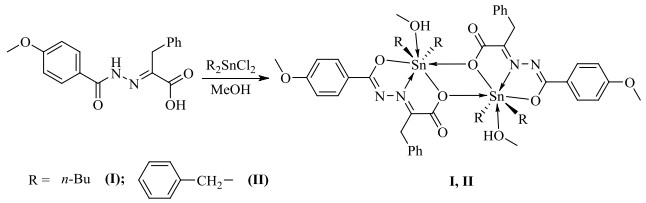
Figure 1.
Syntheses of the complexes
Syntheses, Crystal Structures and Biological Activity of the Diorganotin 2-(2-(4-Methoxybenzoyl)hydrazono)-3-phenylpropanoic Carboxylate Complexes
Man WANG , Fei YU , Wu-Jiu JIANG , Yu-Xing TAN , Fu-Xing ZHANG , Dai-Zhi KUANG

In the late 1960s, the discovery and clinical application of the anti-cancer effect of cisplatin(II) promoted the rapid development of metal anticancer drugs[1]. Therefore, the design and synthesis of new metal anticancer drugs have become a research hotspot. Moreover, with the frequent use of similar anticancer drugs, most cancer cells will develop a certain degree of drug resistance[2, 3], which has forced scientific researchers to continuously develop new anticancer drugs with strong anticancer effects, good targeting, and small toxic and side effects[4-6]. It is well known that diorganotin compounds have attracted scientists' attention because of their excellent properties, and in particular, some compounds have shown superior in vitro anticancer activity to platinum drugs such as cisplatin[7, 8]. Studies have shown that the type, number and ligands of the alkyl groups linked by tin atoms have important effects on the anticancer activity, toxicity and water solubility of diorganotin carboxylate[8, 9]. The alkyl groups attached to the tin atom and the ligand coordinated with the tin atom determine the biological activity of the organotin compound, so changing the type of alkyl groups or the ligand can obtain anticancer complexes with different activities. In recent years, scientists have carried out a lot of work on the hydrocarbon group modification and ligand screening of diorganotin carboxylate, which has become a focus of current research on organotin chemistry[5, 10]. Based on our previous work[11-15], we have employed the diorganotin and 2-(2-(4-methoxybenzoyl)hydrazono)-3-phenylpropanoic acid ligand, obtained two new diorganotin complexes and studied preliminarily their effects on cancer cells and the mechanism of interaction with calf thymus DNA, which laid a foundation for screening new organometallic complexes with high anticancer activity.
Infrared spectrum (KBr) was recorded by the Prestige-21 infrared spectrometer (Japan Shimadzu, 4000~400 cm–1). 1H, 13C and 119Sn NMR spectra were measured with a Bruker AVANCE-500 NMR spectrometer. The elemental analysis was determined by PE-2400(II) elemental analyzer. Crystallographic data of the complexes were collected on a Bruker SMART APEX II CCD diffractometer. HRMS was measured with the Thermo Scientific LTQ Orbitrap XL (ESI source). Melting points were determined using an X4 digital microscopic melting point apparatus without correction (Beijing Tektronix Instrument Co. Ltd.). Thermogravimetric analyses (TGA) were recorded on a NETZSCH TG 209 F3 instrument at a heating rate of 20 ℃/min from 40 to 800 ℃ under air. The UV spectra were determined with the UV-2550 spectrophotometer (Shimazu). Fluorescence spectra were obtained with a Hitachi F-7000 spectrophotometer with quartz cuvette (path length = 1 cm).
2-(2-(4-Methoxybenzoyl)hydrazono)-3-phenylpropanoic acid ligand and dibenzyltin dichloride were prepared according to the literatures[14]. Ethidium bromide (EB), calf thymus DNA and three hydroxymethyl aminomethanes (tris) were from Sigma-Aldrich LLC. Carboplatin was from J & K Scientific Ltd. Dibutyltin oxide was from Alfa Aesar (China) Chemical Co., Ltd. Other chemicals were from Sinopharm Chemical Reagent Co., Ltd. All reagents were of analytical grade obtained from commercial sources and used without further purification. The human lung cancer cells (NCI-H460, ATCC No: HTB-177), human liver cancer cells (HepG2, ATCC No: HB-8065) and human breast cancer cells (MCF-7, ATCC No: HTB-22) were obtained from American Tissue Culture Collection (ATCC). Culture media RPMI-1640 (10.0%) were purchased from USA GIBICO. Ultrapure water (18.2 MΩ·cm) obtained from a Milli-Q water purification system (Millipore Co., USA) was used in all experiments. Tris-HCl (0.01 mol·L–1) buffer solution was prepared by a certain amount of tris dissolved in super-pure water before using, and the pH of the solution was adjusted to 7.40 with hydrochloric acid solution (0.1 mol·L–1). The purity of CT-DNA was determined by comparing the absorbance at 260 and 280 nm (A260/A280 = 1.8~1.9/1). The concentration of CT-DNA was calculated by measuring the absorbance at 260 nm (ε260 = 6600 L·mol–1·cm–1). The reserve solution was stored at 4 ℃. The ethidium bromide solution was prepared by a certain amount of ethidium bromide solid dissolved in tris-HCl (0.01 mol·L–1) buffer solution.
A mixture of 2-(2-(4-methoxybenzoyl)hydrazono)-3-phenylpropanoic acid (1.0 mmol), dibutyltin oxide (1.0 mmol) and CH3OH (20.0 mL) was added in a round-bottomed flask (50.0 mL), and refluxed with stirring for 6.0 h, then the mixture was cooled and filtered. The complex crystals were obtained by controlling solvent evaporation. Complex I was yellow crystal, Yield: 79%. m.p.: 171~173 ℃. Anal. Calcd. (C26H36N2O5Sn): C, 54.28; H, 6.31; N 4.87%. Found: C, 54.31; H, 6.44; N, 4.79%. UV-vis (DMSO+H2O) λmax: 358 nm. FT-IR (KBr, cm–1): 3451 ν(-OH), 3063, 3028 ν(Ar–H), 2959, 2926, 2857 ν(C–H), 1601, 1391 ν(COO), 1584 ν(C=N), 623 ν(Sn–O), 588 ν(Sn–O–Sn), 521 ν(Sn–N), 449 ν(Sn–C). 1H NMR (500 MHz, CDCl3, δ/ppm): 8.21 (d, J = 8.9 Hz, 2H), 7.54 (d, J = 7.2 Hz, 2H), 7.28~7.25 (m, 2H), 7.18~7.20 (m, 1H), 6.99 (d, J = 9.0 Hz, 2H), 4.43 (s, 2H), 3.90 (s, 3H), 3.49 (d, J = 5.2 Hz, 3H), 1.57~1.67 (m, 4H), 1.54~1.48 (m, 4H), 1.21~1.28 (m, 4H), 1.09 (q, J = 5.3 Hz, 1H), 0.79 (t, J = 7.3 Hz, 6H). 13C NMR (126 MHz, CDCl3, δ/ppm): 174.79, 164.24, 163.30, 154.49, 135.28, 130.72, 129.87, 128.63, 126.96, 124.96, 113.81, 55.50, 50.86, 33.02, 26.62, 26.34, 21.20, 13.40. 119Sn NMR (187 MHz, CDCl3, δ/ppm): –154.59. H RMS (ESI) m/z calcd. for C25H33N2O4Sn+ [M-CH3OH+H]+ 545.14568, found 545.14563; calcd. for C50H65N4O8Sn2+ [2M-2CH3OH+H]+ 1087.28349, found 1087.28467.
Complex II was prepared in a similar procedure (Fig. 1) as I by dibenzyltin dichloride (1.0 mmol) in place of dibutyltin oxide. The product was yellow crystal, Yield: 83%. m.p.: 190~192 ℃ (dec). Anal. Calcd. (C32H32N2O5Sn): C, 59.74; H, 5.01; N, 4.35%. Found: C, 59.72; H, 4.98; N, 4.32%. UV-vis (DMSO+H2O) λmax: 384 nm. FT-IR (KBr, cm–1): 3449 ν(-OH), 3059, 3024, 3003 ν(Ar–H), 2936, 2837 ν(C–H), 1599, 1389 ν(COO), 1582 ν(C=N), 623 ν(Sn–O), 590 ν(Sn–O–Sn), 523 ν(Sn–N), 459 ν(Sn–C). 1H NMR (500 MHz, CDCl3, δ/ppm): 8.00 (d, J = 8.7 Hz, 2H), 7.33 (d, J = 7.4 Hz, 2H), 7.25~7.28 (m, 2H), 7.21 (t, J = 7.2 Hz, 1H), 6.96 (d, J = 8.7 Hz, 2H), 6.83 (s, 10H), 3.91 (s, 3H), 3.88 (s, 2H), 3.48 (s, 3H), 3.17 (d, J = 11.6 Hz, 2H), 3.13 (d, J = 11.6 Hz, 2H). 13C NMR (126 MHz, CDCl3, δ/ppm): 174.74, 168.12, 162.92, 150.76, 136.28, 135.33, 130.85, 129.97, 128.44, 128.20, 128.11, 126.59, 125.49, 125.23, 113.50, 55.43, 50.82, 34.99, 32.17. 119Sn NMR (187 MHz, CDCl3, δ/ppm): –633.12. HRMS (ESI) m/z calcd. for C31H29N2O4Sn+ [M-CH3OH+H]+ 613.11438, found 613.11450; calcd. for C62H57N4O8Sn2+ [2M-2CH3OH+H]+ 1223.22089, found 1223.22327.
Suitable single crystals with dimensions of 0.25mm × 0.23mm × 0.23mm (I) and 0.21mm × 0.21mm × 0.20mm (II) were selected for data collection at 296(2) K on a Bruker SMART APEX II CCD diffractometer equipped with graphite-monochromated MoKα radiation (λ = 0.071073 nm) using a φ~ω mode. All the data were corrected by Lp factors and empirical absorbance. The structure was solved by direct methods. All non-hydrogen atoms were determined in successive difference Fourier synthesis, and all hydrogen atoms were added according to theoretical models. All hydrogen and non-hydrogen atoms were refined by their isotropic and anisotropic thermal parameters through full-matrix least-squares techniques. All calculations were completed by the SHELXTL-97[16] program. For complex I, a total of 14266 reflections were obtained in the range of 2.54 < θ < 25.10° with 4909 unique ones (Rint = 0.0173), S = 1.082, (Δρ)max = 0.559 and (Δρ)min = –0.315 e/Å3, max transmission was 0.8096, min transmission was 0.7955, and the completeness was 99.5%. For complex II, a total of 13905 reflections were obtained in the range of 2.39 < θ < 25.10° with 5130 unique ones (Rint = 0.0137), S = 1.046, (Δρ)max = 0.726, (Δρ)min = –0.722 e/Å3, max transmission was 0.8366, min transmission was 0.8294, and the completeness was 99.5%. The selected bond lengths and bond angles for I and II are listed in Table 1.
 DownLoad:
CSV
DownLoad:
CSV
| I | |||||||
| Bond | Dist. | Bond | Dist. | Bond | Dist. | ||
| Sn(1)–C(22) | 2.117(2) | Sn(1)–O(1) | 2.1773(17) | Sn(1)–O(2) | 2.3058(17) | ||
| Sn(1)–C(18) | 2.122(2) | Sn(1)–N(1) | 2.2385(19) | Sn(1)–O(2)i | 2.8017(18) | ||
| Angle | (°) | Angle | (°) | Angle | (°) | ||
| C(22)–Sn(1)–C(18) | 155.92(11) | O(1)–Sn(1)–N(1) | 70.16(7) | C(22)–Sn(1)–O(5) | 82.36(10) | ||
| C(22)–Sn(1)–O(1) | 95.17(9) | C(22)–Sn(1)–O(2) | 91.91(9) | C(18)–Sn(1)–O(5) | 80.30(9) | ||
| C(18)–Sn(1)–O(1) | 97.24(9) | C(18)–Sn(1)–O(2) | 91.82(8) | O(1)–Sn(1)–O(5) | 77.48(7) | ||
| C(22)–Sn(1)–N(1) | 103.55(9) | N(1)–Sn(1)–O(2) | 69.73(6) | O(2)i–Sn(1)–O(5) | 77.96(6) | ||
| O(2)–Sn(1)–O(2)i | 64.75(6) | O(2)i–Sn(1)–C(18) | 79.87(8) | O(2)i–Sn(1)–C(22) | 80.26(8) | ||
| II | |||||||
| Bond | Dist. | Bond | Dist. | Bond | Dist. | ||
| Sn(1)–C(25) | 2.1425(16) | Sn(1)–O(1) | 2.1511(11) | Sn(1)–O(2) | 2.3759(11) | ||
| Sn(1)–C(18) | 2.1463(16) | Sn(1)–N(1) | 2.2630(13) | Sn(1)–O(2)i | 2.6815(13) | ||
| Angle | (°) | Angle | (°) | Angle | (°) | ||
| C(25)–Sn(1)–C(18) | 165.88(7) | O(1)–Sn(1)–N(1) | 70.30(4) | C(25)–Sn(1)–O(2) | 86.86(5) | ||
| C(25)–Sn(1)–O(1) | 93.10(6) | C(25)–Sn(1)–O(5) | 93.73(6) | C(18)–Sn(1)–O(2) | 87.38(5) | ||
| C(18)–Sn(1)–O(1) | 99.70(5) | C(18)–Sn(1)–O(5) | 83.23(6) | N(1)–Sn(1)–O(2) | 68.90(4) | ||
| C(18)–Sn(1)–N(1) | 92.99(6) | O(1)–Sn(1)–O(5) | 77.99(4) | O(2)i–Sn(1)–O(5) | 76.22(4) | ||
| O(2)–Sn(1)–O(2)i | 67.09(4) | O(2)i–Sn(1)–C(18) | 81.15(5) | O(2)i–Sn(1)–C(25) | 84.73(5) | ||
| Symmetry codes: (I) i 2–x, –y, –z; (II) i 2–x, 1–y, 1–z | |||||||
The drug was dissolved in a small amount of DMSO and diluted with water to the required concentration, the final concentration was kept to DMSO < 0.1%. NCI-H460, HepG2 and MCF7 cells were cultured in RPMI-1640 medium supplemented with 10% fetal bovine serum (FBS) and grown at 37 ℃ in a saturated humidified atmosphere in the presence of 5.0% (volume fraction) CO2. The susceptibility test in vitro of anticancer drugs was determined by MTT assay. The cell viability was calculated with Graph Pad Prism version 7.0 programs. The IC50 values of complexes were obtained by fitting the nonlinear regression model with S-shaped doseresponse in the program.
The investigation of the possible binding modes of complex to DNA and the calculation of the corresponding DNA-binding constants (Kb) were carried out by UV-vis spectroscopy. UV-visible absorption spectrometry experiments were carried out with a constant concentration of I (50 μM) by varying the concentration of CT-DNA (0~80 μM) in tris-HCl (0.01 mol·L–1) buffer solution. The intrinsic binding constant (Kb) was calculated according to the following Wolfe-Shimmer equation[17]:
cDNA/(εA – εF) = cDNA/(εB – εF) + 1/Kb(εB – εF)
Where cDNA is the concentration of CT-DNA, εA the observed extinction coefficient at arbitrary DNA concentration, εF the extinction coefficient of the free complex, and εB the extinction coefficient of the complex when fully combined to CT-DNA. The DNA-binding constant Kb was determined by the Wolfe-Shimmer equation and the plot cDNA/(εA – εF) versus cDNA, Kb is given by the ratio of the slope to intercept.
In the fluorescence study, a mixture of CT-DNA (30 μM), EB (30 μM) and different concentration complex I solution (0~80 μM) was placed in a 5 mL volumetric flask in tris-HCl (0.01 mol·L–1) buffer solution. After 3 h, the fluorescence spectra were acquired at 25 ℃. The excitation wavelength was 258 nm, and the emission wavelength is shown in the spectrum. The slit scanning width of emission and excitation is 5.0 nm. Finally, the quenching constant (Ksv) values of I were determined by using the Stern-Volmer equation[18].
The FT-IR spectra of complexes I and II exhibit the characteristic absorption of hydroxyl group that appear at 3451 and 3449 cm–1, which can be ascribed to the presence of coordinated methanol molecules in the structures. In I and II, the asymmetric and symmetric stretching vibrations of carbonyl group are as follows: νas(COO) = 1601 and νs(COO) = 1391 cm–1 (I), νas(COO) = 1599 and νs(COO) = 1389 cm–1 (II), Δν = 210 (I), 210 cm–1 (II), suggesting that both carboxyl groups are coordinated with Sn atoms in a monodentate manner[19]. In I, the characteristic peaks of ν(Sn–O), ν(Sn–O–Sn), ν(Sn–N) and ν(Sn–C) occur at 623, 588, 521 and 449 cm–1, and the same characteristic peaks of II are located at 623, 590, 523, and 459 cm–1[13, 20], respectively, which indicate similar structures for both complexes. The above results are confirmed by single-crystal X-ray diffraction analysis.
In the 1H NMR spectrum, the integral area ratio of each peak is consistent with the number of protons in each group of the expected structure[21]. The hydrogen proton absorption peaks of the aryl ring of complexes I and II are observed at 6.99~8.21 and 6.83~8.00 ppm in the low field, respectively. The methoxyl hydrogen proton absorption peaks of aryl ring in ligand are present at 3.90 and 3.91 ppm, and the benzylic methylene protons in ligand appear at 4.43 and 3.88 ppm. The hydrogen proton of n-butyl in complex I are found at 0.79~1.67 ppm. The benzylic methylene protons connected with Sn in complex II present two double peaks. It is speculated that complex II cannot be freely rotated due to its large steric hindrance, resulting in the geminal coupling[11].
13C NMR spectra show carbon atoms from carboxyl of I and II have same peak positions, and the peaks of other groups are consistent with the number of structural carbon atoms theoretically speculated[21]. It can be inferred from the positions of hydrogen proton and carbon peaks of both complexes that I and II have similar structures, which is consistent with the results of X-ray single-crystal diffraction.
The 119Sn NMR spectrum indicates Sn-core peaks of I and II are a single peak with –154.59 and –633.12 ppm, respectively, showing the existence of a single organotin complex in both complexes.
In HRMS spectra, mass spectral peaks of complex I appear at m/z 545.14563 and 1087.28467, respectively, which can be attributed to the absorption peaks of [M-CH3OH+H]+ and [2M-2CH3OH+H]+. It is worth noting that the [M-CH3OH+H]+ peak is stronger than the [2M-2CH3OH+H]+ peak, which shows that the weak Sn–O bond of complex I can fracture and most of complex I exists as a monomer structure in the solution. The spectrum of II is similar to that of I, and mostly exists as a monomer structure in the solution.
As shown in Fig. 2, both complexes are iso-structural and feature a centrosymmetric structure mode with a fourmembered central Sn2O2 unit. The center of the ring is the center of symmetry of the whole molecule. The Sn atoms are bridged by two O(2) from the carboxyl with μ3-O coordination mode. Furthermore, the distances of Sn–O are different. The bond length of Sn(1)–O(2) is 2.3058(17) Å for I, but that for II is 2.3759(11) Å, which belong to normal Sn–O covalent bond length. However, the bond length of Sn(1)–O(2)i (2.8017(18) Å (I), 2.6815(13) Å (II)) is greater than the covalent one of Sn–O, but less than the sum of Vande radii of Sn and O atoms, which is the similar ones of Sn–O to similar complexes reported in literatures[12, 22]. Since the structures of I and II follow the same pattern, their main difference lies in the alkyl group attached to the tin atom, so we present I as an example. In I, Sn(1) is seven-coordinated by two oxygen atoms (O(1) and O(2)) from the ligand, one imino nitrogen atom (N(1)), one oxygen atom (O(5)) from the coordination alcohol, two methylene carbon atoms (C(18) and C(22)) from two n-butyl groups, and O(2) from another ligand molecule. Atoms O(1), O(2), O(5), N(1) and O(2)i occupy the equatorial plane, while C(18) and C(22) locate at the axial positions to form a seven-coordinated pentagonal dipyramidal configuration. The axial angle C(18)–Sn(1)–C(22) is 155.92(11)°, which deviates from 24.08° by 180°. In addition, the bond lengths and bond angles around Sn(1) are different, so the geometry around Sn(1) can be best described as a distorted pentagonal dipyramidal configuration.
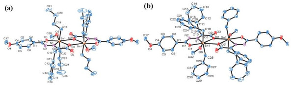
In both complexes, the bond lengths of Sn–N are 2.2385(19) Å (I) and 2.2630(13) Å (II), similar to those observed in literatures[23-25].
Thermal stabilities of both complexes are carried out using a NETZSCH TG 209 F3 thermogravimetric analyzer from 40 to 800 ℃ at a rate of 20 ℃·min–1 under an air atmosphere with a flowing rate of 20.0 mL·min–1. As shown in Fig. 3, the weight loss curves of I and II are relatively similar. Three stages of weight loss can be observed. At the initial stage of 40~200 ℃, the mass loss in the first stage (–5.57% (I), –3.42% (II)) corresponds to the loss of methanol molecules. In the last two stages, both complexes suffer complete decomposition until 800 ℃, corresponding to the removal of ligand and the alkyl group attached to the tin atom. The remaining weight (26.02% (I) and 24.66% (II)) indicates the final products are SnO2 (26.19% (I) and 23.43% (II)). In summary, I and II are rather stable up to about 283 and 214 ℃, respectively.
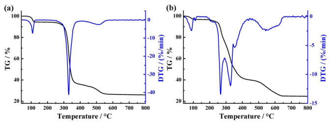
Table 2 lists the values of semi-inhibitory concentrations (IC50) of I, II and carboplatin on cultured cancer cells NCI-H460, HepG2 and MCF-7 in vitro. I and II have obvious inhibitory effects on these three cancer cells. The corresponding inhibitory activity of I is higher than the other two. Among three cancer cells, I is the most sensitive to MCF-7 with an IC50 value of 0.33 ± 0.05 μM. Therefore, I is expected to be further chemically optimized as a candidate complex for anticancer drugs. It can be found that the molecular structure difference between I and II is only the alkyl group attached to the tin atom by crystal structure analysis. Based on this observation, the possible structure-activity relationship can be recognized as follows: dibutyltin complex > dibenzyltin complex. It can be presumed that the style of alkyl-substituted on tin played a key role in killing cancer cells. However, in diorganotin complexes, the difference in cytotoxicity might be attributed to the small steric hindrance or small molecular weight. This trend is in accordance with the results reported in literatures[11].
DownLoad:
CSV
| IC50/μM | |||
| NCI-H460 | HepG2 | MCF-7 | |
| I | 1.19 ± 0.26 | 0.67 ± 0.10 | 0.33 ± 0.05 |
| II | 6.66 ± 0.65 | 5.64 ± 0.49 | 6.12 ± 0.58 |
| Carboplatin | 7.26 ± 0.32 | 7.70 ± 0.25 | 8.22 ± 0.41 |
The UV-vis spectra of I in the absence and presence of CT-DNA are shown in Fig. 4(a). It can be seen from Fig. 4(a) that the decrease in absorbance occurs with the increase of DNA concentration. Hypochromism occurs owning to the breakage of the secondary structure of DNA, whereas hypochromism is due to the stabilization of DNA duplex through a strong interaction such as intercalation. From the absorption spectroscopy tests, Kb values of I are calculated as 1.26 × 10³ L·mol–1 (r2 = 0.98), as can be seen that it is smaller to the reported complexes in literatures[26] and explains the intercalation of I and DNA is relatively weaker. It preliminarily indicates there is intercalation in the interaction between I and DNA.
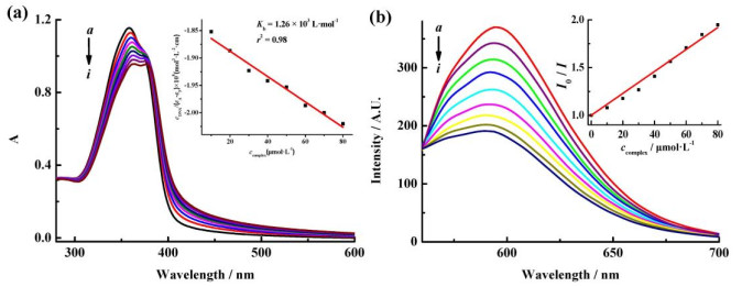
Fig. 4(b) shows the effects of I on the fluorescent spectra of EB-DNA system with different concentrations. With increasing the complexes concentration, the fluorescence of EB-DNA complex system is quenched, so it is speculated the complexes can coordinate with the base groups of DNA molecule, resulting in that EB is squeezed out of the base pairs of the DNA molecule. To study quantificationally the binding capacity of complexes and DNA, we employed the classical Stern-Volmer equation[18] I0/I = 1 + KSVccomplex to obtain the quenching constants KSV of complexes replacing EB and DNA with 1.14 × 10⁴ L·mol–1. This value suggests the complexes insert in DNA to a certain degree. Furthermore, the quenching constants of I are comparable to those of similar reports in literatures[27]. As a result, we can describe the quenching principle as follows: the central Sn atom of complex combined with the bases groups of DNA molecule, the alkyl-substituted on tin is inserted into the DNA base pairs, and then they compete for EB to combine with DNA, resulting in EB to be squeezed out from the base pairs of DNA molecules.
Two diorganotin 2-(2-(4-methoxybenzoyl)hydrazono)-3-phenylpropanoic carboxylate complexes were synthesized. Both complexes show seven-coordination distorted pentagonal dipyramidal configuration with Sn2O2 ring as the center of symmetry. Thermal analysis showed that complexes I and II are stable below 283 and 214 ℃, respectively. The inhibitory activities of I in vitro on cancer cells NCI-H460, HepG2 and MCF-7 were studied. I has obvious inhibitory effects on three cancer cells NCI-H460, HepG2 and MCF-7. I is expected to be further chemically optimized as a candidate compound for anticancer drugs. In tris-HCl buffer solution, the interaction between complex I and calf thymus DNA was studied by UV-vis and fluorescence spectroscopy. The results show that the interaction between complex I and calf thymus DNA is caused by insertion and binding.
Rosenberg, B.; Vancamp, L.; Trosko, J. E.; Mansour, V. H. Platinum compounds: a new class of potent antitumour agents. Nature 1969, 222, 385–386. doi: 10.1038/222385a0
Köberle, B.; Tomicic, M. T.; Usanova, S.; Kaina, B. Cisplatin resistance: preclinical findings and clinical implications. Bba-rev. Cancer 2010, 1806, 172–182.
Amable, L. Cisplatin resistance and opportunities for precision medicine. Pharmacol. Res. 2016, 106, 27–36. doi: 10.1016/j.phrs.2016.01.001
Kenny, R. G.; Marmion, C. J. Toward multi-targeted platinum and ruthenium drugs-a new paradigm in cancer drug treatment regimens? Chem. Rev. 2019, 119, 1058–1137. doi: 10.1021/acs.chemrev.8b00271
Banti, C. N.; Hadjikakou, S. K.; Sismanoglu, T.; Hadjiliadis, N. Anti-proliferative and antitumor activity of organotin(IV) compounds. An overview of the last decade and future perspectives. J. Inorg. Biochem. 2019, 194, 114–152. doi: 10.1016/j.jinorgbio.2019.02.003
Su, W.; Luo, Z. J.; Cui, H.; Tang, Z. F.; Li, P. Y.; Peng, B. H. Synthesis and cytotoxicity of the first arene ruthenium compound containing two thiosemicarbazone ligands. Chin. J. Struct. Chem. 2019, 38, 1543–1548.
Hong, M.; Geng, H. L.; Niu, M. J.; Wang, F.; Li, D. C.; Liu, J. F.; Yin, H. D. Organotin(IV) complexes derived from Schiff base N΄-[(1E)-(2-hydroxy-3-methoxyphenyl)methylidene]pyridine-4-carbohydrazone: synthesis, in vitro cytotoxicities and DNA/BSA interaction. Eur. J. Med. Chem. 2014, 86, 550–561. doi: 10.1016/j.ejmech.2014.08.070
Shang, X. M.; Meng, X. G.; Alegria, E. C. B. A.; Li, Q. S.; Guedes da Silva, M. F. C.; Kuznetsov, M. L.; Pombeiro, A. J. L. Syntheses, molecular structures, electrochemical behavior, theoretical study, and antitumor activities of organotin(IV) complexes containing 1-(4-chlorophenyl)-1-cyclopentanecarboxylato ligands. Inorg. Chem. 2011, 50, 8158–8167. doi: 10.1021/ic200635g
Sirajuddin, M.; Ali, S.; McKee, V.; Akhtar, N.; Andleeb, S.; Wadood, A. Spectroscopic characterizations, structural peculiarities, molecular docking study and evaluation of biological potential of newly designed organotin(IV) carboxylates. J. Photochem. Photobiol. B 2019, 197, 111516. doi: 10.1016/j.jphotobiol.2019.111516
Khan, A.; Parveen, S.; Khalid, A.; Shafi, S. Recent advancements in the anticancer potentials of phenylorganotin(IV) complexes. Inorg. Chim. Acta 2020, 505, 119464. doi: 10.1016/j.ica.2020.119464
Jiang, W. J.; Fan, S. J.; Zhou, Q.; Zhang, F. X.; Kuang, D. Z.; Tan, Y. X. Diversity of complexes based on p-nitrobenzoylhydrazide, benzoylformic acid and diorganotin halides or oxides self-assemble: cytotoxicity, the induction of apoptosis in cancer cells and DNA-binding properties. Bioorg. Chem. 2020, 94, 103402. doi: 10.1016/j.bioorg.2019.103402
Liu, J.; Li, Z. Q.; Yi, Y. Y.; Zhong, Y. X.; Yu, H. T.; Tan, Y. X.; Jiang, W. J. Syntheses, crystal structures and in vitro anticancer activity of four binuclear benzyltin complexes based on acylhydrazone ligand. Chin. J. Inorg. Chem. 2019, 35, 2200–2208.
Yu, H. T.; Tan, Y. X.; Kuang, D. Z.; Zhang, F. X.; Jiang, W. J. Synthesis, structure and biological activity of diphenyltin complexes based on O, N, O-tridentate ligands. Inorg. Chim. Acta 2019, 496, 119044. doi: 10.1016/j.ica.2019.119044
Li, Y. X.; Yu, H. T.; Zeng, H. T.; Liu, M. Q.; Kuang, D. Z.; Tan, Y. X.; Jiang, W. J. Two new dibenzyltin complexes based on the 2-oxo-3-phenylpropionic acid arylformylhydrazone: syntheses, crystal structures and biological activity. Chin. J. Struct. Chem. 2019, 38, 1947–1955.
Jiang, W. J.; Zhou, Q.; Liu, M. Q.; Zhang, F. X.; Kuang, D. Z.; Tan, Y. X. Microwave assisted synthesis of disubstituted benzyltin arylformylhydrazone complexes: anticancer activity and DNA-binding properties. Appl. Organomet. Chem. 2019, 33, e5092.
Sheldrick, G. M. SHELXL-97, a Program for Crystal Structure Refinement. Germany Göttingen: University of Göttingen 1997.
Pyle, A. M.; Rehmann, J. P.; Meshoyrer, R.; Kumar, C. V.; Turro, N. J.; Barton, J. K. Mixed-ligand complexes of ruthenium(II): factors governing binding to DNA. J. Am. Chem. Soc. 1989, 111, 3051–3058. doi: 10.1021/ja00190a046
Yan, C. Q.; Zhang, J. L.; Liang, T. G.; Li, Q. S. Diorganotin(IV) complexes with 4-nitro-N-phthaloyl-glycine: synthesis, characterization, antitumor activity and DNA-binding studies. Biomed. Pharmacother. 2015, 71, 119–127. doi: 10.1016/j.biopha.2015.02.027
Deacon, G. B.; Phillips, R. J. Relationships between the carbon-oxygen stretching frequencies of carboxylato complexes and the type of carboxylate coordination. Coordin. Chem. Rev. 1980, 33, 227–250. doi: 10.1016/S0010-8545(00)80455-5
Zhang, Z. J.; Zeng, H. T.; Liu, Y.; Kuang, D. Z.; Zhang, F. X.; Tan, Y. X.; Jiang, W. J. Synthesis, crystal structure and anticancer activity of the dibutyltin(IV)oxide complexes containing substituted salicylaldehyde-o-aminophenol Schiff base with appended donor functionality. Inorg. Nano-Met. Chem. 2018, 48, 486–494. doi: 10.1080/24701556.2019.1571513
Pretsch, E.; Bühlmann, P.; Badertscher, M. Structure Determination of Organic Compounds. Fourth ed. Berlin Heidelberg: Springer-Verlag 2009, 69–242.
Luo, B.; Yu, H. T.; Liu, M. Q.; Zhang, F. X.; Kuang, D. Z.; Tan, Y. X.; Jiang, W. J. Syntheses, crystal structures and biological activity of dibenzyltin complexes base on the substituted benzoyl hydrazine-pyruvic acid. Chin. J. Inorg. Chem. 2019, 35, 1212–1220.
Tan, Y. X.; Zhang, Z. J.; Feng, Y. L.; Yu, J. X.; Zhu, X. M.; Zhang, F. X.; Kuang, D. Z.; Jiang, W. J. Syntheses, crystal structures and biological activity of the 1D chain benzyltin complexes based on 2-oxo-propionic acid benzoyl hydrazone. J. Inorg. Organomet. P 2017, 27, 342–352. doi: 10.1007/s10904-016-0477-5
Tan, Y. X.; Zhang, Z. J.; Liu, Y.; Yu, J. X.; Zhu, X. M.; Kuang, D. Z.; Jiang, W. J. Synthesis, crystal structure and biological activity of the Schiff base organotin(IV) complexes based on salicylaldehyde-o-aminophenol. J. Mol. Struct. 2017, 1149, 874–881. doi: 10.1016/j.molstruc.2017.08.058
Tian, L. J.; Chen, L. X.; An, W. G.; Liu, X. C. Diorganotin complexes of N-[4-(diethylamino)salicylidene]-(L)-tryptophane: syntheses, structures and properties. Chin. J. Struct. Chem. 2019, 38, 1977–1985.
Liu, Y. Z.; Gao, H. Y.; Yi, X. G.; Li, D. P.; Li, Y. X. Crystal structures and DNA binding properties of 2-naphthoxyacetic acid Cu(II) complexes. Chin. J. Struct. Chem. 2019, 38, 1362–1369.
Zhao, Y.; Li, Z.; Li, H. H.; Wang, S. N.; Niu, M. J. Synthesis, crystal structure, DNA binding and in vitro cytotoxicity studies of Zn(II) complexes derived from amino-alcohol Schiff-bases. Inorg. Chim. Acta 2018, 482, 136–143. doi: 10.1016/j.ica.2018.06.008

Figure 2 Molecular structures of I (a) and II (b). Symmetry codes for I: i 2–x, –y, –z; II: i 2–x, 1–y, 1–z

Figure 4 (a) Electronic spectra of I in tris-HCl buffer upon addition of CT–DNA, ccomplex = 50 μmol·L–1; from a to i, cDNA = 0, 10, 20, 30, 40, 50, 60, 70 and 80 μmol·L–1, respectively. The arrow shows the absorbance changes upon increasing DNA concentrations. Inset: plot of cDNA/(εA − εF) vs. ccomplex; (b) Effects of I on the fluorescent spectra of EB-DNA system cCT–DNA = 30 umol·L–1; cEB = 3 umol·L–1; from a to i, ccomplex = 0, 10, 20, 30, 40, 50, 60, 70 and 80 μmol·L–1, respectively; inset: plot of I0/I vs. ccomplex; λex = 258 nm
Table 1. Selected Bond Lengths (Å) and Bond Angles (°) for I and II
| I | |||||||
| Bond | Dist. | Bond | Dist. | Bond | Dist. | ||
| Sn(1)–C(22) | 2.117(2) | Sn(1)–O(1) | 2.1773(17) | Sn(1)–O(2) | 2.3058(17) | ||
| Sn(1)–C(18) | 2.122(2) | Sn(1)–N(1) | 2.2385(19) | Sn(1)–O(2)i | 2.8017(18) | ||
| Angle | (°) | Angle | (°) | Angle | (°) | ||
| C(22)–Sn(1)–C(18) | 155.92(11) | O(1)–Sn(1)–N(1) | 70.16(7) | C(22)–Sn(1)–O(5) | 82.36(10) | ||
| C(22)–Sn(1)–O(1) | 95.17(9) | C(22)–Sn(1)–O(2) | 91.91(9) | C(18)–Sn(1)–O(5) | 80.30(9) | ||
| C(18)–Sn(1)–O(1) | 97.24(9) | C(18)–Sn(1)–O(2) | 91.82(8) | O(1)–Sn(1)–O(5) | 77.48(7) | ||
| C(22)–Sn(1)–N(1) | 103.55(9) | N(1)–Sn(1)–O(2) | 69.73(6) | O(2)i–Sn(1)–O(5) | 77.96(6) | ||
| O(2)–Sn(1)–O(2)i | 64.75(6) | O(2)i–Sn(1)–C(18) | 79.87(8) | O(2)i–Sn(1)–C(22) | 80.26(8) | ||
| II | |||||||
| Bond | Dist. | Bond | Dist. | Bond | Dist. | ||
| Sn(1)–C(25) | 2.1425(16) | Sn(1)–O(1) | 2.1511(11) | Sn(1)–O(2) | 2.3759(11) | ||
| Sn(1)–C(18) | 2.1463(16) | Sn(1)–N(1) | 2.2630(13) | Sn(1)–O(2)i | 2.6815(13) | ||
| Angle | (°) | Angle | (°) | Angle | (°) | ||
| C(25)–Sn(1)–C(18) | 165.88(7) | O(1)–Sn(1)–N(1) | 70.30(4) | C(25)–Sn(1)–O(2) | 86.86(5) | ||
| C(25)–Sn(1)–O(1) | 93.10(6) | C(25)–Sn(1)–O(5) | 93.73(6) | C(18)–Sn(1)–O(2) | 87.38(5) | ||
| C(18)–Sn(1)–O(1) | 99.70(5) | C(18)–Sn(1)–O(5) | 83.23(6) | N(1)–Sn(1)–O(2) | 68.90(4) | ||
| C(18)–Sn(1)–N(1) | 92.99(6) | O(1)–Sn(1)–O(5) | 77.99(4) | O(2)i–Sn(1)–O(5) | 76.22(4) | ||
| O(2)–Sn(1)–O(2)i | 67.09(4) | O(2)i–Sn(1)–C(18) | 81.15(5) | O(2)i–Sn(1)–C(25) | 84.73(5) | ||
| Symmetry codes: (I) i 2–x, –y, –z; (II) i 2–x, 1–y, 1–z | |||||||
 下载: 导出CSV
Table 2. Inhibition Action of the Complexes to Cancer Cell in vitro
| IC50/μM | |||
| NCI-H460 | HepG2 | MCF-7 | |
| I | 1.19 ± 0.26 | 0.67 ± 0.10 | 0.33 ± 0.05 |
| II | 6.66 ± 0.65 | 5.64 ± 0.49 | 6.12 ± 0.58 |
| Carboplatin | 7.26 ± 0.32 | 7.70 ± 0.25 | 8.22 ± 0.41 |
下载: 导出CSV

 扫一扫看文章

扫一扫关注我们

