College of Biomedical Engineering, Sichuan University, Chengdu 610065, China
b.
State Key Laboratory of Oral Diseases, National Center for Stomatology, National Clinical Research Center for Oral Diseases, West China Hospital of Stomatology, Sichuan University, Chengdu 610041, China
c.
Department of Orthopedics, Orthopedic Research Institute, West China Hospital, Sichuan University, Chengdu 610041, China
d.
Department of Oral Implantology, The Affiliated Stomatological Hospital of Southwest Medical University, Luzhou 646000, China
e.
Sichuan Provincial Engineering Research Center of Oral Biomaterials, Chengdu 610041, China
Received Date:
09 December 2023 Accepted Date:
03 February 2024 Revised Date:
01 February 2024 Available Online:
15 November 2024
Abstract:
Triple-negative breast cancer, due to its aggressive nature and lack of targeted treatment, faces serious challenges in breast cancer treatment. Conventional therapies, such as chemotherapy, are encumbered by a range of limitations, and there is an urgent need for more effective treatment strategies. Ferroptosis, as an iron-dependent form of cell death, has exhibited promising potential in cancer treatment. Combining ferroptosis with other cancer therapies offers new avenues for treatment. Tetrahedral DNA nanostructure (TDN), a novel DNA-based three-dimensional (3D) nanomaterial, is promising drug delivery vehicle and can be utilized for functionalizing inorganic nanomaterials. In this work, we have demonstrated the preparation of Fe3O4-PEI@TDN-DOX nanocomposites and elucidated their antitumor mechanism. The TDN facilitated the enhanced cellular uptake of polyetherimide (PEI)-modified Fe3O4, and the delivery of the chemotherapeutic drug doxorubicin (DOX) further augmented their anti-tumor effect. This novel strategy can destroy the tumor redox homeostasis and produce overwhelming lipid peroxides, consequently sensitizing the tumor to ferroptosis. The integration of ferroptosis with other cancer therapies opens up new possibilities for treatment. This research provides valuable mechanistic insights and practical strategies for leveraging nanotechnology to induce ferroptosis and amplify its impact on tumor cells.
Cancer is a significant global health concern killing approximately 10 million people every year, with breast cancer being the most common form among women [1]. Triple-negative breast cancer (TNBC), as a subtype of breast cancer, is regarded as a significant challenge due to its aggressive nature and lack of targeted treatment options [2,3]. Current treatment approaches for breast cancer mainly include surgery, radiotherapy, chemotherapy, and targeted therapy [4,5].
The discovery of programmed cell death processes has led to advances in cancer treatment that differ from conventional treatments [6]. Numerous pathways for cell death have been unveiled, encompassing apoptosis and pyroptosis, with ferroptosis representing a prominent discovery since reported in 2012 [7,8]. Distinct from other forms of cell death, the key mechanism of ferroptosis is due to the depletion of glutathione (GSH) or the inactivation of the regulatory core glutathione peroxidase 4 (GPX4) under the action of ferrous iron or lipoxygenase [9,10]. This culminates in the peroxidation of unsaturated fatty acids abundant in cell membranes, consequently instigating cell death [11].
In the past few years, ferroptosis has emerged as a promising strategy for tumor treatment, providing a new avenue for tumor treatment [12]. Traditional approaches to induce ferroptosis have focused on using small molecule inducers. For example, Erastin has been reported as an inducer of ferroptosis, which can directly inhibit the system xc− to reduce GSH levels, thereby inhibiting the growth of cervical and ovarian cancer cells [13,14]. However, this type of molecular drug has limitations such as poor water solubility, low cell targeting, and rapid metabolism in the body, making it difficult to exert the desired effect [11,15]. With the in-depth research on ferroptosis and the development of nanotechnology, nanomaterials have been proven to be more effective drugs for treating ferroptosis than biological drugs [16]. Among them, iron-based nanomaterials, such as Fe3O4 [17], can directly release high concentrations of iron ions, increase the level of reactive oxygen species (ROS) in cells, thereby promoting ferroptosis [18,19].
In recent years, efforts have been made to improve the anti-tumor properties of ion-based nanomaterials through the design of multifunctional iron-based materials for enhanced cancer therapy [20]. Fox example, Zen et al. demonstrate the CroFe@BSA nanoparticles for combination cancer therapy by utilizing the mechanisms of ferroptosis and the photothermal effect [21]. Xu et al. reported the development of Tf-LipoMof@PL nanoparticles, which are featured with iron-containing metal-organic frameworks (MOF) and transferrin-mediated iron endocytosis, inducing both ferroptosis and pyroptosis [22]. Among these additional therapies applied, chemotherapy, specifically the use of doxorubicin (DOX), is a commonly employed treatment for cancer [23]. However, DOX is associated with several limitations, including poor drug resistance, limited tissue penetration, and non-selective distribution, leading to unsatisfactory clinical outcomes and various side effects such as immunosuppression, cardiotoxicity, myelosuppression, neuropathy, and myalgia [24-26]. Consequently, the development of an effective drug delivery system for DOX that can selectively target and kill cancer cells presents a significant challenge in cancer treatment.
In order to find a suitable transport platform for iron-based nanomaterials and DOX, we focused on DNA nanomaterials [27]. DNA nanostructures have received widespread attention due to their versatile editability, inherent biocompatibility, and outstanding biomedical application capabilities [28,29]. Among various DNA materials, DNA tetrahedron (tetrahedron DNA nanostructure, TDN) has the advantages of high structural stability in biological environment, good biocompatibility, high cell entry efficiency, strong loading capacity, and easy diversified modification [30-32]. Notably, the anthracene ring structure of DOX allows it to insert between the base pairs of double helix DNA to form a DNA-drug complex [33-35]. Therefore, TDN can be one promising nanodrug delivery system for constructing multi-functional iron-based nanomaterials.
In this study, we demonstrate the fabrication of Fe3O4-PEI@TDN-DOX nanocomposites and explore their role in the synergistic treatment of tumors. TDN was assembled by four specially designed DNA single-strands, then loaded an anti-cancer drug DOX onto the TDN structure. Then it is combined with polyetherimide (PEI)-modified iron-based nanomaterials to form a nanocomposite (Fe3O4-PEI@TDN-DOX) through electrostatic adsorption. Using TDN as a drug delivery media, this allows iron-based nanomaterials as ferroptosis inducers and doxorubicin as anti-cancer drugs to achieve a synergistic approach to cancer therapy. This work provides a promising strategy for using nanotechnology to treat tumor through a synergy of ferroptosis and chemotherapy (Scheme 1).
Scheme 1
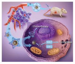
Scheme 1.
Tetrahedral DNA nanostructure serves as a drug delivery medium for the co-delivery of chemotherapeutic drug doxorubicin and iron-based nanomaterial into 4T1 cells, enabling a synergistic treatment approach that combines chemotherapy and the promotion of ferroptosis for effective tumor therapy.
The previously established protocols for the synthesis of TDN were used [36,37]. In brief, four specially designed single-stranded DNA molecules (ssDNA) were added into Tris-MgCl2 buffer (Table S1 in Supporting information), the mixed solution was heated to 95 ℃ for 10 min and then cooled to 4 ℃ for 20 min to obtain TDN. Subsequently, DOX was incorporated into the base pairs of TDN through incubation at room temperature for 24 h to form TDN-DOX. Finally, Fe3O4-PEI@TDN-DOX was synthesized through electrostatic adsorption of PEI-modified iron-based nanomaterials (namely Fe3O4 in this work) and TDN-DOX.
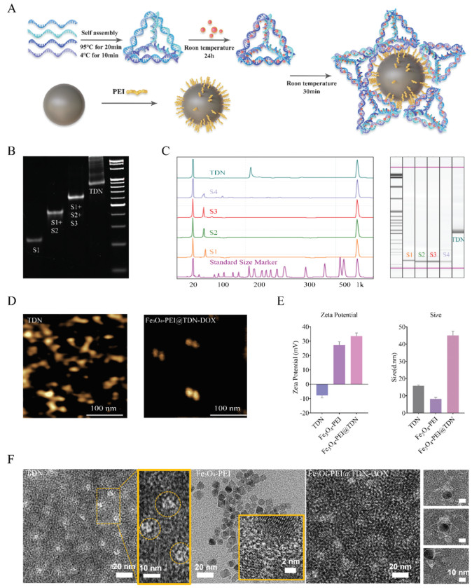
The synthesis of Fe3O4-PEI@TDN-DOX consists of two steps (Fig. 1A). The successful synthesis of TDN was verified by polyacrylamide gel electrophoresis (PAGE) analysis (Fig. 1B). As shown in the gel image, the bands correspond to S1, S1+S2, S1+S2+S3, TDN (from left to right). The synthesized TDN matched the 200 bp band in the gel image, which is consistent with the theoretical value reported in our previous study [38]. In addition, the synthesis of TDN was verified using high performance capillary electrophoresis (HPCE) (Fig. 1C). Subsequently, agarose gel electrophoresis (Fig. S1 in Supporting information) and dynamic light scattering (DLS) were used to measure the zeta potential (Fig. 1E and Fig. S2 in Supporting information), which proved that Fe3O4-PEI and TDN-DOX could be obtained via electrostatic adsorption. Due to the phosphate group on the nucleotide, TDN has a negative charge of approximately −7.85 ± 1.37 mV. In addition, Fe3O4-PEI nanoparticles exhibit a positive charge of approximately 27.4 ± 1.74 mV, making it attracted to TDN-DOX. Additionally, the zeta potential value of Fe3O4-PEI@TDN-DOX nanocomposites can be determined 33.5 ± 1.76 mV, which indicates their successful synthesis. The gel image clearly demonstrates the reversal of electronegativity in Fe3O4-PEI@TDN-DOX compared to TDN-DOX. Furthermore, this reversal also causes the Fe3O4-PEI@TDN-DOX to move in the opposite direction in the gel electrophoresis, providing further evidence of successful synthesis. Additionally, the size distribution was determined by DLS, and the morphology was observed by atomic force microscopy (AFM) and transmission electron microscope (TEM), as displayed in Figs. 1D and F. The images of TDN reveals a triangular shape with dimensions of approximately 15 nm, whereas the Fe3O4-PEI exhibits a circular morphology with a size of about 9 nm. Consequently, Fe3O4-PEI@TDN-DOX is comprised of multiple TDN-DOX surrounding Fe3O4-PEI from the outside, resulting in a larger size of approximately 45 nm. These alterations in size and morphology of the complex confirms the successful synthesis of Fe3O4-PEI@TDN-DOX.
Figure 1
Figure 1.
Characterization of TDN, TDN-DOX and Fe3O4@TDN-DOX. (A) Schematic illustration of Fe3O4-PEI@TDN-DOX fabrication. (B) PAGE result of ssDNA and TDN. (C) Results of HPCE verifying the synthesis of TDN. (D) AFM image of Fe3O4-PEI. (E) Zeta potential and size of TDN, TDN-DOX and Fe3O4-PEI@TDN-DOX measured by DLS. (F) Morphology of TDN, TDN-DOX and Fe3O4-PEI@TDN-DOX by TEM. Data are presented as mean ± standard deviation (SD) (n = 3).
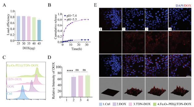
To elucidate the desirable encapsulation efficiency, we calculated the standard curve of absorbance-concentration relationship of DOX and varied the concentration of DOX to interact with TDN (Fig. S3 in Supporting information) [38]. The result indicated that when 3.5 µg of DOX was added to 100 µL (1000 nmol/L), the encapsulation efficiency rate was still greater than 80% (Fig. 2A). Furthermore, the image provides evidence that the loading capacity of DOX on TDN has a maximum limit (Fig. S4 in Supporting information). Subsequently, we conducted tests to confirm the stability of TDN-DOX. As displayed in Fig. S5 (Supporting information), TDN-DOX nanocomposites exhibit strong stability both at room temperature and at 4 ℃ for up to 7 days. Subsequently, the DOX release behavior was measured via adialysis membrane against PBS at different pH values (Fig. 2B). Fe3O4-PEI@TDN-DOX exhibited a negligible DOX release under physiological conditions (pH 7.4) within 30 h, revealing its excellent stability. While changing to acidic conditions (pH 5.5), the complexes showed significant DOX release over time and at 24 h over 80% DOX was released, which is anticipated to have a long-term anti-tumor effect in the acidic tumor microenvironment. Likewise, the behaviors of iron ion release at various pH levels mimics that of DOX, exhibiting faster release under acidic conditions, with 90% released within a short duration of 60–80 min (Fig. S6 in Supporting information). This indicates that Fe3O4-PEI@TDN-DOX can retain stability under physiological conditions, safeguard normal tissue, and promptly release under acidic conditions to facilitate treatment.
Figure 2
Figure 2.
Drug release and cellular uptake of Fe3O4-PEI@TDN-DOX. (A) Encapsulation efficiency of TDN with DOX. (B) Cumulative release of DOX from TDN-DOX at different pH values (pH 5.5 or 7.4). (C) Quantitative analysis of DOX cellular uptake by flow cytometry. (D) Statistical analysis of (C). (E) Confocal laser scanning microscopy (CLSM) images of DOX cellular uptake. Scale bar: 200 µm. Data are presented as mean ± SD (n = 3). ****P < 0.0001. ns, no significance.
DOX exhibited highly therapeutic effect on a variety of solid tumors through combining to the DNA and blocking the DNA synthesis [23,39]. Due to the free diffusion mechanism of small molecules, free DOX exhibits the effect of rapidly enhancing cellular uptake. According to the images (Figs. 2C and E), compared with free DOX, DOX on TDN can also quickly enter cells and thus exert its effect. The quantitative results (Fig. 2D) show that there is no obvious statistical difference compared with free DOX. However, benefiting from remarkable cellular uptake capability of TDN and pH-sensitive release behavior, TDN is expected to delivery DOX and enhance the effects of chemotherapy (Fig. S7 in Supporting information).
The results of the cytotoxicity experiment (Fig. 3A) showed that after 24 h of co-culture, TDN, serving as vehicles had no obvious effect on 4T1 cells in the concentration range of 100–300 nmol/L, suggesting its superior biocompatibility. When Fe3O4-PEI is used individually, it shows stronger toxicity as the concentration increases. However, this does not mean better treatment results, Fe3O4-PEI nanoparticles suffer from limited cellular entry and is unable to effectively aggregate and remain in the target region. Its therapeutic performance will be weakened and high concentrations of Fe3O4-PEI nanoparticles may cause damage to normal tissues. At the same time, Fe3O4-PEI@TDN-DOX nanoparticles demonstrate stronger cytotoxicity compared to Fe3O4-PEI and TDN-DOX when used individually.
Figure 3
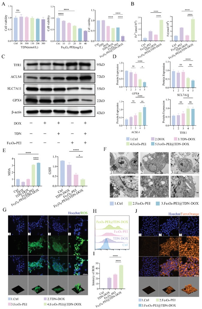
Figure 3.In vitro antitumor efficacy and ferroptosis. (A) Cell ability on 4T1 for 24 h. (B) Changes of Fe2+ and Fe3+on 4T1. (C, D) Protein expression levels and semiquantitative analysis. (E) Changes in intracellular GSH and MDA levels. (F) Ferroptosis cell morphological changes by TEM. Scale bar: 4 µm and 1 µm. (G) Fluorescence microscopy assay detected the ROS level. Scale bar: 200 µm. (H) Quantitative analysis of ROS level. (I) Statistical analysis of (H). (J) Fluorescence microscopy assay detected the FerroOrange staining. Scale bar: 200 µm. Data are presented as mean ± SD (n = 3). P < 0.05, **P < 0.01, ****P < 0.0001.
Iron is essential in a variety of physiological processes, including electron transfer, oxygen transport, and DNA biosynthesis [40]. Normally, iron is stored and transported in the form of Fe3+, while Fe2+ serves as an electron donor catalyzing reactions in the Fenton reaction [41]. Under conditions of iron overload, excessive iron participates in the generation of free radicals and generates ROS through the iron-dependent Fenton reaction, thereby promoting lipid peroxidation and ultimately triggering ferroptosis [11,42]. Therefore, by monitoring the dynamic changes of intracellular iron ions during this process, the extent of ferroptosis can be evaluated. After 24 h of drug exposure to the cells, we detected the concentrations of intracellular iron and ferrous ions through colorimetric analysis (Fig. 3B). Both iron ions and ferrous ions were found to have significantly higher concentrations in the Fe3O4-PEI@TDN-DOX groups compared to the Fe3O4-PEI group. This can be primarily attributed to the mediation of TDN, which facilitates the increased entry into the cells. Additionally, the synergistic effects of chemotherapy drugs stimulated the release of endogenous iron from tumor cells, allowing it to participate in the Fenton cycle and further promoting tumor death. Simultaneously, consistent results were obtained through FerroOrange staining and observation under confocal microscopy (Fig. 3J).
System xc− plays an important regulatory role in the process of ferroptosis, and its main function is to mediate the exchange between cystine and cysteine [43]. Cystine serves as an important antioxidant and fights oxidative stress by maintaining intracellular GSH levels [44]. In ferroptosis, the activity of System xc− is critical for maintaining intracellular GSH levels [45]. When System xc− is inhibited or inactivated, intracellular GSH levels decrease, leading to an increase in oxidative stress, thereby promoting lipid peroxidation and ultimately promoting ferroptosis [46]. GPX4 not only maintains the redox homeostasis but also converts lipid ROS into lipid alcohols, earning it the designation of the "gatekeeper" of ferroptosis [8,43]. Direct inactivation of GPX4 has been demonstrated to drive ferroptosis. SLC7A11, by maintaining the GSH level, acts against oxidative stress and collaborates with GPX4 to maintain the cellular redox balance [15]. Acyl-CoA synthetase long chain family member 4 (ACSL4) indirectly participates in the lipid peroxidation process by regulating lipid composition [44,47]. In addition, transferrin receptor protein 1 (TFR1) also plays a certain role in mediating cellular iron uptake, and TFR1 can also be used as a marker for ferroptosis [48,49]. In this work, the changes in relevant proteins during the process were assessed through Western blot experiments to determine the extent of ferroptosis (Figs. 3C and D). After 24 h of interaction with cells, the Fe3O4-PEI@TDN-DOX group exhibits stronger inhibition of System xc−, accompanied by a significant decrease in the expression levels of GPX4 and SLC7A11. This suggests that the intracellular redox balance is significantly disturbed. At the same time, the expression of ACSL4 was increased. Given that the activity of ACSL4 is related to sensitivity to lipid peroxidation, its upregulation indicates an increase in cells sensitivity to lipid peroxidation, making cells more susceptible to oxidative stress. It is worth noting that the expression level of TFR1 did not change significantly in group Fe3O4-PEI@TDN-DOX and group Fe3O4-PEI. This suggests that TDN plays a key role in helping Fe3O4-PEI enter the cell. The decrease in GSH during the process of ferroptosis signifies a compromised cellular redox balance and an escalation in oxidative stress. GSH, being a pivotal antioxidant, its reduction implies a diminished capacity of cells to resist oxidative pressure, providing a conducive environment for the occurrence of ferroptosis. Simultaneously, malondialdehyde (MDA), as the end product of lipid peroxidation, serves as a detectable biomarker [50]. In ferroptosis, the generation of MDA is induced by lipid peroxidation, and its increased levels reflect the intensification of lipid peroxidation reactions. Through the assessment of intracellular GSH and MDA levels (Fig. 3E), we observed a significant reduction in GSH and a notable increase in MDA in the Fe3O4-PEI@TDN-DOX group. This further confirms the pronounced development of ferroptosis within the cells. This outcome not only substantiates the occurrence of ferroptosis at the biological level but also furnishes observable biological indicators, aiding in the in-depth investigation of this cell death pathway. In ferroptosis, the generation of ROS is a key step. ROS are highly active oxidizing molecules including superoxide ions, hydrogen peroxide, hydroxyl radicals, etc. Excess iron can catalyze the production of ROS through the Fenton reaction. The Fenton reaction involves iron ions participating in the oxygen reduction reaction to generate highly active hydroxyl radicals [51]. These ROS trigger oxidative stress in cells, leading to the destruction of cell structures such as lipid peroxidation, protein damage, and DNA damage. The accumulation of ROS during ferroptosis is the result of oxidative stress in cells, which is a key feature of ferroptosis. In the Fe3O4-PEI@TDN-DOX group, the production of ROS was significantly higher than in the other groups (Figs. 3G–I). This indicates that the cells are in a more vulnerable state, experiencing elevated levels of intracellular oxidative stress, ultimately leading to cell death. The changes of cell morphology after Fe3O4-PEI and Fe3O4-PEI@TDN-DOX treatment were observed by TEM (Fig. 3F). It can be observed that the mitochondria of cells become smaller, the membrane density increases, and the cristae decrease after Fe3O4-PEI and Fe3O4-PEI@TDN-DOX culture with 4T1 for 24 h, and additionally, partial cell membrane rupture occurs. The results indicated a notable occurrence of ferroptosis in the cells, with the Fe3O4-PEI@TDN-DOX group displaying a more intense response.
The experimental plan for in vivo anti-tumor research in 4T1 tumor-bearing mice is illustrated in Fig. 4A. All animal experiments were approved by the Research Ethics Committee of West China Hospital of Stomatology, Sichuan University, and performed in accordance with the laws on experiment animals. After transplanting 4T1 cells for 10 days (when the tumor volume reached approximately 100 mm3), tumor-bearing mice were treated through tail vein injection (every 2 days). Tumor volume comparison is the most direct indicator for assessing anti-tumor efficacy. In Fig. 4B, it can be observed that, compared to the saline group, free DOX exhibited unsatisfactory non-selective distribution in vitro, resulting in suboptimal cytotoxicity. Additionally, appropriately sized TDN-DOX facilitated increased accumulation of DOX in tumors, demonstrating superior anti-tumor effects compared to free DOX. However, to mitigate the toxicity of chemotherapy drugs, a lower drug concentration was chosen. Although the anti-tumor effects were not ideal, the Fe3O4-PEI@TDN-DOX group, which combined iron death treatment for tumors, significantly enhanced the tumor treatment efficacy. Consistent trends in tumor volume changes (Fig. 4C) and tumor weight (Fig. 4E) among different groups indicate that Fe3O4-PEI@TDN-DOX has a notable anti-tumor effect. Meanwhile, there was no significant difference in body weight of 4T1 tumor-bearing mice among all groups (Fig. 4D). Changes in major organs and tumors were observed through histology and immunohistochemical staining before and after treatment. Hematoxylin and eosin (H&E) staining (Fig. 4F) showed dense arrangement and intact morphology of tumor cells in the saline group, while the Fe3O4-PEI@TDN-DOX group exhibited evident nuclear fragmentation and condensation. Compared to saline free DOX and TDN-DOX, terminal deoxynucleotidyl transferase-mediated dUTP nick end labeling (TUNEL) staining exhibited obvious apoptosis in the Fe3O4-PEI@TDN-DOX group (Figs. 4G and H). The cell adhesion molecule-1 (CD31) was used to evaluate the level of tumor angiogenesis, where the presence of a deep brown color indicated positive results. The immunohistochemical analysis of CD31 showed a noteworthy decrease in the quantity of tumor microvessels in the Fe3O4-PEI@TDN-DOX group, suggesting a marked inhibitory impact on tumor angiogenesis (Fig. S11 in Supporting information). Ki67 reflected the level of cellular proliferation or growth activity. The Ki67 staining results indicated that the brownish-yellow positive cells in the Fe3O4-PEI@TDN-DOX group were significantly fewer than in the other groups, signifying a marked inhibition of tumor cell proliferation by Fe3O4-PEI@TDN-DOX (Fig. S11). These results suggest that Fe3O4-PEI@TDN-DOX demonstrated excellent efficacy in suppressing tumor growth. Simultaneously, we assessed the biocompatibility of the drugs and potential side effects (Figs. S12 and S13 in Supporting information). After ten administrations, abnormalities in specific indicators of kidney and liver function were detected in both the free DOX and TDN-DOX groups, while the indicators in the Fe3O4-PEI@TDN-DOX group were more similar to those in the saline group, suggesting favorable biocompatibility of Fe3O4-PEI@TDN-DOX.
Figure 4
Figure 4.In vivo antitumor effect in 4T1 tumor-bearing mice. (A) Experiment plan for the in vivo antitumor study in 4T1 tumor-bearing mice. (B) Visible 4T1 tumor excised from 4T1 tumor-bearing mice after 20 days. (C) Tumor volume−time graph. (D) Mouse weight−time graph. (E) Tumor weight. (F) H&E staining of tumor tissue. Scale bar: 200 µm. (G) TUNEL staining of tumor excised from 4T1 tumor-bearing mice. Scale bar: 400 µm and 100 µm. (H) Statistical analysis of TUNEL staining. Data are presented as mean ± SD (n = 4). **P < 0.01, ***P < 0.001, ****P < 0.0001.
This study successfully synthesized the Fe3O4-PEI@TDN-DOX nanocomposite, providing an innovative strategy for chemical and ferroptosis synergistic cancer therapy. The nanocomposite exhibited excellent biocompatibility and potent cytotoxicity at the cellular level, particularly demonstrating significant anti-tumor effects in ferroptosis-based treatments. Intracellular iron ion concentration monitoring revealed the inherent therapeutic mechanism of the composite, highlighting the essential role of the tetrahedral carrier in this process. Further investigations into cellular biological indicators, such as antioxidant capacity and lipid peroxidation, deepened our understanding of the ferroptosis process. Ultimately, the practical anti-tumor performance of Fe3O4-PEI@TDN-DOX nanocomposite was confirmed by a murine tumor model, demonstrating a significantly enhanced tumor therapeutic efficacy and outstanding biocompatibility. This study presents an innovative synergistic anti-tumor strategy for delivering exogenous iron into cells to promote ferroptosis and providing additionally chemotherapy drugs delivered by TDN, offering valuable insights for the future development of cancer therapies.
Declaration of competing interest
The authors declare that they have no known competing financial interests or personal relationships that could have appeared to influence the work reported in this paper.
Acknowledgments
This work was supported by the National Key R&D Program of China (No. 2019YFA0110600), the National Natural Science Foundation of China (Nos. 82370929 and 81970916), the Sichuan Science and Technology Program (Nos. 2022NSFSC0002 and 2023YFG022), Sichuan Province Youth Science and Technology Innovation Team (No. 2022JDTD0021), Research and Develop Program, West China Hospital of Stomatology Sichuan University (No. RD03202302), Q. S. acknowledges the Research Funding from West China School/Hospital of Stomatology Sichuan University (No. QDJF2022-2). The authors would like to thanks to Liying Hao (State Key Laboratory of Oral Diseases, West China Hospital of Stomatology, Sichuan University) for help in characterizing AFM.
Supplementary materials
Supplementary material associated with this article can be found, in the online version, at doi:10.1016/j.cclet.2024.109620.
Scheme 1
Tetrahedral DNA nanostructure serves as a drug delivery medium for the co-delivery of chemotherapeutic drug doxorubicin and iron-based nanomaterial into 4T1 cells, enabling a synergistic treatment approach that combines chemotherapy and the promotion of ferroptosis for effective tumor therapy.
Figure 1
Characterization of TDN, TDN-DOX and Fe3O4@TDN-DOX. (A) Schematic illustration of Fe3O4-PEI@TDN-DOX fabrication. (B) PAGE result of ssDNA and TDN. (C) Results of HPCE verifying the synthesis of TDN. (D) AFM image of Fe3O4-PEI. (E) Zeta potential and size of TDN, TDN-DOX and Fe3O4-PEI@TDN-DOX measured by DLS. (F) Morphology of TDN, TDN-DOX and Fe3O4-PEI@TDN-DOX by TEM. Data are presented as mean ± standard deviation (SD) (n = 3).
Figure 2
Drug release and cellular uptake of Fe3O4-PEI@TDN-DOX. (A) Encapsulation efficiency of TDN with DOX. (B) Cumulative release of DOX from TDN-DOX at different pH values (pH 5.5 or 7.4). (C) Quantitative analysis of DOX cellular uptake by flow cytometry. (D) Statistical analysis of (C). (E) Confocal laser scanning microscopy (CLSM) images of DOX cellular uptake. Scale bar: 200 µm. Data are presented as mean ± SD (n = 3). ****P < 0.0001. ns, no significance.
Figure 3In vitro antitumor efficacy and ferroptosis. (A) Cell ability on 4T1 for 24 h. (B) Changes of Fe2+ and Fe3+on 4T1. (C, D) Protein expression levels and semiquantitative analysis. (E) Changes in intracellular GSH and MDA levels. (F) Ferroptosis cell morphological changes by TEM. Scale bar: 4 µm and 1 µm. (G) Fluorescence microscopy assay detected the ROS level. Scale bar: 200 µm. (H) Quantitative analysis of ROS level. (I) Statistical analysis of (H). (J) Fluorescence microscopy assay detected the FerroOrange staining. Scale bar: 200 µm. Data are presented as mean ± SD (n = 3). P < 0.05, **P < 0.01, ****P < 0.0001.
Figure 4In vivo antitumor effect in 4T1 tumor-bearing mice. (A) Experiment plan for the in vivo antitumor study in 4T1 tumor-bearing mice. (B) Visible 4T1 tumor excised from 4T1 tumor-bearing mice after 20 days. (C) Tumor volume−time graph. (D) Mouse weight−time graph. (E) Tumor weight. (F) H&E staining of tumor tissue. Scale bar: 200 µm. (G) TUNEL staining of tumor excised from 4T1 tumor-bearing mice. Scale bar: 400 µm and 100 µm. (H) Statistical analysis of TUNEL staining. Data are presented as mean ± SD (n = 4). **P < 0.01, ***P < 0.001, ****P < 0.0001.

 DownLoad:


 下载:
下载:


