Assessing the synergy effect of additive and matrix on single-crystal growth: Morphological revolution resulted from gel-mediated enhancement on CIT-calcite interaction
Citation:
Liu Yujing, Tan Ying, Ren Jie, Chen Hongzheng, Li Hanying. Assessing the synergy effect of additive and matrix on single-crystal growth: Morphological revolution resulted from gel-mediated enhancement on CIT-calcite interaction[J]. Chinese Chemical Letters,
2018, 29(8): 1296-1300.
doi:
10.1016/j.cclet.2018.07.005
Assessing the synergy effect of additive and matrix on single-crystal growth: Morphological revolution resulted from gel-mediated enhancement on CIT-calcite interaction
Assessing the synergy effect of additive and matrix on single-crystal growth: Morphological revolution resulted from gel-mediated enhancement on CIT-calcite interaction
MOE Key Laboratory of Macromolecular Synthesis and Functionalization, Department of Polymer Science and Engineering, State Key Laboratory of Silicon Materials, Zhejiang University, Hangzhou 310027, China
Received Date:
25 June 2018 Accepted Date:
03 July 2018 Revised Date:
30 June 2018 Available Online:
22 August 2018
Abstract:
It is well known that in biomineralization, the inorganic solids crystallized in the presence of organic phases, which are generally recognized as additives and matrix, leading to the crystal morphology modification. However, the synergy effects of both soluble additive and insoluble matrix on regulating the morphology of synthetic single-crystals are less studied. Here, we examine the morphological revolution of calcite single crystals induced by the additive, citrate (CIT), or/and the matrix, agarose gel network. The agarose gel matrix is inert to the crystal morphology in the sense that the agarose gelgrown calcite crystals maintain in characteristic rhombohedra. In contrast, CIT additives are active in crystal morphology modification and crystals begin to exhibit curved rough surfaces when grown in solution with the concentration of CIT coated Au nanoparticles ([CIT-Au NPs]) of more than 2.25 mg/mL. Interestingly, once agarose gel and CIT-Au NPs are simultaneously introduced, the curved morphological feature emerges at a much lower[CIT-Au NPs] of around 0.2 mg/mL. Increasing the gel concentrations further reduce the[CIT-Au NPs] needed to trigger calcite morphological modification, suggesting that the gel networks reduce the CIT diffusion and thereby enhance the kinetic effects of CIT on crystallization. As such, this work may have implications for understanding the mechanism of hierarchical biominerals construction and provide rational strategy to control single-crystal morphologies.
Crystalline materials with designed morphologies are intensively demanded towards their specific structural, mechanical, optical and optoelectronic properties [1-4]. In nature, organisms exert exact control over biomineralization, resulting in inorganic/organic hybrid single-crystals (e.g., sea urchins and Atrina rigida and Pinna nobilis) with their structures and morphologies regulated to realize optimized functions from a materials chemistry perspective [5-8]. It is widely believed that biological minerals are endowed with a remarkable range of morphologies through the crystallization in the presence of various organic phases including soluble additives and insoluble matrix, spurring investigations of biomimetic morphogenesis in the sense of introducing additives and/or matrix to control the nucleation, growth, and alignment of single-crystal [9-13].
In recent years, the chemical compositions and complex structures of additives and matrix have been tremendously broadened to affect the crystallization kinetically [14-23]. The inorganic ions, such as Mg2+, Li+ and SO42-, have been demonstrated to be incorporated into the lattice of calcite, thus influencing the morphology of calcite crystals [24-26]. Especially, relevant investigations revealed that the Mg2+ is preferentially adsorbed onto the surface of growing calcite and inserted inside the step edge in molecular scale[27]. Together with the mechanism of stabilizing the amorphous precursor phase, Mg2+ effectively control the morphology of calcite [28]. Apart from inorganic ions, the organic components including amino acids [29-31], peptides [32, 33], diblock copolymers [34] as well as proteins [35] also contributed to shape crystals while acquiring the in-depth understanding of crystal-additive interactions. In these cases, the crystal shape is generally controlled by step-specific interactions between flexible impurities and individual step edges incorporated in preexisting crystal faces [36]. Particularly, the electrostatic adsorption between functional groups and growing surface [37], accompanied with the helical configuration[33] of organic additivesare also of significance in recognizing specific crystal plane to control the crystal growth.
In addition to the additives, the insoluble matrix can also regulate the morphologies of crystal, especially generating the complex internal structure. The calcite single-crystal crystallized in the presence of polymer membranes, formed by templating sea urchin skeletal plates, exhibited sponge-like morphologies [38]. Furthermore, the ordered three dimensional (3D) matrix, such as ordered structured colloidal crystals [39] and gyroid structured block copolymer [40], were introduced to endow the calcite singlecrystal with correspondingly specific internal structure, respectively. Moreover, the gels matrix as random polymer networks were widely demonstrated to affect the crystal morphology, as the nucleation and crystal growth could be controlled in a hydrogel medium through the diffusion-controlled effect [41-44]. In a silica gel, the crystals were shaped into sheet and helical morphologies originating from the adsorption of silicate anions [45, 46]. In gelatin matrix, the as-grown calcite in ellipsoidal shape and the vaterite with porous hexagonal columns were obtained [47, 48]. As to the agarose gels, the calcite crystallized inside resulted in star-like [49] and spherical shapes [44] under controlled growth conditions. Particularly, the combination of gel matrix and soluble polymeric components [50] or even the inorganic nanoparticles [51, 52] could induce the crystal morphological transformation. Spurred by these unique phenomena, it is desired to know, whether or not the additives cooperate with matrix to govern the morphological revolution of single-crystal. Although a wide range of crystalshaping effects and mechanisms were investigated, the synergistic effects of additives together with matrix in controlling crystal morphology were less frequently considered. Here, we shaped calcite single-crystal using citrate (CIT) introduced agarose gel media to address this issue (Fig. 1). By monitoring the morphological revolution of as-grown calcite, the mechanism of gel-mediated enhancement on CIT-calcite interaction has been identified.
Figure 1
Figure 1.
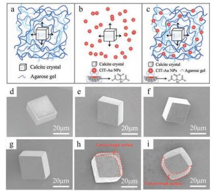
Schematic diagram of the calcite crystals grown in (a) agarose gel; (b) CIT-Au NPs solution; (c) agarose gel containing CIT-Au NPs. (d-i) SEM images of calcite crystals grown in varied media: (d) 1 w/v% agarose gels; (g) 3 w/v% agarose gels; (e) 0.5 mg/mL CIT-Au NPs solution; (h) 3 mg/mL CIT-Au NPs solution; (f, i) 1 w/v% agarose gels containing CIT-Au NPs (f) 0.1 mg/mL and (i) 2 mg/mL CIT-Au NPs. The red dashed lines highlight the curved rough surface.
First, we investigated the influence of agarose gels (Fig. 1a) or CIT exerted on calcite morphologies, respectively. On one hand, calcite crystals were grown at a fixed concentration(5 mmol/L)of Ca2+ using the ammonium carbonate method as previously reported [53, 54]. Initially, we imaged the morphologies of calcite crystals grown in agarose gels by scanning electron microscopy (SEM). As the agarose concentration increased from 1 w/v% to 3 w/v%, calcite crystals retained the characteristic rhombohedral morphology (Figs. 1d and g), consistent with the previous reports [53, 55-56]. On the other hand, we introduced the 20 nm CITcoated Au nanoparticles (CIT-Au NPs) to investigate the interaction between CIT additive and calcite crystal (Fig. 1b). Rhombohedral morphology was retained as the calcite crystallized in aqueous solution containing 0.5 mg/mL CIT-Au NPs (Fig. 1e). However, when the concentration of CIT-Au NPs ([CITAu NPs]) reached 3 mg/mL, the as-grown crystals exhibited curved rough surfaces that are approximately parallel to the c-axis, capping with smooth rhombohedral planes (Fig. 1h). According to previous investigations, the caps were identified as{104}faces, and the newlyoccurred irregular curved surfaces were generated by the step growth inhibition originating from CIT adsorption at{104}steps[57]. In addition, CIT could contact with the surface carbonate simultaneously, by virtue of its hydroxylic group [58]. In this way, CIT selectively favors the faces of calcite crystals to affect the crystal growth process, and further regulates the crystal morphology. Therefore, the agarose gel matrix is inert to the crystal morphology, while the CIT additives are active in crystal morphology modification as the [CIT-Au NPs] is high.
Next, agarose gels and CIT-Au NPs were both introduced into the crystallization media through crystallizing in agarose gels containing dispersed CIT-Au NPs (Fig. 1c). When the [CIT-Au NPs] was 0.1 mg/mL in gel, the as-grown crystals showed the characteristic rhombohedral morphology (Fig. 1f). Conversely, as [CIT-Au NPs] in agarose gels was increased to 2 mg/mL, the crystals showed the curved morphological features (Fig. 1i), demonstrating that CIT-Au NPs could shape the calcite crystal in either solution or agarose gel media.
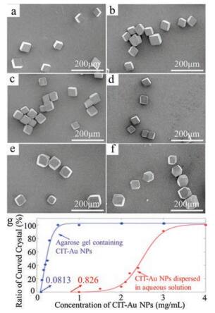
Furthermore, a closer examination of how the simultaneously introduced agarose gel and CIT-Au NPs affect the calcite morphology was performed. As a control experiment, the crystallization in the presence of CIT-Au NPs in solution, instead of gel media, was first examined. Representative SEM images of calcite crystals grown in CIT-Au NPs aqueous solution are shown in Figs. 2a, c, e. The crystals grown in 0.5 mg/mL CIT-Au NPs solution maintained in characteristic rhombohedra (Fig. 2a), while the calcite crystals in 2.25 mg/mL CIT-Au NPs solution began to exhibit rough, curved surfaces (Fig. 2c). With the increase of [CIT-Au NPs] from 2.25 mg/mL to 4 mg/mL, the number of curved calcite crystals progressively increased. These results are consistent with the previous study where the increasing [CIT] led to remarkable influence on calcite growth, favoring the formation of irregular vicinal faces [59]. Then we grew calcite inside agarose gels (1 w/v%) containing CIT-Au NPs and examine the morphologies of crystal (Figs. 2b, d, f). In sharp contrast, at an extremely low [CIT-Au NPs] of 0.2 mg/mL in agarose gel, the curved morphological feature of calcite crystals emerged (Fig. 2d). Similar to the crystals grown in CIT-Au NPs solutions, the calcite crystallized in the presence of agarose gel and CIT-Au NPs exhibited an increase in the number of curved crystals as the [CIT-Au NPs] increased from 0.1 mg/mL to 4 mg/mL. To acquire an accurate evaluation of morphologies change, the ratio of curved crystal (RCC) was defined as the ratio of the curved crystals' number to the total number of crystals. And the RCC were plotted as a function of [CIT-Au NPs] with lines fitted by the logistic regression model (Fig. 2g) [60]. We subsequently used the fitted curve to calculate its x-intercept termed as the "critical [CIT-Au NPs]" where the [CIT-Au NPs] triggers the calcite morphological revolution with statistical significance. For CIT-Au NPs in agarose gel, the critical [CIT-Au NPs] was 0.0813 mg/mL, while for CIT-Au NPs in solution, the critical [CIT-Au NPs] was 0.826 mg/mL. The logistic fitting curves reconfirmed that the introduction of agarose gel media significantly reduced the [CIT-Au NPs] to transform calcite morphology.
Figure 2
Figure 2.
(a-f) SEM images of calcite crystals grown in (a, c, e) solution and (b, d, f) 1 w/v% agarose gel containing CIT-Au NPs with the concentrations of (a) 0.5 mg/mL; (c) 2.25 mg/mL; (e) 3 mg/mL; (b) 0.1 mg/mL; (d) 0.2 mg/mL; (f) 2 mg/mL. (g) The morphological revolution diagram: the RCC was plotted as a function of [CIT-Au NPs], with lines showing the fits of the logistic regression model. The critical [CIT-Au NPs] were also highlighted. Red curve: CIT-Au NPs dispersed solution; blue curve: gel containing CIT-Au NPs.
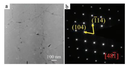
Furtherly, we explore the internal structures of the curved crystals. The thin section cut from a calcite crystal grown in the presence of agarose gel and CIT-Au NPs was imaged by high-angle annular-dark-field scanning transmission electron microscopy (HAADF-STEM), exhibiting the gel fibers and CIT-Au NPs incorporated inside the crystal, which was similar to the previous report exemplifying the gel and Au NPs incorporation inside crystal (Fig. 3a) [61]. As a supporting evidence, the calcite crystals exhibited colored, further suggesting the CIT-Au NPs incorporation inside crystal (Fig. S1 in Supporting information). Besides, the selected-area electron diffraction (SAED) image of a large area (3 μm in diameter), showed a single set of calcite diffraction spots (Fig. 3b), indicating that the calcite crystal maintained the nature of single-crystal although incorporating gel fibers and CIT-Au NPs.
Figure 3
Figure 3.
(a) A HAADF-STEM image of a thin section cut from the calcite crystal grown in agarose gel containing CIT-Au NPs (1 mg/mL CIT-Au NPs, 1 w/v%, agarose); (b) The SAED pattern of a region (3 μm in diameter) containing calcite crystal, gel fibers as well as CIT-Au NPs.
Previous literature have reported the diffusion-controlled CaCO3 crystallization in the gel matrix [11, 48, 49, 62], which led to irregular star-shaped [49] and ellipsoidal [62] morphologies of calcite under disequilibrium conditions. In these cases, gel molecules did not directly affect the crystallization but served as a growth matrix limiting the diffusion of reactant solutes. Inspired by this theory, we inferred that the diffusion limited originating from the gel matrix might be a possible mechanism to explain the calcite shaping in this work.
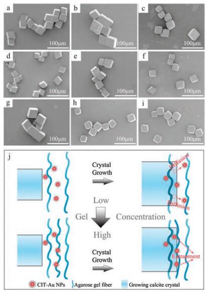
In order to verify the mechanism for morphological transformation associated with gel diffusion control, we further increase the gel concentration to reduce the diffusivity in the gels. The morphologies of calcite crystals grown in agarose gels containing CIT-Au NPs with increased concentrations were examined (Figs. 4a-i). While the concentration of agarose gels was 1 w/v%, the increase of [CIT-Au NPs] from 0.01 mg/mL to 0.12 mg/mL exerted no obviously impact on calcite morphologies (Figs. 4a, d, g) and the calcite crystals remained rhombohedral in morphology. As we increased the concentration of agarose gels to 2 w/v% (Figs. 4b, e, h), the curved crystal morphology feature emerged when [CIT-Au NPs] was 0.12 mg/mL (Fig. 4h). With further increase of the concentration of agarose gels to 3 w/v% (Figs. 4c, f, i), the calcite crystals exhibited morphological revolution at an even lower [CITAu NPs] of 0.04 mg/mL (Fig. 4f). Particularly, the denser gel networks originating from higher gel concentration favors the entrapment of CIT-Au NPs for a given size of nanoparticle. These results suggest the possible mechanism as follows: The growing calcite crystal repels the CIT-Au NPs additive and the CIT-Au NPs accumulates on the growth front [63] resulting in the CIT-Au NPs diffusing away from the crystal surface to surroundings. The gel media reduces the CIT-Au NPs diffusion to strengthen the interaction between CIT-Au NPs and calcite, and even to drive the CIT-Au NPs to be incorporated inside calcite single-crystal (Fig. 4j). Consequently, the crystal morphologies are regulated through the kinetic effects induced by the CIT additive. Therefore, the gel matrix amplifies the effect of CIT additives on morphological modification by reducing their diffusivity and enhancing their interaction with growing crystals.
Figure 4
Figure 4.
SEM images of calcite crystals grown in (a, d, g) 1 w/v%, (b, e, h) 2 w/v%, and (c, f, i) 3 w/v% agarose gels with CIT-Au NPs of (a, b, c) 0.01 mg/mL, (d, e, f) 0.04 mg/mL, and (g, h, i) 0.12 mg/mL). (j) Schematic representation of the gel effect of diffusion control on CIT-Au NPs.
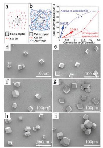
To further verify this mechanism, we grew calcite single-crystal in the presence of CIT ions (Figs. 5a and b). Compared to the CIT-Au NPs, the dispersing CIT ions are much more difficult to be confined in the sense of diffusion motion since the large CIT-Au NPs are easily to be trapped in gel network. Calcite crystals were grown in aqueous solution with the concentration of CIT ions ([CIT ions]) ranging from 0.02 mmol/L to 0.2 mmol/L (Figs. 5d, f, h and Figs. S2a, c, e in Supporting information), and the crystal with curved morphological features emerged as the [CIT ions] reached 0.1 mmol/L (Fig. 5f). Instead of solutions, crystals were also grown in agarose gels (1 w/v %) containing CIT ions with the concentration from 0.01 mmol/L to 0.25 mmol/L (Figs. 5e, g, i and Figs. S2b, d, f in Supporting information), and the crystal morphology revolution occurred (Fig. 5g) as the [CIT ions] reached just 0.02 mmol/L which was much lower than that (0.1 mmol/L) in the case of solution. The discrepancy between the solution-grown and gel-grown crystals indicates that the gel matrix contribute to the morphological revolution by enhancing the effect of CIT additives. According to the fitting method discussed above, the related fitting curves (Fig. 5c) were obtained and the critical [CIT ions] was calculated to be 0.0052 mmol/L in gel and 0.0183 mmol/L in solution. Also, the incorporation of gel fibers inside calcite single crystals was demonstrated by HAADF-STEM and SAED imaging (Fig. S3 in Supporting information). Consequently, we conclude that the agarose gel networks, as the media for crystallization, affect the crystal growth by entrapping the CIT species to enhance the interaction between CIT and growing crystal faces. Thus, the gel matrix indirectly regulated crystal morphology with the mechanism of diffusion control on additives.
Figure 5
Figure 5.
Schematic diagram of the calcite crystal grown in (a) solutions of CIT ions or (b) agarose gels containing CIT ions. (c) The morphological revolution diagram: the RCC was plotted as a function of [CIT ions], with lines showing the fits of the logistic regression model. The critical [CIT ions] calculated were also highlighted. (Red curve: CIT ions dispersed solution; blue curve: gel containing CIT ions. (d–i) SEM images of calcite crystals grown in CIT ions solution with the concentration of (d) 0.08 mmol/L, (f) 0.10 mmol/L, and (h) 0.12 mmol/L and calcite crystals grown in 1 w/v% agarose gel containing CIT ions with the concentration of (e) 0.01 mmol/L, (g) 0.02 mmol/L, and (i) 0.12 mmol/L.
In summary, we have demonstrated the synergy effects of the additive, CIT, and the matrix, agarose gel, on morphology regulation of synthetic calcite single-crystals. CIT-Au NPs dispersed in solution media triggered the calcite morphological transformation from characteristic faceted rhombohedra to morphologies with curved rough surfaces, as the [CIT-Au NPs] reached a critical value. The introduction of the alternative growth media, the agarose gel, even significantly promoted the morphological revolution of calcite single-crystal by reducing the critical [CITAu NPs] needed for morphological modification. Furthermore, increasing agarose gel concentrations resulted in a decrease of the critical [CIT-Au NPs]. This morphological regulation enhanced by the gel media is attributed to the gel effect in limiting the CIT diffusion, which strengthens the CIT-calcite interaction during crystallization. By showing the synergy effects, this work has implications for understanding the shaping of biominerals where often both additives and matrix exist.
Acknowledgments
This work was supported by the 973 Program (No. 2014CB643503) and the National Natural Science Foundation of China (Nos. 51625304, 51461165301).
Figure 2
(a-f) SEM images of calcite crystals grown in (a, c, e) solution and (b, d, f) 1 w/v% agarose gel containing CIT-Au NPs with the concentrations of (a) 0.5 mg/mL; (c) 2.25 mg/mL; (e) 3 mg/mL; (b) 0.1 mg/mL; (d) 0.2 mg/mL; (f) 2 mg/mL. (g) The morphological revolution diagram: the RCC was plotted as a function of [CIT-Au NPs], with lines showing the fits of the logistic regression model. The critical [CIT-Au NPs] were also highlighted. Red curve: CIT-Au NPs dispersed solution; blue curve: gel containing CIT-Au NPs.
Figure 3
(a) A HAADF-STEM image of a thin section cut from the calcite crystal grown in agarose gel containing CIT-Au NPs (1 mg/mL CIT-Au NPs, 1 w/v%, agarose); (b) The SAED pattern of a region (3 μm in diameter) containing calcite crystal, gel fibers as well as CIT-Au NPs.
Figure 4
SEM images of calcite crystals grown in (a, d, g) 1 w/v%, (b, e, h) 2 w/v%, and (c, f, i) 3 w/v% agarose gels with CIT-Au NPs of (a, b, c) 0.01 mg/mL, (d, e, f) 0.04 mg/mL, and (g, h, i) 0.12 mg/mL). (j) Schematic representation of the gel effect of diffusion control on CIT-Au NPs.
Figure 5
Schematic diagram of the calcite crystal grown in (a) solutions of CIT ions or (b) agarose gels containing CIT ions. (c) The morphological revolution diagram: the RCC was plotted as a function of [CIT ions], with lines showing the fits of the logistic regression model. The critical [CIT ions] calculated were also highlighted. (Red curve: CIT ions dispersed solution; blue curve: gel containing CIT ions. (d–i) SEM images of calcite crystals grown in CIT ions solution with the concentration of (d) 0.08 mmol/L, (f) 0.10 mmol/L, and (h) 0.12 mmol/L and calcite crystals grown in 1 w/v% agarose gel containing CIT ions with the concentration of (e) 0.01 mmol/L, (g) 0.02 mmol/L, and (i) 0.12 mmol/L.

 DownLoad:

 下载:
下载:


