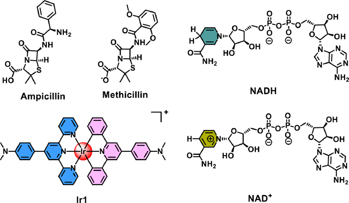
Scheme 1.
The chemical structure of antibiotics, NADH, NAD+ and the bis-tridentate Ir(Ⅲ) photocatalyst used in this study.
Photo-catalytic Staphylococcus aureus inactivation and biofilm destruction with novel bis-tridentate iridium(Ⅲ) photocatalyst
Shihao Chen , Zhishang Zhang , Li Wei , Zhongxian Fan , Yue Li , Xuezhi Wang , Tongming Feng , Huaiyi Huang

The discovery of organic antibiotics several decades ago has dramatically reduced the death caused by bacterial infections [1]. However, due to the abuse of antibiotics, multidrug-resistant bacteria have emerged and threatened thousands of human’s health around the world nowadays [2]. For example, the methicillin-resistant Staphylococcus aureus (S. aureus) is a common type of hospitalization infectious bacteria that shows resistance to several antibiotics [3]. Thus, it is important to investigate antimicrobial agents with novel mechanism of action (MoA).
All the time, transition metal complexes are widely used in cancer diagnosis and treatment [4-7], which arises people to explore their effectiveness to antibacterial. Therefore, in addition to the traditional organic antibiotic, transition metal complexes which exhibit inherent phosphorescent and readily tunable photo-physicochemical properties have been reported to combat the severe drug-resistant bacterial. Collins’s group discovered several series of polynuclear complexes Ru(Ⅱ) complexes for antimicrobial application via intercalating with nucleic acids [8,9]. On the other hand, iridium complexes have also been discovered as potential antibacterial agents. In 2018, Sadler et al. synthesized Ir(Ⅲ) complexes with metformin which showed antimicrobial activity [10]. In addition, Sasmal et al. reported a series of cyclometalated Ir(Ⅲ) complexes with simultaneous ultrasensitive detection properties to eliminate drug-resistant bacteria [11]. However, the above metal complexes function as antimicrobial chemotherapeutic drugs lacking spat-temporal selectivity and thus may induce side effects.
Antimicrobial photodynamic therapy (APDT) has been recognized as a promising therapeutic, providing patio-temporal control over drug activation to inactive bacteria. Upon light activation, APDT uses photosensitizers (PSs) to generate diverse reactive oxygen species (ROS) to induced intracellular oxidative damage via two main pathways. The type Ⅰ pathway includes electron transfer between the excited state PS with substrate and oxygen at the ground states, generating diverse radicals and superoxide anion. Type Ⅱ pathway involves direct energy transfer from excited state PS to oxygen, producing the highly reactive singlet oxygen [12]. Currently, photosensitizers applied for antibacterial mainly include type Ⅱ organic photosensitizers, such as porphyrin [13], 4,4-difluoro-boradiazaindacene (BODIPY) [14] and aggregation-induced emission PSs analogues [15]. However, the poor water solubility and high oxygen dependent MoA limited their applications. It is important to mention that metal PSs are rarely investigated in antimicrobial photo-therapy. Only until recently, Sadler and co-workers reported a photoactivatable Ru(Ⅱ)-isoniazid complex consist of the labile coordinated antimicrobial drug isoniazid for mycobacteria [16]. Chen and coworkers reported Ir(Ⅲ) complexes targeting negatively charged bacterial membrane, despite extremely high light dose required (white light, 484.2 J/cm2) [17]. It is necessary to mention that the above mentioned metal PSs did not or can only slightly inhibit the capability of antibiofilm. Therefore, developing highly efficient metal complexes with the ability to destruct biofilms are urgently needed.
Our group previously reported tri-dentate Ir(Ⅲ) photocatalysts exhibited high photocatalytic activity for coenzyme I, the reduced nicotinamide adenine di-nucleotide (NADH), as novel photocatalytic mechanism for phototherapeutics. NADH participates in the maintenance of intracellular redox balance, and as a coenzyme for cellular oxidoreductases [18]. However, the APDT application of bis-tridentate Ir(Ⅲ) photocatalysts were seldom reported. Herein, we reported a novel Ir(Ⅲ) photocatalyst (Ir1) (Scheme 1) for synergistically photodynamic and photocatalytic antimicrobial therapy. Ir1 effectively inactivated Gram-positive S. aureus via generating diverse ROS and photocatalytic oxidation of NADH under low light dose (21.1 J/cm2). Importantly, Ir1 damaged the cell membrane of S. aureus leading to protein leakage and showed stronger ability to damage the biofilm capability than the clinical used antibiotic.
The synthetic routes for the tridentate ligands and Ir1 were shown in Scheme S1 (Supporting information) with slightly modification from literature [19]. Ir1 was characterized by 1H NMR, 13C NMR and HRMS (Figs. S1 and S2 in Supporting information). Notably, the positive-charged Ir1 may improve the potent interaction with the negatively charged bacterial membrane [11]. The absorption and photoluminescent spectra of Ir1 were depicted in Figs. 1a and b. Differ from the widely studied tri-bidentate Ir(Ⅲ) complexes, Ir1 exhibited extended absorption bands tailing to 600 nm and emit intense deep-red phosphorescence (λmax = 685 nm) [20]. Importantly, the phosphorescent intensity enhanced about 3-fold in the absence of oxygen, indicating strong interaction between the excited state of Ir1 and the molecular oxygen (Fig. 1b). In view of the absorbance spectrum of Ir1, the white light source was employed to trigger the photo-induced activity in phosphate buffer solution (PBS) buffer solution.
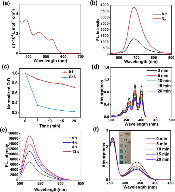
The dark- and photo-stability are two important factors for a potent photosensitizer candidate to avoid off-target dark toxicity and photo-degradation. Ir1 exhibited higher photostability than the widely used organic PS chlorin e6 (Ce6), as evaluated by the time-dependent UV–vis spectra under continuous light irradiation (Fig. 1c). The singlet oxygen generation ability (type Ⅱ energy transfer pathway) of Ir1 was detected by 9,10-anthracenediyl-bis(methylene) dimalonic acid (ABDA) [21]. Upon white light irradiation, the absorbance of ABDA decreased readily (Fig. 1d), compared with the standard metal PS [Ru(bpy)3]2+ (Fig. S7 in Supporting information). Since superoxide anion can inactive bacteria via type Ⅰ electron transfer pathway [22]. Thus we detected the superoxide anion generation efficiency with a fluorescent sensor dihydrorhodamine 123 (DHR123) [23]. As shown in Fig. 1e, after extremely short white light irradiation (4 s), DHR123 was rapidly oxidized and generated the highly fluorescent rhodamine 123. Especially, when irradiated for 12 s, the fluorescent intensity enhanced 3-fold, indicating high superoxide anion generation ability of Ir1.
We previously reported that Ir(Ⅲ) PSs can catalyze cellular NADH oxidation upon light irradiation and induce intracellular redox imbalance [24,25]. Thus, we explored the NADH catalytic activity of Ir1. In the absence of light, the absorbance of NADH remained unchanged with Ir1 (Fig. S6 in Supporting information). After 5 min white light irradiation, significant decrease of absorbance at 339 nm was observed, indicating effective depletion of NADH (TOF = 219.9 h−1, Fig. 1f). This property might be a critical factor for antibacterial activity, since NADH plays an important role in cell metabolism of living bacteria [18]. Moreover, H2O2 generation was detected by H2O2 test paper during NADH photocatalysis (Fig. 1f, inserted photograph). The above results indicated that Ir1 can generate diverse ROS and oxidized NADH upon white light activation.
The excellent photosensitization and photocatalytic activity of Ir1 promoted us to investigate the antimicrobial activity toward typical bacteria such as the Gram-positive bacteria (S. aureus), the Gram-negative bacteria E. coli (E. coli) and Mycolicibacterium smegmatis (M. smegmetis). The antibiotic used in clinic such as ampicillin, methicillin and streptomycin (Scheme 1) were tested as well. The minimum inhibitory concentration (MIC90) was defined. After incubating with Ir1 for 4 h, the bacteria were either kept in the dark or received white light irradiation (21.1 J/cm2), following by 14 h incubation in the dark. The survival rates of bacteria after dark or light treatment were shown in Figs. 2a and b and Fig. S8 (Supporting information). The light MIC90 of Ir1 against S. aureus was 3.13 µmol/L while there was no obvious inactivation ability toward Gram-negative bacteria. Notably, Ir1 also exhibited obvious dark toxicity against S. aureus. These results illustrated that Ir1 exhibited synergistic effects against S. aureus, functioning as chemotherapeutic agent in the dark and phototherapeutic agent upon light activation. The dark and light MIC90 values of Ir1 and several clinical antibiotics were summarized in Fig. S8 and Table 1. Moreover, light MIC90 of Ir1 against S. aureus reaches to 1.56 µmol/L when administrated with 3-fold intensity of light power (63.3 J/cm2, Fig. S10 in Supporting informaton), indicating excellent photo-stability of Ir1 and the antimicrobial activity can enhance via increasing the light dose. Since the fact that low light dose can decrease side effects, further experiments were performed with white light power of 21.1 J/cm2.
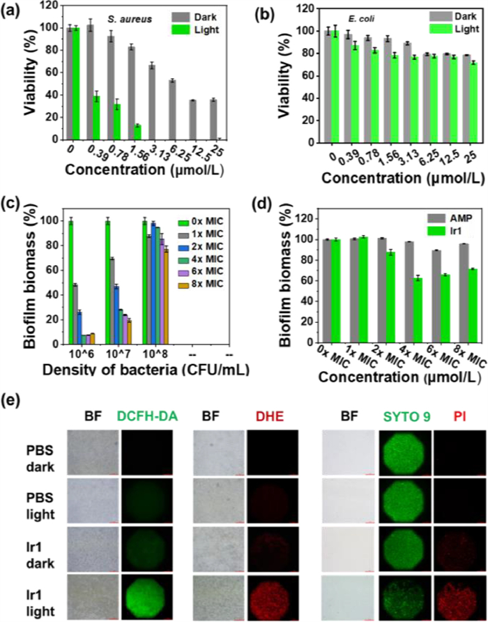
Bacteria can generate biofilm, a stronger barrier, to protect themselves from antibiotics. As a result, though the clinical used antibiotics exhibit strong antimicrobial activity toward bacteria, the therapeutic effect will be significantly inhibited treating with biofilm. Given the excellent activity of Ir1 against S. aureus, the capability of biofilm destruction was further investigated. We employed the crystal violet staining to explore the efficacy of biofilm inhibition and biofilm destruction, respectively [26]. The inhibition of biofilm formation was detected 44 h after dark or light treatment with S. aureus at 37 ℃. As shown in Fig. 2c, the inhibition efficiency of biofilm formation strongly depends on the density of bacteria (106–108 CFU/mL) and the dose of Ir1. For 106 CFU/mL S. aureus, 4 × MIC90 concentration of Ir1 can inhibit ~90% of biofilm biomass upon light irradiation. When the bacterial density increased, Ir1 can still inhibit biofilm formation but was less effective than ampicillin (Fig. S11 in Supporting informaton). It is important to mention that ampicillin exhibited inconspicuous mature biofilm elimination capability (Fig. 2d), which promoted us to investigate the mature S. aureus biofilm destruction ability of Ir1. As shown in Fig. 2d, Ir1 can effectively damage the mature S. aureus biofilm (destruction rate, ~35%−40%, over 4 × MIC90 concentration) after white light treatment.
The excellent ability of Ir1 towards both inhibition of biofilm formation as well as the mature biofilm destruction promoted us to further investigate the potent mechanism of action towards S. aureus. Firstly, the positive-charged nature of Ir1 may enhance the drug interaction with the negatively charged bacterial membrane. In order to figure out the interaction mode, we measured the change of zeta potential of S. aureus surface. However, the zeta potential of S. aureus just changed slightly in the presence or absence of Ir1 (Fig. 3a). Thus, Ir1 did not locate at the surface of bacteria due to negligible impact on zeta potentials change of the bacteria [27,28]. Considering the inconspicuous change of zeta potential of S. aureus and strong inactivation effect of Ir1, it is expected that Ir1 was able to insert into the poriferous cell wall of S. aureus. Besides, ROS produced in biofilm is able to destroy component of biofilm containing DNA, proteins, lipids, polysaccharide, etc. As expected, Ir1 can provide ROS upon biofilm, causing significant death of S. aureus biofilm (Fig. 2e). Herein, ROS is very important for biofilm destruction, which may explain why ampicillin exhibits low effect on biofilm.
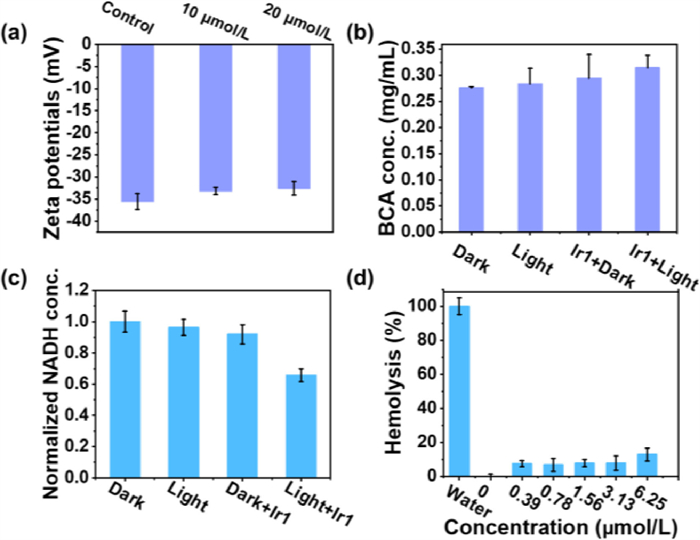
Subsequently, the enhanced bicinchoninic acid (BCA) protein assay kit was employed to evaluate the bacterial cell membrane integrity after treatment [26]. As shown in Fig. 3b, the BCA protein released from Ir1 and light treated group was higher than the other groups, indicating that a certain degree of bacterial cell membrane was ruptured after treatment. Additionally, NADH in S. aureus was also eliminated effectively when bacteria incubated with Ir1 was irradiated with white light (21.1 J/cm2) (Fig. 3c).
Hemolysis, which involves the disruption of erythrocyte membranes, is an obstacle for potent pre-clinical compounds. We explored the hemolysis of Ir1 preliminary by treating 10% erythrocyte stock solution with diverse concentrations of Ir1 (0–6.25 µmol/L). As shown in Fig. 3d, Ir1 caused less than 10% hemolysis after 1 h of incubation at 37 ℃, suggesting that Ir1 exhibited high biocompatibility.
Compare with Ir(Ⅲ) photocatalysts published previously [20,21,24,25,29], we may conclude the structure activity relationship that Ir(Ⅲ) photocatalysts with bis-tridentate system can significantly extend the light absorption wavelength as well as enhance the molar extinction coefficient. Thus, bis-tridentate Ir(Ⅲ) photocatalysts may be a suitable platform to develop long wavelength light excited Ir(Ⅲ) photocatalyst for the treatment of bacterial infection in deep tissue. However, it should also be concerned that multiple side product may occur during synthesis which results in low synthetic yield due to the high reaction temperature (200 ℃).
In summary, we designed and synthesized a novel bis-tridentate Ir(Ⅲ) photocatalyst and evaluated the antimicrobial photodynamic therapy activity. Ir1 generated diverse ROS and induced photocatalytic oxidation of NADH upon low-dose white light irradiation. Ir1 exhibited excellent S. aureus inactivation activity via synergistic type Ⅰ and type Ⅱ mechanisms of action. Additionally, Ir1 showed promising biofilm formation inhibition and mature biofilm destruction activity, indicating relatively low antimicrobial drug resistance. The antibacterial and anti-biofilm mechanisms of Ir1 may be attributed to high lipophilicity, excellent photocatalytic ROS production and NADH oxidation activity. These results suggest that Ir1 may be a promising antimicrobial PDT candidate for the treatment of Gram-positive bacterial infection. As one of the few studies about iridium complexes for antimicrobial PDT, this work would further facilitate the exploration of transition metal antibacterial materials to combat the severe bacterial infection.
The authors declare that they have no known competing financial interests or personal relationships that could have appeared to influence the work reported in this paper.
This work was financially supported by the “Summit Plan” High-Level Hospital Construction Project of Foshan (No. FSSYKF-2020002), the Medical Scientific Research Projects of Foshan Health Bureau (No. 20210358), the National Natural Science Foundation of China (NSFC, Nos. 22277153, 22007104), Guangdong Basic and Applied Basic Research Foundation (No. 2021B1515020050), Science, Technology and Innovation Commission of Shenzhen Municipality Project (No. JCYJ20190807152616996) and the Fundamental Research Funds for the Central Universities (No. 22lgqb37).
Supplementary material associated with this article can be found, in the online version, at doi:
G.A. Durand, D. Raoult, G. Dubourg, Int. J. Antimicrob. Agents 53 (2019) 371–382. doi: 10.1016/j.ijantimicag.2018.11.010
M.Y. Cao, Z.S. Chang, J.S. Tan, et al., ACS Appl. Mater. Interfaces 14 (2022) 13025–13037. doi: 10.1021/acsami.1c23676
A.S. Lee, H. Lencastre, J. Garau, et al., Nat. Rev. Dis. Primers 4 (2018) 18033. doi: 10.1038/nrdp.2018.33
L.N. Xie, Z.D. Luo, Z.N. Zhao, et al., J. Med. Chem. 60 (2017) 202–214. doi: 10.1021/acs.jmedchem.6b00917
Z.N. Zhao, P. Gao, L. Ma, et al., Chem. Sci. 11 (2020) 3780–3789. doi: 10.1039/d0sc00862a
M.K. Chen, X.T. Huang, J. Lai, et al., Chin. Chem. Lett. 32 (2021) 158–161. doi: 10.1016/j.cclet.2020.11.050
J.J. Li, H.J. Luo, X.Q. Zhu, et al., Chin. Chem. Lett. 33 (2022) 788–792. doi: 10.1016/j.cclet.2021.08.088
F.F. Li, J.G. Collins, F.R. Keene, Chem. Soc. Rev. 44 (2015) 2529. doi: 10.1039/C4CS00343H
A.K. Gorle, M. Feterl, J.M. Warner, et al., Dalton Trans. 43 (2014) 16713. doi: 10.1039/C4DT02139H
F. Chen, J. Moat, D. McFeely, et al., J. Med. Chem. 61 (2018) 7330–7344. doi: 10.1021/acs.jmedchem.8b00906
A. Gupta, P. Prasad, S. Gupta, et al., ACS Appl. Mater. Interfaces 12 (2020) 35967–35976. doi: 10.1021/acsami.0c11161
Q.Y. Jia, Q. Song, P. Li, et al., Adv. Healthc. Mater. 8 (2019) 1900608. doi: 10.1002/adhm.201900608
J. Hynek, J. Zelenka, J. Rathousky, et al., ACS Appl. Mater. Interfaces 10 (2018) 8527–8535. doi: 10.1021/acsami.7b19835
D.O. Frimannsson, M. Grossi, J. Murtagh, et al., J. Med. Chem. 53 (2010) 7337–7343. doi: 10.1021/jm100585j
M.M. Kang, C.C. Zhou, S.M. Wu, et al., J. Am. Chem. Soc. 141 (2019) 16781–16789. doi: 10.1021/jacs.9b07162
N.A. Smith, P.Y. Zhang, S.E. Greenough, et al., Chem. Sci. 8 (2017) 395. doi: 10.1039/C6SC03028A
P.Y. Ho, S.Y. Lee, C. Kam, et al., Adv. Healthc. Mater. 10 (2021) 2100706. doi: 10.1002/adhm.202100706
A. Chiarugi, C. Dölle, R. Felici, et al., Nat. Rev. Cancer 12 (2012) 741–752. doi: 10.1038/nrc3340
A. Sil, U. Ghosh, V.K. Mishra, et al., Inorg. Chem. 58 (2019) 1155–1166. doi: 10.1021/acs.inorgchem.8b02440
H.Y. Huang, S. Banerjee, K.Q. Qiu, et al., Nat. Chem. 11 (2019) 1041–104. doi: 10.1038/s41557-019-0328-4
C. Huang, C. Liang, T. Sadhukhan, et al., Angew. Chem. Int. Ed. 60 (2021) 9474–9479. doi: 10.1002/anie.202015671
X. Yang, J. Li, T. Liang, et al., Nanoscale 6 (2014) 10126–10133. doi: 10.1039/C4NR01965B
M. Yazdani, Toxicol. In Vitro 30 (2015) 578–582. doi: 10.1016/j.tiv.2015.08.010
Z.X. Fan, J.E. Xie, T. Sadhukhan, et al., Chem. Eur. J. 28 (2022) e02103346.
Z.X. Fan, Y. Rong, T. Sadhukhan, et al., Angew. Chem. Int. Ed. 61 (2022) e202202098. doi: 10.1002/anie.202202098
J.W. Zhu, J. Tian, C. Yang, et al., Small 17 (2021) e2101495. doi: 10.1002/smll.202101495
H.X. Yuan, Z. Liu, L.B. Liu, et al., Adv. Mater. 26 (2014) 4333–4338. doi: 10.1002/adma.201400636
H.T. Bai, H.X. Yuan, C.Y. Nie, et al., Angew. Chem. Int. Ed. 54 (2015) 13208–13213. doi: 10.1002/anie.201504566
L. Wei, R. Kushwaha, A.Y. Dao, et al., Chem. Commun. 59 (2023) 3083–3086. doi: 10.1039/d2cc06721h

Scheme 1 The chemical structure of antibiotics, NADH, NAD+ and the bis-tridentate Ir(Ⅲ) photocatalyst used in this study.

Figure 1 Photophysical and photochemical properties of Ir1. (a) Absorption spectra of Ir1 in CH2Cl2. (b) Phosphorescence spectra of Ir1 in CH2Cl2 at 298 K, λex = 525 nm. (c) Photo-stability of Ir1 and Ce6. (d) ABDA photodegradation by Ir1. (e) Change of fluorescent intensity of DHR123 by Ir1. (f) Photocatalytic oxidation of NADH by Ir1. Insert: H2O2 production after white light exposure.

Figure 2 Photo-inactivation of S. aureus (a) and E. coli (b) with Ir1 under white light (21.1 J/cm2). (c) Biofilm formation inhibition experiments, and S. aureus are incubated with Ir1 for 44 h after 21.1 J/cm2 white light treatment. (d) Mature biofilm destruction experiments, and mature S. aureus biofilm are incubated with Ir1 for 3 h after 21.1 J/cm2 white light treatment. (e) The first two pictures are ROS and superoxide anion produced in bacteria of biofilm treated with various conditions, detected by DCFH-DA and DHE. On the right is S. aureus biofilm staining with SYTO 9 and PI.

Figure 3 (a) Zeta potentials of S. aureus in H2O pretreated with or without Ir1 for 60 min. (b) BCA released from bacteria following receiving various treatments. The amount of BCA released from Ir1-treated bacteria versus from that of other formulations-treated bacteria. (c) NADH deletion within S. aureus following receiving various treatments. (d) Hemolysis test results of Ir1.

 扫一扫看文章

扫一扫关注我们
 DownLoad:

 下载:
下载: