Figure 1.
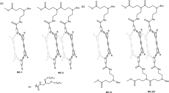
Structures of (a) Single-stranded macrocyclic monomer MC-1 and dimer MC-2, (b) double-stranded monomer MC-O and dimer MC-2O.
Enhancing self-assembly efficiency of macrocyclic compound into nanotubes by introducing double peptide linkages
Cheng-Yan Wu , Yi-Nan Gao , Zi-Han Zhang , Rui Liu , Quan Tang , Zhong-Lin Lu

Tubular structures, especially those with nanoscale inner pores, have attracted widespread interest in many fields [1–3]. Pores with small diameters and sub-nanometer pore sizes exhibit many unusual and fascinating properties [4–7]. The hydrophobic pores of carbon nanotubes have been found to mediate mass transport [5]. However, due to the unique structure of carbon nanotubes, it is difficult to introduce functional groups on both the inner and outer surfaces [8,9].
Designing and synthesizing molecules with specific structures that self-assemble into hollow cylindrical nanostructures, known as organic nanotubes (ONTs), has attracted widespread attention for decades [10,11]. Organic nanotubes can provide us with a spatially selective microreactor and have broad applications in asymmetric catalysis and other fields [12,13]. Due to their molecular size space, they also exhibit unique application values in areas such as molecular storage materials and molecular recognition [14,15]. In addition, the inner and outer surfaces of organic nanotubes can be chemically or physically modified to have different functions [16].
The building blocks of self-assembly have gradually shifted from simple amphiphilic molecules to complex biological molecules [17], π-conjugated systems [18], supramolecular polymers [19,20], and multi-component systems [21]. The Gong's group designed and synthesized four generations of macrocyclic compounds that were able to self-assemble into nanotubes in non-polar environments, exhibiting excellent ion and water molecule transport functions [22–25]. By changing the internal cavity substituents, selective ion transport was achieved [16]. The Zeng's group designed and synthesized a series of folded aromatic amide molecules containing "sticky" groups. These molecules were connected end-to-end through the sticky groups to further assemble into tubular structures with excellent substance transport functions [26–29].
According to our knowledge, most self-assembling organic nanotubes are made up of non-rigid components, resulting in deformable inner pores that are difficult to precisely control [2,30–33]. Furthermore, the stability of these nanotubes varies with temperature, solvent conditions, and other factors such as length due to their dynamic nature. As a result, developing organic nanotubes or molecular nanotubes with controllable pore sizes and lengths as well as modifiable inner and outer walls remains a significant challenge in this field.
Recently, our group constructed molecular nanotubes with precise structures through covalent bonding [34]. We introduced single β-alanine fragments on the outer side of hexakis(m-phenylene ethynylene) (m-PE) macrocycle and further synthesized macrocyclic tetramers by peptide chain synthesis (Fig. 1a). In non-polar solvents, macrocyclic tetramers showed stronger assembly stability compared to dimers and monomers. However, in highly polar solvents such as DMF, the molecular assembly of single-chain oligomers may be disrupted due to their structural characteristics, leading to macrocycle misalignment. To further improve the stability of their assembly, in this work we added one more connecting site at the para position of m-PE macrocycles to obtain monomer MC-O, and then connected them into dimer MC-2O by two peptide linkages (Fig. 1b). The introduction of a double peptide chain fixed the relative positions between macrocycles, and reduced their misalignment. Spectral study demonstrated that dimer MC-2O can form more stable self-assembled nanotubes than monomer MC-O.
The synthesis route of the macrocycle basically followed the reported "3+3" synthetic strategy [34,35]. To be noticed, 3‑bromo-5-iodobenzamide was synthesized as intermediate instead of 3,5-diiodobenzamide, which greatly improved the yield of the semi-cyclic fragment. The C2 symmetric structure of macrocycle MC-O allows the two half-cycle fragments to share the same intermediates, which reducing the total synthetic steps. The macrocyclic diamine intermediate was obtained by deprotecting Boc under acidic conditions, and the macrocyclic dicarboxylic acid intermediate was obtained by hydrolyzing the two methyl esters under basic conditions. Then, the two fragments were condensed to afford the macrocyclic dimer MC-2O. To be noticed, synthesizing dimers is also a process of forming a new macrocycle structure, and the yield is much lower than that of synthesizing single peptide chain dimers [34]. All of the key intermediates and target molecules were characterized by 1H NMR, 13C NMR and MS data (Supporting information).
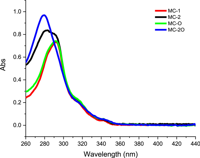
For face-to-face π-π aggregation, when the angle α between the connecting line of the chromophore centers and the transition dipole moment is greater than 54.7°, H-aggregates are formed, resulting in a blue shift of the UV–vis absorption band. The greater the blue shift, the stronger the face-to-face π-π interaction [36]. By comparing the UV–vis absorption spectra of double-stranded macrocyclic dimer MC-2O and single-stranded macrocyclic dimer MC-2, it was found that introducing one more strand resulted in a single absorption band (Fig. 2), while two mixed absorption bands were observed in the single-stranded macrocyclic dimers [34]. Obviously, this is due to stronger chromophore π-π interactions in the double-stranded dimer MC-2O, which may be caused by closer distances between adjacent chromophores or smaller helical angles of the macrocycles.
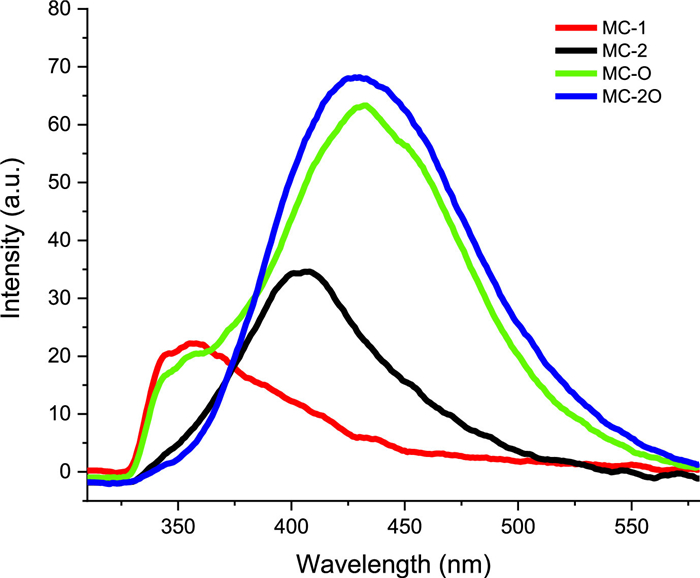
In the fluorescence spectra, single-stranded macrocyclic monomer MC-1 showed fluorescence quenching in CCl4 solution, and no obvious excimer-like emissions peak was observed [37,38]. However, we found a clear excimer-like emissions peak for the double-stranded macrocyclic monomer MC-O in CCl4 solution (Fig. 3). Moreover, the emission peak of the dimer did not further red-shift. Based on the above experimental results and analysis, we believe that the centrally symmetric structure is conducive to a more regular arrangement of the macrocyclic molecules, which in turn is conducive to the appearance of excimer-like emissions. In the variable-temperature fluorescence spectral, the maximum emission wavelength of both MC-O and MC-O2 showed slower change towards shorter wavelengths along the temperature increase, when compared to those of the single-stranded structures (Fig. 4).

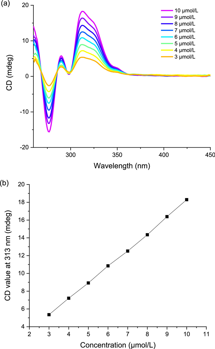
The assemblies of m-PE macrocycles with C6 rotational symmetry can transfer the chiral information of the side chains to the macrocyclic backbone through hydrogen bonding, forming an ordered unidirectional helical assembly that places the chromophores in a chiral environment. The helical structure of the assembly can be deduced by circular dichroism experiments [35,39]. The macrocyclic monomer MC-O has a C2 rotational symmetry with the axis in the direction perpendicular to the plane of the macrocycle, but its self-assembly in CCl4 solution did not show reliable CD signals. The double-stranded macrocyclic dimer MC-2O, on the other hand, can form an ordered intramolecular helix due to the fixed relative position of the side chains, thus obvious CD signals were observed (Fig. 5a). Moreover, a comparison of the CD spectra of the single-stranded macrocyclic dimer and the double-stranded dimer indicated that the CD signal intensity of the double-stranded dimer gave a better linear relationship with concentration (Fig. 5b). This demonstrated that the introduction of the double-stranded structure led to a more regular arrangement of the two macrocycles. In the temperature-dependent CD experiment, the melting temperature of MC-2O was found to be 327 K, higher than that of the single-stranded macrocyclic dimer MC-2 at 323 K. Even at a temperature close to the boiling point of CCl4, such as 70 ℃, a strong CD signal can still be detected (Fig. S1 in Supporting information). The unidirectional helical conformation of MC-2O brings the super-strong thermal stability in CCl4.
The tubular assembly of m-PE macrocycles can facilitate the transport of water and ions through lipid bilayers. Mass transport mediated by the tubular assemblies of m-PE macrocycles was evaluated based on lipid bilayer assays. The macrocycles were added to solutions of large unilamellar vesicles (LUVs) of dipalmitoylphos-phatidylcholine (DPPC) lipid, partitioning into the hydrophobic and nonpolar interior of the lipid bilayer and assembling into tubular stacks. Given the typical aromatic stacking distance of ~3.6 Å, it takes approximately 9–10 m-PE macrocyclic molecules to form a tubular stack capable of spanning the hydrophobic core of a lipid bilayer with a thickness of 3–4 nm [35]. Therefore, it was expected that 9–10 molecules of MC-O are needed to form an effective transmembrane channel. In contrast, MC-2O is more favorable to form transmembrane channels as only 4–5 MC-2O were required.
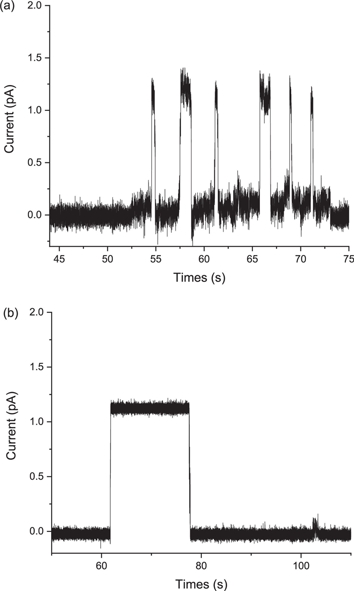
Lipid bilayer assays were used to detect the proton transport activity of MC-O and MC-2O. Clearly single-channel current traces were recorded in the presence of MC-O and MC-2O, indicating their effective proton transport activity. In comparison, the proton channel formed by MC-2O showed an open state lasting an average of 20 s (Fig. 6b), which is much longer than the open state of the self-assembling channels formed by MC-O (about 1 s, Fig. 6a). The enhanced stability of transmembrane channels formed by MC-2O is attributed to the reduced entropic barrier resulting from the covalent tethering of multiple stacked macrocyclic units. Compared to single-stranded dimers MC-2, the double-stranded dimers MC-2O formed ion channels with similar open duration times [34], possibly due to the similar stability in the weakly polar environment of the lipid bilayer.
The results indicate that covalently linked multiple macrocycles, especially those with double peptide chains at the para position of the macrocycles, provide a reliable strategy for the efficient construction of organic nanotubes with defined length and enhanced thermodynamic stability.
To summarize, we covalently linked two m-PE macrocyclic units at the para position using double oligo(β-alanine) linkers to afford the macrocyclic dimer MC-2O. Studies based on HNMR, UV absorption, fluorescence, and CD spectral showed that both the monomeric macrocycle MC-O and the dimeric macrocycle MC-2O can form tubular self-assemblies in nonpolar solvents. Compared to the ones with single linker, the double linkers at the para position reduced the swing of the unfixed end of the macrocycle, making the relative position between adjacent macrocycles more stable. Proton transport experiments also showed that the self-assemblies of the macrocyclic dimer MC-2O can form ion channels with longer open times. Although the introduction of two linkers increases the difficulty of synthesis, it provides a beneficial exploration for the synthesis of more stable molecular nanotubes.
The authors declare that they have no known competing financial interests or personal relationships that could have appeared to influence the work reported in this paper.
We thank the financial support by the National Natural Science Foundation of China (Nos. 91227109 and 21778012 to Z.-L. Lu., No. 21801020 to R. Liu) and Bohai University.
Supplementary material associated with this article can be found, in the online version, at doi:
S.P. Zheng, L.B. Huang, Z. Sun, M. Barboiu, Angew. Chem. Int. Ed. 60 (2021) 566–597. doi: 10.1002/anie.201915287
J. Montenegro, M.R. Ghadiri, J.R. Granja, Acc. Chem. Res. 46 (2013) 2955–2965. doi: 10.1021/ar400061d
B. Gong, Z. Shao, Acc. Chem. Res. 46 (2013) 2856–2866. doi: 10.1021/ar400030e
D. Xia, P. Wang, X. Ji, et al., Chem. Rev. 120 (2020) 6070–6123. doi: 10.1021/acs.chemrev.9b00839
R.H. Tunuguntla, R.Y. Henley, Y.C. Yao, et al., Science 357 (2017) 792–796. doi: 10.1126/science.aan2438
J. Shen, G. Liu, Y. Han, W. Jin, Nat. Rev. Mater. 6 (2021) 294–312. doi: 10.1038/s41578-020-00268-7
H. Ma, R. Ye, L. Jin, et al., Chin. Chem. Lett. 34 (2023) 108355. doi: 10.1016/j.cclet.2023.108355
J.R. Sanchez-Valencia, T. Dienel, O. Groning, et al., Nature 512 (2014) 61–64. doi: 10.1038/nature13607
F. Yang, M. Wang, D. Zhang, et al., Chem. Rev. 120 (2020) 2693–2758. doi: 10.1021/acs.chemrev.9b00835
T. Shimizu, M. Masuda, H. Minamikawa, Chem. Rev. 105 (2005) 1401–1443. doi: 10.1021/cr030072j
T. Shimizu, W. Ding, N. Kameta, Chem. Rev. 120 (2020) 2347–2407. doi: 10.1021/acs.chemrev.9b00509
K.I. Assaf, W.M. Nau, Chem. Soc. Rev. 44 (2015) 394–418. doi: 10.1039/C4CS00273C
S.J. Barrow, S. Kasera, M.J. Rowland, et al., Chem. Rev. 115 (2015) 12320–12406. doi: 10.1021/acs.chemrev.5b00341
V. Sgarlata, V.G. Organo, D.M. Rudkevich, Chem. Commun. (2005) 5630–5632.
B. Hua, L. Shao, Z. Zhang, et al., J. Am. Chem. Soc. 141 (2019) 15008–15012. doi: 10.1021/jacs.9b08257
X. Wei, G. Zhang, Y. Shen, et al., J. Am. Chem. Soc. 138 (2016) 2749–2754. doi: 10.1021/jacs.5b12698
K. Matsuura, Bull. Chem. Soc. Jpn. 90 (2017) 873–884. doi: 10.1246/bcsj.20170133
J.C. Nelson, J.G. Saven, J.S. Moore, P.G. Wolynes, Science 277 (1997) 1793–1796. doi: 10.1126/science.277.5333.1793
T. Aida, E.W. Meijer, S.I. Stupp, Science 335 (2012) 813–817. doi: 10.1126/science.1205962
L. Yuan, P. Jiang, J. Hu, et al., Chin. Chem. Lett. 33 (2022) 2026–2030. doi: 10.1016/j.cclet.2021.09.089
T. Shimizu, N. Kameta, W. Ding, M. Masuda, Langmuir 32 (2016) 12242–12264. doi: 10.1021/acs.langmuir.6b01632
L. Yuan, W. Feng, K. Yamato, et al., J. Am. Chem. Soc. 126 (2004) 11120–11121. doi: 10.1021/ja0474547
L. He, Y. An, L. Yuan, et al., Proc. Natl. Acad. Sci. U. S. A. 103 (2006) 10850–10855. doi: 10.1073/pnas.0602912103
W. Feng, K. Yamato, L. Yang, et al., J. Am. Chem. Soc. 131 (2009) 2629–2637. doi: 10.1021/ja807935y
J.S. Ferguson, K. Yamato, R. Liu, et al., Angew. Chem. Int. Ed. 48 (2009) 3150–3154. doi: 10.1002/anie.200900584
J. Shen, J. Fan, R. Ye, et al., Angew. Chem. Int. Ed. 59 (2020) 13328–13334. doi: 10.1002/anie.202003512
F. Chen, J. Shen, N. Li, et al., Angew. Chem. Int. Ed. 59 (2020) 1440–1444. doi: 10.1002/anie.201906341
J. Shen, R. Ye, Z. Liu, H. Zeng, Angew. Chem. Int. Ed. 61 (2022) e202200259. doi: 10.1002/anie.202200259
Y. Huo, H. Zeng, Acc. Chem. Res. 49 (2016) 922–930. doi: 10.1021/acs.accounts.6b00051
G.W. Gokel, Chem. Commun. (2000) 1–9.
M.B. Patel, A. Stavri, N.S. Curvey, G.W. Gokel, Chem. Commun. 50 (2014) 11562–11564. doi: 10.1039/C4CC04769A
A. Cazacu, C. Tong, A. van der Lee, et al., J. Am. Chem. Soc. 128 (2006) 9541–9548. doi: 10.1021/ja061861w
C. Ren, J. Shen, H. Zeng, J. Am. Chem. Soc. 139 (2017) 12338–12341. doi: 10.1021/jacs.7b04335
C.Y. Wu, S.L. Su, X. Zhang, et al., Angew. Chem. Int. Ed. 62 (2023) e202303242. doi: 10.1002/anie.202303242
X. Zhou, G. Liu, K. Yamato, et al., Nat. Commun. 3 (2012) 949. doi: 10.1038/ncomms1949
E.G. McRae, M. Kasha, The molecular exciton model, in: L. Augenstein, R. Mason, B. Rosenberg (Eds. ), Physical Processes in Radiation Biology, Academic Press, New York and London, 1964, pp. 23–42.
Y. Zhong, Y. Yang, Y. Shen, et al., J. Am. Chem. Soc. 139 (2017) 15950–15957. doi: 10.1021/jacs.7b09647
R.B. Prince, J.G. Saven, P.G. Wolynes, J.S. Moore, J. Am. Chem. Soc. 121 (1999) 3114–3121. doi: 10.1021/ja983995i
Q. Wang, Y. Zhong, D.P. Miller, et al., J. Am. Chem. Soc. 142 (2020) 2915–2924. doi: 10.1021/jacs.9b11536

Figure 1 Structures of (a) Single-stranded macrocyclic monomer MC-1 and dimer MC-2, (b) double-stranded monomer MC-O and dimer MC-2O.

Figure 2 Absorption spectra of MC-1, MC-2, MC-O and MC-2O (solvent, CCl4; concentration of each compound, 10 µmol/L; light path, 4 mm; 25 ℃).

Figure 3 Emission spectra of MC-1, MC-2, MC-O and MC-2O in CCl4 (concentration of each compound, 5 µmol/L; excitation wavelength, 295 nm; light path, 1 cm; 25 ℃).

Figure 4 Temperature-dependent fluorescence spectra of (a) MC-O and (b) MC-2O in CCl4 (~5 µmol/L). Plot of (c) I433/I350 (MC-O) and (d) I430/I350 (MC-2O) against temperature.

Figure 5 (a) CD spectra of MC-2O in CCl4. (b) The CD intensity (at 313 nm) of MC-2O in CCl4 varies with concentration at 25 ℃ (light path, 4 mm).

 扫一扫看文章

扫一扫关注我们
 DownLoad:
 下载:
下载:
