Citation:
Wenhao Wang, Siyuan Peng, Zhengwei Huang, Xin Pan. Tuning amino/hydroxyl ratios of nanovesicles to manipulate protein corona-mediated in vivo fate[J]. Chinese Chemical Letters,
2024, 35(11): 110134.
doi:
10.1016/j.cclet.2024.110134
Tuning amino/hydroxyl ratios of nanovesicles to manipulate protein corona-mediated in vivo fate
English
Tuning amino/hydroxyl ratios of nanovesicles to manipulate protein corona-mediated in vivo fate
With the rapid advancement of nanotechnology, nanocarriers for drug delivery have created promising prospects for efficient drug administration and precise disease therapy. However, only a limited number of these nanocarriers were successfully processed into the clinic or market [1]. The unclear fate of these nanocarriers when administrated in vivo is believed to be the one to blame. When nanocarriers are administrated in vivo, they will encounter complicated environmental factors including biomacromolecules, diversified pH, functional immune cells and hydromechanical forces, which might make the tracking of nanocarriers unpredictable. Among them, the formation of protein corona, a phenomenon where proteins inevitably are adsorbed on the surface of nanocarriers in biological fluids, has garnered significant attention. The formation of protein corona significantly affects drug absorption, distribution, metabolism, and excretion by causing substantial alterations in the surface properties of nanocarriers. Thus, understanding the formation and mechanism of protein corona represents a promising avenue for advancing the development of nanocarriers.
Although various side effects of protein corona formation have been documented, there is also potential for leveraging precise manipulation of protein corona formation to enhance drug delivery performance. Further elucidation of the influencing factors and mechanisms is required for specific strategies to manipulate the formation of protein corona on nanocarriers. Surface chemistry of nanocarriers has been proven to play a crucial role, and surface modification has been established as an efficient strategy for regulating the in vivo fate. Polyethylene glycol (PEG) and block copolymers are the most widely applied surface modification approaches to minimize the protein corona formation, thereby extending blood circulation time. However, the production of anti-PEG and the subsequently accelerated blood clearance (ABC) phenomenon hindered further application of PEGylation [2]. Thus, there is an urgent need to develop an alternative surface modification approach to regulate the formation of protein corona and the subsequent in vivo fate. Up to now, several other polymers have been reported as alternatives for PEG to avoid ABC effects, such as poly(ethyl ethylene phosphate) (PEEP), polydopamine, poly(vinyl alcohol), poly(2-alkyl-2-oxazoline) and glycosylated polyhydroxy polymers [3]. However, there is still a lack of in-depth mechanism studies on the impact of their structure on protein corona formation.
Recently, in Nat. Commun., Gan et al. explored the effect of amino/hydroxyl ratios on the protein corona formation and evolution of glycosylated polyhydroxy polymer (CSO-g-TCP)-modified lipid nanovesicles (CP-LVs) (Fig. 1) [4]. The findings demonstrated that the CP-LVs with an amino/hydroxyl ratio of about 0.4 (CP1-LVs) exhibited the desired protein corona formation performance. On one hand, the authors found that CP1-LVs reduced the adsorption of immunoglobulin and albumin in blood and livers, leading to prolonged circulation. On the other hand, the highest adsorption of tumor distinctive proteins like CD44 and osteopontin (OPN) was revealed in CP1-LVs at the tumor site, which promotes tumor cellular internalization. The improved anti-tumor efficiency of paclitaxel (PTX)-loaded CP1-LVs was justified both in the A549 and HeLa tumor models.
Figure 1
Figure 1.
Schematic diagram describing the design of nanovesicles modified with different ratios of amino and hydroxyl groups to explore the dynamic protein corona compositions on nanovesicle surfaces and protein corona-affected in vivo behaviors. Copied with permission [4]. Copyright 2024, Nature Publishing Group.
To obtain the lipid nanovesicles with different amino/hydroxyl ratios, the amino-modified hydrophilic polymers CSO-g-TCP were synthesized and hydrated with 1,2-dipalmitoyl-sn‑glycero-3-phosphorylcholine (DPPC). The CP-LVs with amino/hydroxyl ratios of 0.2, 0.3, and 0.4 were designated as CP4-LVs, CP2-LVs, and CP1-LVs, respectively (Fig. 2A).
Figure 2
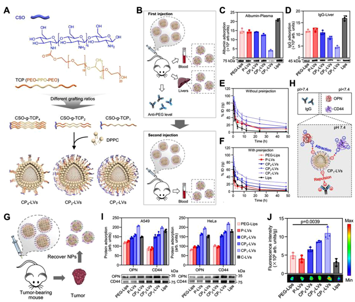
Figure 2.
(A) Schematic diagram of the structure of CP-LVs with different amino/hydroxyl ratios. (B) Schematic diagram describing the characterization of the protein corona in mice with single and repeated administrations (C, D) Quantitative analysis of albumin. (C) and IgG (D) adsorbed on nanovesicles. (E, F) Blood circulation curves in mice (E) with or (F) without preinjection. (G) Schematic illustration describing the characterization of protein coronas on nanovesicles recovered from tumors in mice injected with different nanovesicles. (H) Schematic diagram of CP1-LVs adsorbing IgG, CD44, and OPN affected by protein pI. (I) Quantitative analysis of CD44 and OPN adsorbed on nanovesicles recovered from A549 and HeLa tumors. (J) Ex vivo fluorescence images and quantitative analysis of tumors at 48 h postinjection. Reproduced with permission [4]. Copyright 2024, Nature Publishing Group.
Subsequently, the protein corona formation on CP-LVs was systematically studied. During incubation with plasma, a decrease in total protein adsorption on CP-LVs was observed as the amino group density increased. CP1-LVs with the highest amino density exhibited the lowest protein adsorption amounts. In addition, a notable reduction in the adsorption of immune response opsonins such as IgG and IgM was revealed on CP1-LVs. Similar results were also evident in the case of liver tissue interstitial fluid incubation, where the lowest IgG and liver metabolic enzyme adsorption were found in CP1-LVs. These findings suggested that increasing the density of amino groups can enhance the capacity to prevent protein adsorption. Further, the dynamic protein corona formation was evaluated in vitro and in vivo. The nanovesicles were incubated with plasma, phosphate buffer (PBS) and liver tissue interstitial fluid in turn to mimic the trafficking process. The nanovesicles were administered intravenously and subsequently collected for the investigation of in vivo dynamic protein corona formation (Fig. 2B). The results consistently showed that CP1-LVs exhibited the lowest adsorption of IgG, IgM, and albumin, leading to reduced internalization in Kupffer cells and weakened ABC effects caused by anti-PEG antibody binding (Figs. 2C and D). The blood circulation curve further justified the conclusion. As the amino/hydroxyl ratios increased, the retention in blood circulations of CP-LVs was enhanced, and CP1-LVs exhibited the best blood circulation time (Figs. 2E and F). The mechanism can be attributed to the weakened ABC effect as well as decreased recognition and phagocytosis of macrophages.
Having proved the prolonged blood circulation time and its associated protein corona mechanism, the dynamic protein coronas of CP-LVs with different amino/hydroxyl ratios in the transportation from blood to tumor were evaluated. Similarly, the in vitro and in vivo studies were conducted (Fig. 2G). Surprisingly, CP-LVs with elevated amino/hydroxyl ratios exhibited heightened protein adsorptions in tumor tissue interstitial fluid. Notably, CP1-LVs showed the highest protein corona formation both in vitro and in vivo. This result is opposite to the findings in the liver and plasma. The authors speculated that the different pI of proteins might result in the distinctive adsorption behavior (Fig. 2H). The pI of IgG is about 8.0 while that of CD44 and OPG are below 5.5, which makes them opposite charged under normal physiological environment (pH 7.4). Thus, the CP1-LVs with dense cationic amino groups tend to interact with proteins with low pI, such as CD44 and OPG, but not high pI proteins like IgG. The protein compositions of the tumor protein corona were subjected to further analysis. The amount of OPN and CD44 were profoundly abundant in the protein coronas, which also showed an increasing tendency with higher amino/hydroxyl ratios (Fig. 2I). OPN is a tumor extracellular matrix component while CD44 is a kind of transmembrane protein, which are known for the involvement in adhesion and uptake of tumor cells. Thus, the OPN-mediated integrin recognition and CD44-mediated ligand-receptor binding contribute to enhanced tumor cellular uptake and tumor accumulation (Fig. 2J). Finally, PTX@CP1-LVs featuring an optimal amino/hydroxyl ratio of approximately 0.4 demonstrated remarkable ability in suppressing tumors in both A549 or HeLa tumor-bearing mice.
In summary, the study proposed an effective protein corona regulating strategy by tuning the amino/hydroxyl ratio on nanovesicles to affect the blood circulation time and tumor accumulation, finally benefiting the antitumor therapy efficiency. Of note, there are some limitations that should be improved in the future study, and we would like to provoke some discussion herein. (1) There are some critical issues regarding the employment of CSO-g-TCP. On the one hand, the synthesis of CSO-g-TCP was involved with the introduction of multiple organic solvents. On the other hand, CP1-LVs were prepared using the classical thin-film hydration approach. However, the industrial translation of these lab-scale methods presents a significant challenge; (2) The optimization of the amino/hydroxyl ratio of nanovesicles remains a topic for future exploration. In the current study, the highest ratio of 0.4 demonstrated superior delivery performance. This suggests that nanovesicles with an amino/hydroxyl ratio exceeding 0.4 may present as an even more favorable alternative. Therefore, additional research could be pursued by trying to further increase the amino/hydroxyl ratio. It is important to note that it is crucial to consider maintaining a uniform nanostructure and stable surface properties to adhere to the principle of single-variable control; (3) The mouse model was employed in this work. Considering the different biological fluid compositions and physiological performance between mice and humans, further studies on patient-derived samples or non-human primates might be helpful. Similarly, further studies on orthotopic models were recommended; (4) Further investigation into the influence of protein pI on the adsorption capacity of their nanocarriers warrants consideration. Other interactions between proteins and nanovesicles, including hydrogen bonds, van der Waals' force and hydrophobic interaction, are recommended to be studied systematically to figure out the protein-nanovesicle interaction models more in-depth.
In addition, on the basis of the current encouraging outcome, more efforts should be put into the study to promote the clinical translation or deep investigations of this well-established strategy. Herein, we would like to provide some perspectives and future development directions. (Ⅰ) Except for the optimization of polymers, the study on the impact of cholesterol is arising. Recent studies have shown that the substitution of cholesterol by other phytosterols in nanovesicles, especially ginsenoside Rg3, improved the delivery performance by alleviating the ABC phenomenon [5]. The combinative or comparative study is encouraged to further optimize the delivery performance; (Ⅱ) The potential of the proposed CP1-LVs on loading other drugs with different molecular weight, solubility and electrical property could be evaluated, such as photosensitizers, immune checkpoint inhibitors or novel chemotherapy drugs; (Ⅲ) The extensive applications of the constructed nanovesicles in multiple diseases with unique microenvironments and biomarkers, such as infections and inflammations, are worth of carrying out; (Ⅳ) Encouragement for further "bench to bedside" studies is emphasized. Effective collaboration between chemical scientists and the production sector is crucial in advancing the translation process. Additionally, adherence to standards set forth by relevant governmental agencies can be beneficial.
Overall, the proposed strategy based on amino/hydroxyl ratio regulation exhibit bright prospect in the field of drug delivery. Further clinical translation and extensive applications would be expected.
The authors declare that they have no known competing financial interests or personal relationships that could have appeared to influence the work reported in this paper.
Figure 1
Schematic diagram describing the design of nanovesicles modified with different ratios of amino and hydroxyl groups to explore the dynamic protein corona compositions on nanovesicle surfaces and protein corona-affected in vivo behaviors. Copied with permission [4]. Copyright 2024, Nature Publishing Group.
Figure 2
(A) Schematic diagram of the structure of CP-LVs with different amino/hydroxyl ratios. (B) Schematic diagram describing the characterization of the protein corona in mice with single and repeated administrations (C, D) Quantitative analysis of albumin. (C) and IgG (D) adsorbed on nanovesicles. (E, F) Blood circulation curves in mice (E) with or (F) without preinjection. (G) Schematic illustration describing the characterization of protein coronas on nanovesicles recovered from tumors in mice injected with different nanovesicles. (H) Schematic diagram of CP1-LVs adsorbing IgG, CD44, and OPN affected by protein pI. (I) Quantitative analysis of CD44 and OPN adsorbed on nanovesicles recovered from A549 and HeLa tumors. (J) Ex vivo fluorescence images and quantitative analysis of tumors at 48 h postinjection. Reproduced with permission [4]. Copyright 2024, Nature Publishing Group.


 DownLoad:

 下载:
下载:


