Citation:
WU Hao, WANG Yuan, ZHANG Ling, KANG Rui-Fang, WU Wei-Na. Synthesis, Crystal Structure and DNA-Binding Property of Ag(Ⅰ) Complex with 1-(3-Ethylpyrazin-2-yl)ethylidene-4-phenylsemicarbazide[J]. Chinese Journal of Inorganic Chemistry,
2018, 34(6): 1137-1142.
doi:
10.11862/CJIC.2018.108
Henan Key Laboratory of Coal Green Conversion, College of Chemistry and Chemical Engineering, Henan Polytechnic University, Jiaozuo, Henan 454000, China
Received Date:
05 December 2017 Revised Date:
18 January 2018 Available Online:
10 June 2018
Abstract:
The title complex[Ag2(HL)(NO3)2]n (1) (HL=1-(3-ethylpyrazin-2-yl)ethylidene-4-phenylsemicarbazide) has been synthesized and characterized by single crystal X-ray diffraction, elemental analysis and IR spectroscopy.X-ray diffraction analysis results show that in 1, HL acts as a neutral tetradentate ligand to connect with two Ag(Ⅰ) centers, one of which adopts a distorted square pyramid coordination geometry involving one HL ligand with ON2 donor set (carbonyl O, imine N, and pyrazine N1 atoms) and two monodentate nitrate anions; while the other one is four-coordinated with one monodentate nitrate anion, one bidentate nitrate anion and pyrazine N4 atom, thus giving a planar square coordination geometry.Adjacent Ag(Ⅰ) ions are further connected by bridged nitrate anions to form a 2D framework; Moreover, the fluorescence spectra indicate that the interaction of the complex 1 with DNA is stronger than that of the ligand HL.
In recent years, semicarbazones have become increasingly important as fine chemical intermediates[1-3], which could also form complexes with the transition metal ions, producing amazing biological activities, such as anti-bacterial, antitumor, antivirus[4-6].
On the other hand, as one of the most important heterocycles, pyrazine is of significant interest in the pharmacological field[7].Our previous work has shown that several Cu(Ⅱ)/Zn(Ⅱ) complexes with semicarba-zones bearing pyrazine unit have potential antitumor activities[8-10].However, the investigation on the Ag(Ⅰ) complexes of such type of ligands are relatively scarce[11].In fact, Ag(Ⅰ) complexes have been known for centuries, especially with respect to medical prop-erties[12].Meanwhile, the polynuclear metal complexes are highly desired, because they usually possess higher biological activities than the mononuclear ones[13-15].As the continuation of our work, the complex [Ag2(HL)(NO3)2]n (1) derived from 1-(3-ethylpyrazin-2-yl)ethylidene-4-phenylsemicarbazide (HL) has been synthesized and structural determined by single-crystal X-ray diffraction.In addition, the interactions of HL and 1 with ct-DNA have been studied by ethidium bromide (EB) fluorescence probe.
1.
Experimental
1.1
Materials and measurements
Solvents and starting materials for synthesis were purchased commercially and used as received.The ligand HL was synthesized according to the reference[10].Elemental analysis was carried out on an Elemental Vario EL analyzer.The IR spectra (ν=4 000~400 cm-1) were determined by the KBr pressed disc method on a Bruker V70 FT-IR spectrophotometer.The UV spectra were recorded on a Purkinje General TU-1800 spectrophotometer.The interactions between the compounds and ct-DNA are measured using literature method[9] via emission spectra on a Varian CARY Eclipse spectrophotometer.
1.2
Preparation of complex 1
The complex 1 was generated by reaction of the ligand HL (5 mmol) with AgNO3 (molar ratio 1:2) in methanol solution (10 mL).Crystals suitable for X-ray diffraction analysis were obtained by evaporating the corresponding reaction solutions at room temperature.Colorless blocks.Anal.Calcd.for C15H17Ag2N7O7(%):C 27.86, H 2.67, N 15.59; Found(%):C 28.91, H 2.75, N 15.73.FT-IR (cm-1):ν(C=O) 1 685, ν(C=N) 1 600, ν(C=N)pyrazine 1 537, ν1(NO3) 1 498, ν4(NO3) 1 382 and 1 311.
1.3
X-ray crystallography
The X-ray diffraction measurement for HL and complex 1 was performed on a Bruker SMART APEX Ⅱ CCD diffractometer equipped with a graphite monochromatized Mo Kα radiation (λ=0.071 073 nm) by using φ-ω scan mode.Semi-empirical absorption correction was applied to the intensity data using the SADABS program[16].The structures were solved by direct methods and refined by full matrix least-square on F2 using the SHELXTL-97 program[17].All non-hydrogen atoms were refined anisotropically.All the H atoms were positioned geometrically and refined using a riding model.Details of the crystal parameters, data collection and refinements for complex is summarized in Table 1.
表 1
表 1
Crystal data and structure refinement for HL and complex 1
Table 1.
Crystal data and structure refinement for HL and complex 1
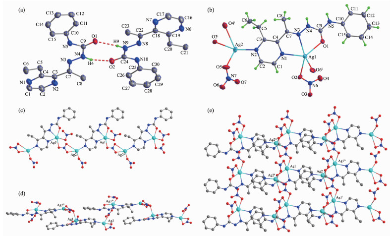
Selected bond distances and angles of HL and complex 1 are listed in Table 2.As shown in Fig. 1a, there are two independent molecules in the asymmetry unit of HL, in which the bond lengths of C9-O1 (0.122 6(3) nm) and C24-O2 (0.122 6(3) nm) are comparable to those of some reported semicarbazone ligands[18].Pairs of intermolecular N-H…O hydrogen bonds link two molecules into a centrosymmetry dimer in the solid state (N4-H4…O2, with D…A distance being 0.286 9(3) nm, D-H…A angle being 162.0°; N9-H9…O1, with D…A distance being 0.294 0(3) nm, D-H…A angle being 162.0°).
表 2
表 2
Selected bond lengths (nm) and angles (°) of HL and complex 1
Table 2.
Selected bond lengths (nm) and angles (°) of HL and complex 1
As illustrated in Fig. 1b, the asymmetry unit of complex 1 contains two independent Ag(Ⅰ) ions.One of them is surrounded by one HL ligand with ON2 donor set, two O atoms from two nitrate anions, giving a distorted square pyramid coordination geometry; while its counterpart with a planar square coordination geometry is coordinated with three O atoms from two adjacent nitrate anions and terminal pyrazine N4 atom of HL.It is worth noting that O4 and O6 atoms also participate coordination, since the bond lengths of Ag1-O6 (0.266 7(3) nm) and Ag2-O4 (0.279 9(4) nm) are within the scope of normal inspection and matches with the Ag-O bond in some Ag(Ⅰ) complexes in the literature[19].The neighboring Ag(Ⅰ) ions are linked by two types of nitrate ions into a 1D chain along a axis (in the case of N7/O5-O7, Fig. 1c), and a 1D zig-zag chain along c axis (in the case of N6/O2-O4, Fig. 1d), respectively, thus giving a 2D framework paralleling to (101) plane (Fig. 1e).In the crystal, intermolecular N-H…O hydrogen bonds between the semicarbazone ligands and nitrate anions are also presented (N5-H5…O7ⅳ, with D…A distance being 0.289 5(5) nm, D-H…A angle being 162.0°; N4-H4…O6ⅳ, with D…A distance being 0.307 1(5) nm, D-H…A angle being 165.0°, Symmetry codes:ⅳx, y, z+1).
图 1
图 1
Diamond drawing of HL (a) and 1 (b) with 30% thermal ellipsoids; (c) Chain-like structure along a axis in complex 1;
(d) Zig-zag chain-like structure along c axis in complex 1; (e) Extended 2D framework structure in complex 1
Figure 1.
Diamond drawing of HL (a) and 1 (b) with 30% thermal ellipsoids; (c) Chain-like structure along a axis in complex 1;
(d) Zig-zag chain-like structure along c axis in complex 1; (e) Extended 2D framework structure in complex 1
The infrared spectral bands most useful for deter-mining the mode of coordination of the semicarbazone ligand are the ν(C=O), ν(C=N), and ν(C=N)pyrizine vibrations.As our previous work shows, such three bands of the ligand HL are at 1 705, 1 609 and 1 595 cm-1[10], while they shift to 1 685, 1 600 and 1 537 cm-1 in the complex 1, respectively, indicating that the carbonyl O, imine N and pyrizine N atoms take part in the coordination[8].In addition, the intense absorption bands in the spectra of complex 1 associated with the asymmetric stretching appear at 1 382, 1 311 cm-1 (ν4) and 1 498 cm-1 (ν1), clearly establishing the existence of monodentate and bidentate NO3- ligands[8, 10, 20].It is in accordance with the crystal structure study.
2.3
UV spectra
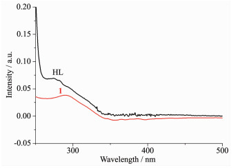
The UV spectra of complex 1 in methanol solution (concentration:10 μmol·L-1) were measured at room temperature (Fig. 2).The spectra of HL features one main band located around 275 nm (ε=6 929 L·moL-1·cm-1) and a shoulder at 282 nm (ε=6 567 L·moL-1· cm-1), which could be assigned to characteristic π-π* transition of benzene and pyrazine units, respectively[10].In complex 1, such two peaks are merged to 291 nm (ε=3 813 L·moL-1·cm-1) with concomitant hypochromic effect, confirming the coordination of ligand HL in complex 1.
图 2
图 2
UV spectra of the ligand HL and complex 1 in the methanol solution at room temperature
Figure 2.
UV spectra of the ligand HL and complex 1 in the methanol solution at room temperature
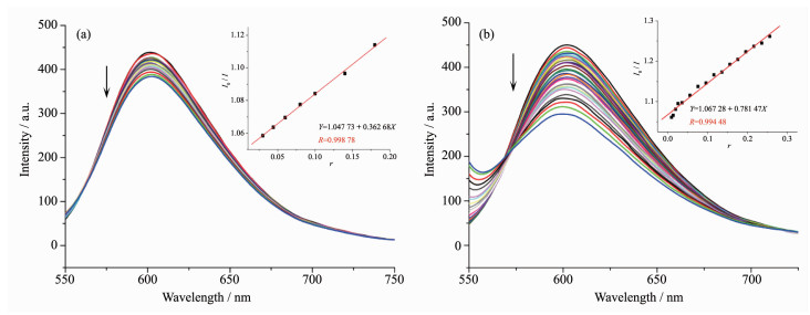
It is well known that EB can intercalate nonspe-cifically into DNA, which causes it to fluoresce strongly.Competitive binding of other drugs to DNA and EB will result in displacement of bound EB and a decrease in the fluorescence intensity[9].As shown in Fig. 3, the fluorescence intensities of EB bound to ct-DNA at about 600 nm show remarkable decreasing trends with the increasing concentration of the tested samples, indicating that some EB molecules are exchanged by the tested compounds.The quenching of EB bound to DNA by the compounds is in agreement with the linear Stern-Volmer equation:I0/I=1+Ksqr[8], where I0 and I represent the fluorescence intensities in the absence and presence of quencher, respectively, Ksq is the linear Stern-Volmer quenching constant, r is the ratio of the concentration of quencher and DNA.In the quenching plots of I0/I versus r, Ksq values are given by the slopes.The Ksq values of complex 1 is tested to be 0.781, which is much higher than that of the ligand HL (0.363).The results indicate that inter-actions of the complex 1 with DNA are stronger than that of the ligand HL.This is probably due to the structure rigidity and metal-ligand synergism effect of the complex 1[8-9].In addition, 2D framework structure of the complex 1 may be also responsible for its DNA interaction ability in some content.
图 3
图 3
Emission spectra of EB-DNA system in the presence of HL (a) and complex 1 (b), respectively
Figure 3.
Emission spectra of EB-DNA system in the presence of HL (a) and complex 1 (b), respectively
Nakamoto K.Infrared and Raman Spectra of Inorganic and Coordination Compounds.4th Ed.New York:Wiley, 1986:257
Figure 1
Diamond drawing of HL (a) and 1 (b) with 30% thermal ellipsoids; (c) Chain-like structure along a axis in complex 1;
(d) Zig-zag chain-like structure along c axis in complex 1; (e) Extended 2D framework structure in complex 1
Symmetry codes:ⅰx+1, y, z; ⅱ-x,-y,-z+1;ⅲx-1, y, z; ⅳx, y, z+1 for 1




 下载:
下载:

 下载:
下载:



