Scheme 1.
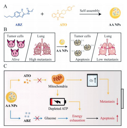
(A) The fabrication of AA NPs. (B) The effect of AA NPs to increase tumor cells apoptosis and reduce lung metastasis. (C) The mechanism of AA NPs to inhibit cellular ATP synthesis and alleviate hypoxia.
Dual inhibition of glucose uptake and energy supply synergistically restrains the growth and metastasis of breast cancer
Yuan Xu , Liling Huang , Yuyang Bi , Qi Song , Mengmeng Zhang , Lingfeng Zhang , Tianjiao Zhou , Lei Xing , Hulin Jiang

Metastatic breast cancer (MBC) has the highest mortality rate in breast cancer, which poses severe threats to female population [1, 2]. Recently, high viability and metastasis have been widely acknowledged as its main characteristics, as well as major obstacles of incurability [3, 4]. In clinical treatment, surgery is the first choice for MBC therapy, which usually combined adjuvant therapy to enhance curative efficiency, such as chemotherapy, radiotherapy, endocrine therapy and targeted therapy [5-7]. However, the mortality rate of MBC still remains high and new approaches urgently need to be explored.
Over the decades, many attempts have been made to eliminate cancer cells proliferation and migration, particularly, disturbing cancer cells metabolism has attracted broad attention [8-10]. As a metabolism disease, a large amount of glucose is required to provide "energy fuel" for cancer cells malignant growth and metastasis, which also makes great contributions to the synthesis of adenosine triphosphate (ATP) [11, 12]. Therefore, many strategies were conceived to combat cancer cells via the regulation of glucose, such as delivering glucose oxidase, glycolysis inhibitor and glucose analogue into tumor site [13-15], which initiated us the expectation of developing glucose uptake inhibitor for anti-tumor therapy.
Unfortunately, since cancer cells could compensatively utilize substances, such as lactate, glutamine and amino acid in microenvironment as substituted "energy fuels" to support cancer cells survival [16-18], only relying on curbing glucose uptake is inadequate to achieve clinical application. Hence, to augment the therapeutic efficiency, the energy supply of cancer cells needs to be suppressed more comprehensively. Although the energetic materials of cancer cells are extensive, mitochondrion is the main site that converted "energy fuels" into ATP through oxidative phosphorylation (OXPHOS) [19]. Therefore, the inhibition of OXPHOS could be expected to simultaneously block multiple energy supply and improve the efficacy of energy metabolism therapy. What is more, as the site of aerobic respiration, the restriction of OXPHOS could also help to elevate cellular oxygen content, which is beneficial for alleviating tumor hypoxic environment and inhibiting metastasis [20, 21]. Therefore, we hypothesize that dual inhibition of glucose uptake and OXPHOS might play synergetic effects on restraining cancer cells proliferation and provide an ingenious method to diminish metastasis of breast cancer.
In this study, we constructed a simple carrier-free self-assembled nanosystem (AA NPs) consisted of albendazole (ABZ) and atovaquone (ATO) to inhibit glucose uptake and energy supply for restraining the growth and metastasis of breast cancer (Scheme 1A). ABZ could evidently decrease glucose uptake, thus reducing the main "energy fuel" of cancer cells [22]. In addition, as a mitochondrial OXPHOS blocker, ATO could concurrently block ATP synthesis and reduce endogenous oxygen consumption (Scheme 1C) [23, 24]. As a result, with the dual-regulation of ABZ and ATO, AA NPs exhibited synergistically intensified ATP depletion and caspase-3 expression effects both in vitro and in vivo, leading to greatly increased tumor cells apoptosis. Besides, the oxygen content assay demonstrated that AA NPs exerted outstanding oxygen elevation performance, which was conducive to obviously diminish the metastasis of lung (Scheme 1B). These results prompt us to believe that dual-inhibition of glucose uptake and energy supply could improve therapeutic efficiency of energy metabolism therapy, which also provides a handy and effective method for better treatment of MBC.
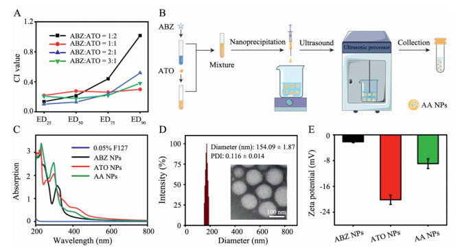
Through the investigation of combination index (CI) value, we found various ratios of ABZ and ATO had synergistic effect on 4T1 cells (ABZ/ATO = 1:1, 2:1, 3:1), demonstrating its potential for individualized therapy with different ratios of drugs. Then, the ABZ/ATO molar ratio of 1:1 was chosen as our final preparation because it exerted optimum efficiency at the 90% effective dose (ED90) (Fig. 1A). Based on that, AA NPs were fabricated by nanoprecipitation method without carrier (Fig. 1B) [25, 26]. The ultra violet (UV)-spectra (Fig. 1C) and Fourier transform infrared spectroscopy (FT-IR) spectra (Fig. S1 in Supporting information) revealed that the characteristic peaks of AA NPs were synchronized with ABZ and ATO, confirming the successful fabrication of AA NPs [27]. According to the dynamic light scattering (DLS) measurements in Fig. 1D and Fig. S2 (Supporting information), the dynamic size of AA NPs was 154 ± 1.87 nm, which was smaller compared with ABZ NPs and ATO NPs, indicating that the combination of ABZ and ATO could compress nanoparticles (NPs). Moreover, transmission electron microscope (TEM) image revealed that AA NPs exhibited monodisperse homogeneous spherical structure. Then, the zeta potentials of ABZ NPs, ATO NPs and AA NPs were −2.15, −20.15 and −8.94 mV, respectively, meaning that they would not cause hematolysis (Fig. 1E) [28]. In addition, ABZ NPs, ATO NPs and AA NPs could maintain stable size in saline, PBS and DMEM at 4 ℃ for 7 days, implying they could achieve long-term stability (Fig. S3 in Supporting information). Then, we prepared uniform NPs with different feed molar ratios of ABZ and ATO (Fig. S4 in Supporting information) and evaluated drug encapsulation efficiency (DEE) by high-performance liquid chromatography (HPLC) (Fig. S5 in Supporting information). The results showed that AA NPs possessed an ideal DEE (> 98%), which was higher than that of ABZ NPs and ATO NPs, proving the advantage of this self-assembled system over ABZ and ATO (Table S1 in Supporting information). Then, to explore the formation mechanism of NPs, various concentrations of NaCl, Triton X-100, SDS-Na, Urea and Tween-20 were added and mixed with NPs, respectively. According to the changed diameters of Triton X-100, SDS-Na and Tween-20 mixed NPs, we inferred that the main force to keep NPs stable was hydrophobic force (Fig. S6 in Supporting information) [29].
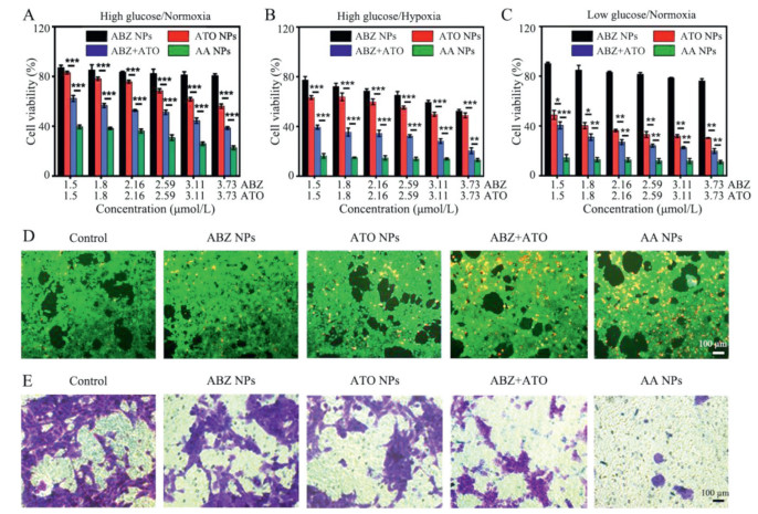
Afterwards, we detected cell viability of 4T1 and MCF-7 cells after treated with different formulations at 24 and 48 h, respectively. As shown in Fig. 2A, Figs. S7 and S8 (Supporting information), the therapeutic efficiency of AA NPs was better than ABZ NPs and ATO NPs, meaning that ABZ and ATO could possess synergistic antitumor effects. In addition, AA NPs performed superior therapeutic effects than ABZ+ATO (free ABZ and ATO mixture) with time dependent manner, which exhibited great potential in cancer therapy. Furthermore, we explored cell viabilities of 4T1 cells in hypoxia and glucose deficient environment, respectively. Hypoxic condition inhibits OXPHOS progression, and low-glucose environment means the role of glycolysis is limited. As shown in Figs. 2B and C, hypoxia environment could elevate the cytotoxicity of ABZ and low-glucose environment would increase the cytotoxicity of ATO, signifying that dual-inhibition of glucose uptake and OXPHOS could improve the efficacy of energy metabolism on anti-tumor. Besides, we also found that the cytotoxicity of ABZ+ATO and AA NPs in hypoxia or low-glucose environment was enhanced than in normoxia and high-glucose environment, demonstrating that intensified energy suppression could further facilitate energy metabolism therapy. Besides, the calculated half maximal inhibitory concentration (IC50) value and Calcein-AM/PI staining were used to further illustrate the desired therapeutic effect of AA NPs (Table S2 in Supporting information and Fig. 2D). Afterwards, we observed the anti-metastasis capability of AA NPs through transwell migration assay [30]. As shown in Fig. 2E and Fig. S9 (Supporting information), compared with ABZ NPs or ATO NPs treated cells, there were fewer 4T1 cells traversed polycarbonate membrane of transwell after AA NPs treatment, inferring that ABZ and ATO played synergistic anti-metastasis effect in vitro and AA NPs could have positive effect on MBC therapy. The same conclusion could also be obtained from the result of wound healing assay in Fig. S10 (Supporting information). These results not only declare that AA NPs have excellent anti-tumor and anti-metastasis efficiency in vitro, but also demonstrate the necessity of dual-regulation in energy metabolism therapy.
Furthermore, we elucidated the therapeutic mechanism of AA NPs. As a glucose uptake inhibitor, ABZ could promote the deficiency of energy. Simultaneously, as an OXPHOS inhibitor, ATO could curb endogenous ATP generation and elevate cellular oxygen content during treatment (Fig. 3A) [24]. To demonstrate the effect of AA NPs to inhibit ATP synthesis and alleviate hypoxia, a series of experiments were conducted. As shown in Fig. 3B, glucose content was greatly decreased after ABZ NPs treatment, demonstrating the strong glucose uptake inhibition ability of ABZ. In addition, ATP content assay was exhibited to evaluate energy exhaustion effect of AA NPs. Expectedly, the synthesis of ATP was eliminated after treated with ABZ or ATO, and their combination displayed favorable synergistic action, implying AA NPs could efficiently reduce cellular energy supply through the synergetic effect of ABZ and ATO (Fig. 3C). After that, we investigated the impact of AA NPs on improving oxygen content. As revealed in Fig. 3D, the dissolved oxygen content was tremendously supplemented under the treatment of ATO NPs, ABZ+ATO and AA NPs, demonstrating the effect of ATO to reduce oxygen consumption. In contrast, when treated with ABZ NPs, the oxygen content was evidently diminished, presumably because down-regulated glycolysis amplified OXPHOS and intensified hypoxia, emphasizing the necessity of dual-regulation on energy metabolism [31].

As vital organelles to modulate endogenous ATP synthesis and oxygen consumption, the structure and function of mitochondria were further studied. First, we detected the expression of mitochondrial-related apoptotic protein caspase-3 and results showed that both ABZ NPs and ATO NPs could promote its expression, especially ABZ+ATO and AA NPs groups, indicating that the depletion of cellular energy could facilitate tumor cells apoptosis (Fig. 3E). In addition, mitochondrial membrane potential (MMP) was further investigated by JC-1 dye to evaluate the damage of mitochondria [32]. As shown in Fig. 3F, MMP was decreased after being treated with ABZ NPs and ATO NPs, which presumably because it has been reported that ABZ could increase cellular ROS level and ATO could inhibit OXPHOS [33, 34]. Therefore, both of them could lead to the damage of mitochondrial membrane. Besides, the combination of ABZ and ATO declined MMP more visibly, which implied that the mitochondrial membrane permeability was further increased and would be more conducive to activate mitochondrial apoptosis pathway [35]. Furthermore, the serious damage of mitochondria was investigated in a more intuitive manner by TEM images. As shown in Fig. 3G, the mitochondria of Control group were integrated, of which the inner boundary membranes appeared fenestrated laminar cristae and were invaginated into tubular periodically. However, after being treated with AA NPs, the mitochondria membrane was broken and the structure of cristae was disappeared, demonstrating the severe disruption of AA NPs to mitochondria and its potential for energy metabolism therapy.
Motivated by favorable experimental results in vitro, more assays were performed on 4T1 subcutaneous tumor-bearing mice via intratumoral injection with various formulations. The animal experiments were approved by the Ethics Committee of China Pharmaceutical University. The treatment process followed the protocol described in Fig. S11 (Supporting information). After treatments, the body weight of all groups had no obvious change, illustrating that all preparations possessed preferable safety (Fig. 4A). Consistent with results in vitro, both ABZ NPs and ATO NPs played anti-tumor activity and AA NPs exhibited an enhanced efficiency (Fig. 4B and Fig. S12 in Supporting information). As revealed in Fig. 4C, the tumor growth inhibition rates with each treatment were 26.2% (ABZ NPs), 46.4% (ATO NPs), 49.7% (ABZ+ATO) and 67.7% (AA NPs), respectively. In addition, as shown in Figs. 4D and E, compared with Control group, the anti-metastasis effect was elevated after treating with ABZ NPs or ATO NPs, meaning that both ABZ and ATO could diminish lung metastasis in vivo. Specially, AA NPs further lessened lung nodules under the combination effect of ABZ and ATO, showing its superior therapeutic efficacy for MBC therapy. Furthermore, to uncover the anti-tumor and anti-metastasis mechanisms generated by AA NPs, immunofluorescence analysis of TUNEL (apoptosis related), Ki-67 (proliferation related) and E-cadherin (metastasis related) were conducted on harvested tumor sections. As illustrated in Fig. S13 (Supporting information), TUNEL positive and E-cadherin positive cells were more easily observed in treated groups, especially in AA NPs treated tumor. In addition, Ki-67 positive cells were thimbleful detected after treated with AA NPs. These results implied that AA NPs had pro-apoptosis, anti-proliferation and anti-metastasis capacities to MBC. Then, to assess the safety of the treatments in vivo, we evaluated the morphology of major organs via H & E staining and detected serum biochemical indexes of liver and kidneys. As displayed in Figs. S14 and S15 (Supporting information), the results showed that there had no apparent histological damage in major organs and the serological indices relevant to the kidney and liver functions had no notable variation, demonstrating its negligible systemic toxicity as expected.
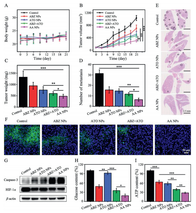
Then, in vivo hypoxia improvement effect was detected by Hypoxyprobe [36]. As shown in Fig. 4F, compared with Control group, the green fluorescence was decreased after treated with ATO, which proved the function of mitochondria to reduce endogenous oxygen consumption and alleviate hypoxia. Notably, the hypoxia environment was also alleviated after treating with ABZ NPs. To explore the mechanism, we detected the expression of hypoxia-inducible factor (HIF-1α). As shown in Fig. 4G and Fig. S16 (Supporting information), the expression of HIF-1α was obviously down-regulated after being treated with ABZ NPs, presumably because it has been reported that ABZ could reduce the expression of HIF-1α in hypoxia environment [37]. In addition, we also evaluated the expression of caspase-3, which was consistent with in vitro assay and demonstrated that AA NPs possessed preferable apoptosis promotion ability in vivo. Furthermore, Figs. 4H and I manifested the significantly reduced glucose and ATP content after being treated with AA NPs. Taken together, these results displayed that AA NPs exhibited satisfactory synergistic effect of exhausting energy and alleviating hypoxia in vivo, which further facilitated the efficacy on combating tumor cells proliferation and metastasis.
In this work, a simple carrier-free self-assembled nanosystem named AA NPs was rationally engineered to boost energy exhaustion and hypoxia alleviation efficiency via a handy preparation process with ABZ and ATO. Notably, the combination of ABZ and ATO could reduce the diameter of nanoparticles and enhance DEE, which was beneficial for anti-tumor therapy. After being ingested by tumor cells, the released ABZ would inhibit glucose uptake, thus reducing the main "energy fuel" of tumor cells. Meanwhile, the released ATO could concurrently restrict ATP production and alleviate hypoxic environment due to its destruction on mitochondria. Ultimately, AA NPs significantly increased tumor cells apoptosis by energy-deficiency and obviously diminished lung metastasis by hypoxia microenvironment alleviation, performing outstanding therapeutic effect on MBC therapy. Moreover, because ABZ and ATO have been approved by the U. S. Food and Drug Administration (FDA), which possess well-known safety and pharmacokinetic profiles of drugs, they have more economically attractiveness compared with de novo drug discovery and development. Overall, this study not only proposes a dual-regulation strategy of energy metabolism via simultaneously inhibition of glucose uptake and OXPHOS, but also puts forward a facile hypoxia improvement tactic and provides an excellent candidate for clinically therapeutic applications in MBC therapy.
The authors declare that they have no known competing financial interests or personal relationships that could have appeared to influence the work reported in this paper.
This work was financially supported by the National Key R&D Program of China (No. 2021YFE0198400) and the National Natural Science Foundation of China (Nos. 82020108029 and 82073398). This work was also supported by the Project of State Key Laboratory of Natural Medicines, China Pharmaceutical University (No. SKLNMZZ202021) and China Postdoctoral Science Foundation (No. 2021M703598). The authors also thank the Cellular and Molecular Biology Center of China Pharmaceutical University for assistance with confocal microscopy work.
Supplementary material associated with this article can be found, in the online version, at doi:
H.M. Fang, X.F. Zhao, X.L. Zhao, et al., Biomacromolecules 21 (2020) 104–113. doi: 10.1021/acs.biomac.9b01012
E.B. Rankin, A.J. Giaccia, Science 352 (2016) 175–180. doi: 10.1126/science.aaf4405
X.Q. Zhang, Y.M. Huang, H.L. Song, et al., J. Control. Release 328 (2020) 454–469. doi: 10.1016/j.jconrel.2020.08.066
T.J. Zhou, L. Xing, Y.T. Fan, et al., J. Control. Release 309 (2019) 82–93. doi: 10.1016/j.jconrel.2019.07.028
E.S. McDonald, A.S. Clark, J. Tchou, et al., J. Nucl. Med. 57 (2016) 9S–16S. doi: 10.2967/jnumed.115.157834
X. Wang, J. Veeraraghavan, C.C. Liu, et al., Clin. Cancer Res. 27 (2021) 2648–2662. doi: 10.1158/1078-0432.CCR-20-2961
S.B. Diego, P.G. José L, P.S. Mariano, et al., Cancer Discov. 11 (2021) 1353–1367. doi: 10.1158/2159-8290.CD-20-1312
X.Y. Chen, B.W. Li, X.H. Chen, et al., Chin. Chem. Lett. 31 (2020) 1153–1158. doi: 10.1016/j.cclet.2019.06.022
Y. Tian, W.L. Jiang, W.X. Wang, et al., Biomaterials 271 (2021) 120736. doi: 10.1016/j.biomaterials.2021.120736
F. Tian, S.Y. Wang, K. Shi, et al., Adv. Sci. 8 (2021) e2102595. doi: 10.1002/advs.202102595
R.H. Jin, Z.N. Liu, T. Liu, et al., Chin. Chem. Lett. 32 (2021) 3076–3082. doi: 10.1016/j.cclet.2021.03.084
M.G. Vander Heiden, L.C. Cantley, C.B. Thompson, et al., Science 324 (2009) 1029–1033. doi: 10.1126/science.1160809
Y.Y. Deng, P.Y. Song, X.H. Chen, et al., ACS Nano 14 (2020) 9711–9727. doi: 10.1021/acsnano.0c01350
Y.X. Guo, H.R. Jia, X.D. Zhang, et al., Small 16 (2020) e2000897. doi: 10.1002/smll.202000897
G.J. Leclerc, J.F. Du, J. DeSalvo, et al., Blood 21 (2012) 2439.
I. Martinez-Reyes, N.S. Chandel, Cell Metab. 26 (2017) 803–804. doi: 10.1016/j.cmet.2017.11.005
S. Sivanand, M.G. Vander Heiden, Cancer Cell 37 (2020) 147–156. doi: 10.1016/j.ccell.2019.12.011
S. Tohyama, J. Fujita, T. Hishiki, et al., Cell Metab. 23 (2016) 663–674. doi: 10.1016/j.cmet.2016.03.001
J.S. Brunner, L.W.S. Finley, Mol. Cell 81 (2021) 3738–3878e1.
L.P. Zuo, W.D. Nie, S.M. Yu, et al., Angew. Chem. Int. Ed. 60 (2021) 25365–25371. doi: 10.1002/anie.202109258
T.J. Zhou, L. Xing, Y.T. Fan, et al., J. Control. Release 307 (2019) 44–54. doi: 10.1016/j.jconrel.2019.06.016
M.C. Vinaud, C.S. Ferreira, S.L. Junior Rde, et al., Exp. Parasitol. 120 (2008) 221–226. doi: 10.1016/j.exppara.2008.07.008
H.Q. Zhu, B. Zhang, N.L. Zhu, et al., Chin. Chem. Lett. 32 (2021) 1220–1223. doi: 10.1016/j.cclet.2020.09.003
L.P. Zhao, R.R. Zheng, H.Q. Chen, et al., Nano Lett. 20 (2020) 2062–2071. doi: 10.1021/acs.nanolett.0c00047
H.Y. Kim, R. Li, T.S.C. Ng, et al., ACS Nano 12 (2018) 12015–12029. doi: 10.1021/acsnano.8b04338
D.R.B. Nora Graf, N. Kolishetti, C. Muus, et al., ACS Nano 5 (2012) 4530–4539.
L. Liang, Y. Peng, L. Qiu, et al., J. Control. Release 337 (2021) 117–131. doi: 10.1016/j.jconrel.2021.07.023
Y.T. Fan, T.J. Zhou, P.F. Cui, et al., Adv. Funct. Mater. 29 (2019) 1806708. doi: 10.1002/adfm.201806708
L. Xing, C.X. Yang, D. Zhao, et al., J. Control. Release 331 (2021) 460–471. doi: 10.1016/j.jconrel.2021.01.037
C. Wang, W.R. Liu, S. Tan, et al., Mol. Cancer 21 (2022) 63. doi: 10.1186/s12943-022-01546-4
M. Elgendy, M. Ciro, A. Hosseini, et al., Cancer Cell 35 (2019) 798–815. doi: 10.1016/j.ccell.2019.03.007
Y. Wang, L.F. Hu, P.F. Cui, et al., Adv. Mater. 33 (2021) 2103307. doi: 10.1002/adma.202103307
R. Martínez-Espinosa, R. Argüello-García, E. Saavedra, et al., Front. Microbiol. 6 (2015) 800.
M. Skwarski, D.R. McGowan, E. Belche, et al., Clin. Cancer Res. 27 (2021) 2459–2469. doi: 10.1158/1078-0432.CCR-20-4128
H. Singh, D. Sareen, J.M. George, et al., Coordin. Chem. Rev. 452 (2022) 214283. doi: 10.1016/j.ccr.2021.214283
T.J. Zhou, Y. Xu, L. Xing, et al., Adv. Mater. 33 (2021) e2100114. doi: 10.1002/adma.202100114
F. Zhou, J. Du, J.J. Wang, et al., Mol. Cell Biochem. 428 (2017) 171–178. doi: 10.1007/s11010-016-2927-3

Scheme 1 (A) The fabrication of AA NPs. (B) The effect of AA NPs to increase tumor cells apoptosis and reduce lung metastasis. (C) The mechanism of AA NPs to inhibit cellular ATP synthesis and alleviate hypoxia.

Figure 1 (A) Comparative analysis of CI values with different molar ratios of ABZ and ATO. (B) Preparation method of AA NPs. (C) UV spectra of 0.05% F 127, ABZ NPs, ATO NPs and AA NPs. (D) Size distribution and TEM image (inserted) of AA NPs (n = 3). (E) Zeta potentials of ABZ NPs, ATO NPs and AA NPs.

Figure 2 Synergistic anti-tumor activity against 4T1 cells in (A) high glucose/normoxia, (B) high glucose/hypoxia and (C) low glucose/normoxia (n = 3). (D) Calcein AM/PI double staining of 4T1 cells after different treatments. (E) Cell migration assays of 4T1 cells (n = 3, *P < 0.05, **P < 0.01, ***P < 0.001).

Figure 3 (A) Schematic illustration of the mechanism to exhaust energy and alleviate hypoxia by AA NPs. The content determination of (B) glucose and (C) ATP (n = 3, *P < 0.05, **P < 0.01, ***P < 0.001). (D) The dissolved oxygen content determination in media. (E) Western blot analysis of caspase-3. (F) MMP detection with JC-1 fluorescent probe. (G) TEM images of mitochondria before and after AA NPs treatment. The yellow arrows represent for the damaged mitochondrial membrane.

Figure 4 (A) Body weight changes, (B) tumor growth curves and (C) tumor inhibition rates of 4T1 tumor-bearing mice treated with different formulations (n = 5). (D) The numbers of lung metastasis nodules (n = 5). (E) Representative images of H & E staining in lung sections. The black circles represent for the metastatic area. (F) Immunofluorescence of tumor sections with Hypoxyprobe. (G) Relative expression of HIF-1α and caspase-3 in tumor tissue (n = 3). (H) Determination of glucose content in tumor tissue (n = 3). (I) Determination of ATP content in tumor tissue (n = 3). *P < 0.05, **P < 0.01, ***P < 0.001.

 扫一扫看文章

扫一扫关注我们
 DownLoad:
 下载:
下载: