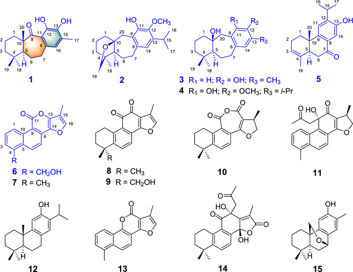
Figure 1.
Structures of 1–15.
Abietane derived diterpenoids as Cav3.1 antagonists from Salvia digitaloides
Jianjun Zhao , Shuzong Du , Kun Hu , Yali Hu , Fan Xia , Yansong Ye , Jian Yang , Yin Nian , Gang Xu

Salvia, one of the largest genera in the economically and medicinally important family Lamiaceae, is distributed worldwide and arises the folkloric belief of its “magical” therapeutic properties for various indispositions [1]. Investigations on Salvia plants have led to the discovery of a number of secondary metabolites, mainly diterpenoids and polyphenols [1-5]. Of these, abietanes and clerodanes have been identified as typical metabolites with diverse structures and significant bioactivities, such as salvicine (a significant antitumor agent), tanshinone IIA (a cardioprotective agent), salvinorin A (a potent agonist at the κ-opioid receptor), and neo-tanshinlactone (a significant and selective inhibitor against two ER human breast cancer cells) [1-5].
Our group has been investigating the diterpenoid constituents of Salvia since 2000, and has discovered many diterpenoids with attractive bioactivities [6-12]. Salvia digitaloides is an indispensable ingredient of a special traditional “red wine” by local Tibetans to strengthen their physical health [13]. Previously, twelve iridoids, four isoprenylated flavonoids, and fifteen abietane diterpenoids have been discovered from its roots [14-17]. Recently, several diterpenoids such as salpratlactones A and B, a pair of cis-trans tautomeric abietanes isolated from S. prattii, have been identified as the first Cav3.1 agonists [10]. In order to explore more diterpenoids with bioactivities on Cav3.1, the roots of S. digitaloides were phytochemically investigated in this study.
In our continuous investigation on S. digitaloides, another nineteen diterpenoids including five new ones, saldigitins A–E (1–3, 5, and 6) were isolated and identified (Fig. 1). Notably, saldigitin A (1) was elucidated to possess an unprecedented 10-methylated 6/7/6 carbon ring system. And the skeleton of 1 should be biogenetically derived through cleavage and re-cyclization of the B/C rings of normal abietane skeleton, which was distinct from the biopathway for icetexane type diterpenoids with a methyl-20-incorporated 6/7/6 carbon scaffold such as compounds 2–4 in this study. The structures of isolated compounds were assigned by extensive analysis of their spectroscopic data, and the absolute configuration of 1–3, and 5 were determined either by X-ray crystallography or theoretical calculations of their electronic circular dichroism (ECD) spectra. In addition, we initially disclosed that 1–5 notably inhibited the peak currents of Cav3.1 low voltage-gated Ca2+ channel (LVGCC), an attractive therapeutic target for neuropathic pain, absence epilepsy, insomnia, and Parkinson's tremors [18-20], in a dose-dependent manner with IC50 values of 7.08, 10.09, 11.70, 4.97, and 3.43 µmol/L, respectively. In addition, it is the first report of icetexane and related diterpenoids possessing 6/7/6 carbon rings system with antagonistic activities on Cav3.1. Reported herein are the isolation, structure characterization, proposed biogenetic pathways, and biological evaluation of these compounds.
Saldigitin A (1) was isolated as a colorless needle crystal and its molecular formula was determined as C19H26O3 by HRESIMS (m/z 301.1814 [M – H]–, calcd. for C19H25O3, 301.1809), including 7 degrees of unsaturation. The IR spectrum displayed bands for hydroxy (3472 cm−1) and carbonyl (1614 cm−1) groups. Besides, the 13C NMR spectrum presented 19 carbon signals ascribed to eight quaternary carbons (including one ketone and five olefinic), two methines (including an olefinic one), five methylenes, and four methyls. The three double bonds and one keto moiety accounted for four degrees of unsaturation, suggesting the tricyclic skeleton of 1. In addition, the characteristic signals for abietane type diterpenoid including three singlet methyls [δH 0.84, δC 33.2, Me-18; δH 0.93, δC 23.1, Me-19; and δH 1.28, δC 16.9, Me-20], five typical methylenes [δH 1.94 (m), 1.23 (m), δC 40.0, H2-1; δH 1.63 (m), 1.47 (m), δC 18.9, H2-2; δH 1.32 (m), 1.09 (m), δC 42.6, H2-3; δH 1.95 (m), 1.79 (m), δC 25.1, H2-6; δH 2.64 (m), 2.56 (m), δC 31.0, H2-7], and two aliphatic quaternary carbons [δC 34.3, C-4; δC 50.8, C-10] indicated that 1 should be an abietane derivative [1-12].
Normally, the chemical shifts of C-10 and Me-20 for an abietane diterpenoid were presented at δC 36–43 (s) and δC 22–28 (q), respectively, in the 13C NMR spectrum [21,22]. Whereas in 1, these two characteristic signals were replaced by two distinct signals at δC 50.8 (s) and 16.9 (q), respectively (Table 1). In addition, a carbonyl group at δC 219.0 (C-9) and the typical methylene (CH2-1) were presented simultaneously. These observations showed that the structure of 1 was quite different from those of common abietanes with ordinary 6/6/6-membered ring system.
 DownLoad:
CSV
DownLoad:
CSV

|
The HMBC correlations of Me-20 with C-9, and the mentioned carbonyl carbon at δC 219.0 led to the assignment of the ketone carbonyl group at C-9. The connection of C-5/C-6/C-7 was accomplished by the proton spin system of H-5/H2-6/H2-7 observed in the 1H-1H COSY spectrum. Furthermore, the HMBC correlations of H-5 with C-7, C-9, and C-10, H-7 and H-16 with C-8, C-9 and C-11 further established the linkage of C-5/C-10 (Me-20)/C-9/C-11/C-8/C-7, which evidently showed the unprecedented 10-methylated seven-membered ring B. The six-membered ring A was established by the HMBC associations of the geminal Me-18 and Me-19 with C-3, C-4, and C-5, of Me-20 with C-1, C-5, and C-10, conjugated with the proton spin system H2-1/H2-2/H2-3 (Fig. S1 in Supporting information). Similarly, the HMBC couplings from the singlet Me-17 (δH 2.22) to C-13, C-15, and C-16, from the unsaturated methine signal at δH 6.40 (1H, s, H-16) to C-11, C-13, C-17, C-8, and C-15, confirmed the existence of aromatic C ring. Thus, the 2D structure of 1 was established.
Assigning H-5 as α-oriented, the large coupling constant (3JH-5/H-6b = 12.2 Hz) indicated that H-6b (δH 1.79, m) adopted β-orientation [23]. The correlations of H3-18/H-5α, H3-19/H-6β, and H3-20/H3-19 observed in the ROESY spectrum revealed that Me-20 was β-oriented. Finally, a fine single crystal of 1 was obtained and the X-ray crystallographic data [Flack parameter = 0.01(6), CCDC: 2120506] further confirmed its absolute configuration as 5S, 10S (Fig. 2).

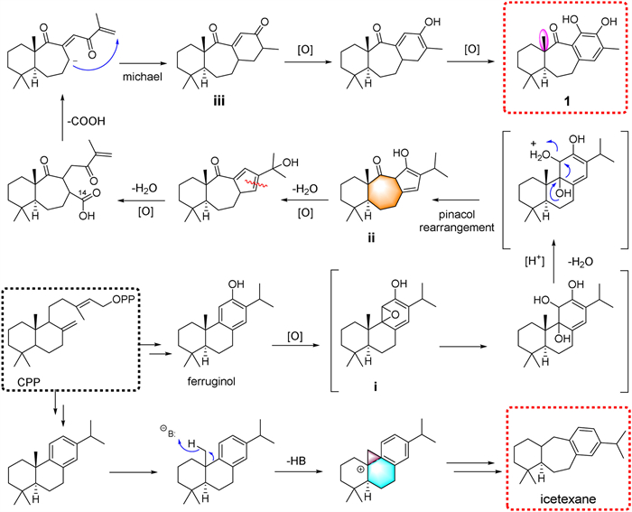
Biogenetically, compound 1 might rationally be generated from a common abietane precursor ferruginol (12) undergoing epoxidation to form 9,11-epoxide i [24]. A pinacol rearrangement occurs from i with acidic conditions lead to a B-homo-C-nor-abietane with a rearranged 6/7/5 ring system (ii) [11]. Meanwhile, the C-13/C-14 double bond further be oxidized to form two ketones (C-13 and C-14). Subsequently, decarboxylation of C-14 proceeded to form a carbanion (C-8), which attached the terminal olefinic link (CH2-16) via a Michael reaction to build up the six-membered ring C (iii), and then undergoes oxidation reaction to form the structure of 1 [10,25,26]. Biogenetically, icetexane diterpenoids, with a similar 6/7/6 skeleton, were believed to arise from a rearrangement of normal abietane and chemically belong to the 9(10→20) abeo-abietane framework with a 6/7/6-membered ring system [1,27,28]. Accordingly, 1 possesses a unique methyl at C-10 that derived from a distinct way of the cleavage of both rings B and C, and then re-cyclization to form the unprecedented 6/7/6 ring system which should be distinct from that for the normal icetexane (Scheme 1).
Saldigitin B (2) was obtained as a white powder, and gave rise to a [M + H]+ peak at m/z 331.1165 (calcd. 331.2268) in the positive-ion-mode HRESIMS, corresponding to the molecular formula C21H30O3. Detailed analysis of its 1D NMR data revealed that the signals for the characteristic aliphatic quaternary carbon [C-10 (δC 36–43)] and the methyl group [Me-20 (δC 22–28)] of normal abietanes were absent in 2 [25,26]. Besides, the observation that H-20a and H-20b appeared as an AB system at δH 3.08, 2.97 (1H each d, J = 14.3 Hz) further confirmed that 2 is an icetexane-type compound with a typical seven-membered ring B [25,26]. Structurally, 2 is quite similar to salvicanol (4) except for the replacement of a methyl with an oxygenated methylene in 2 [29,30]. The long-range HMBC correlations (Fig. S2 in Supporting information) from H2-19 (δH 3.71, 3.57, d, J = 7.9 Hz) to C-3, C-4 (δC 44.2), C-5 (δC 54.8), C-10 (δC 84.9), and C-18 (δC 19.3), and from H2-1 (δH 1.74, 1.54, dd, J = 12.7, 5.7 Hz) to C-5, C-10, and C-20 (δC 34.3) confirmed the presence of an ether bridge between C-10 and C-19. For biogenetic reasons, H-5 and Me-18 were expected to be cofacial and adopt an α-orientation as established by the ROESY correlations of H-7α/H-5/H3-18/H-2α. To determine the absolute configuration of saldigitin B (2), (4S,5S,10S)-2 was subjected to TDDFT ECD calculations at two independent levels of theory: CAM-B3LYP-SCRF/6-311+G(2d,p) (methanol, IEFPCM solvent model) and PBE0-SCRF/6-311+G(2d,p) (methanol, IEFPCM solvent model). At both levels of theory, the calculated ECD spectra matched well with their experimental counterpart (Fig. S4 in Supporting information). Consequently, the structure of saldigitin B (2) was assigned and its absolute configuration was determined as 4S, 5S, and 10S.
Saldigitin C (3), isolated as a white amorphous powder, and showed a molecular ion at m/z 273.1866 [M – H]– in the HRESIMS (calcd. 273.1860), which correlated to the molecular formula C18H26O2. Comparison of its 1D NMR spectroscopic data (Table 1) with those of pisiferanol indicated they were similar to each other except that the isopropyl group at C-13 in pisiferanol was replaced by a methyl group in 3 [28]. This deduction was confirmed by the HMBC correlations from Me-15 (δH 2.03, s) to C-12 (δC 152.7), C-13 (δC 120.2), and C-14 (δC 130.1) [28]. As in the case of 2, Me-19 of icetexane diterpenoid was expected to be β-oriented, while H-5 and Me-18 were α-oriented. Furthermore, the ROESY cross-peaks of HO-10/H3-19, H-1a, H-2b, H-20b, and H-7a confirmed that they were spatially close and can be assigned as β-oriented. Consequently, the correlations of H-5/H3-18, H-7b, H-20a, and H-1b indicated that they were cofacial and adopt an α-orientation. The absolute configuration of saldigitin C (3) was also established by TDDFT ECD calculations. At both levels of theory, the calculated ECD spectra of (5S,10S)-3 were found to be consistent with the experimental spectrum (Fig. S4). Accordingly, the absolute configuration of 3 was determined as 5S and 10S.
Saldigitin D (5) was isolated as a white needle crystal and was assigned the molecular formula C19H24O2, based on HRESIMS (m/z 285.1844 [M + H]+, calcd. C19H25O2, 285.1849). Analysis of its 1D and 2D NMR data revealed that 5 possessed similar scaffold to that of 7-keto-sempervirol (18) [31-33]. Their key difference was the presence of a trisubstituted olefin at the C-3 and C-4 in 5 instead of the saturated single bond in 7-keto-sempervirol. This elucidation was confirmed by the HMBC correlations from Me-18 (δH 1.66, s) to C-3, C-4, and C-5, from H-5 (δH 2.77, br d, J = 14.6 Hz) to C-3, C-4, and C-18, and the 1H-1H COSY spin system H2-1/H2-2/H-3. In addition, the location of the isopropyl moiety at C-12 was supported by the HMBC correlations from Me-16 and Me-17 to C-12. Furthermore, a single-crystal X-ray analysis (CCDC: 2120507) successfully confirmed the established structure (Fig. 2 and Fig. S3 in Supporting information), however, the Flack parameter (–0.35(8)) was insufficient to unambiguously determine its absolute configuration. Then, TDDFT ECD calculations were run on (5S,10S)-5 and revealed the absolute configuration of 5 to be 5S and 10S (Fig. S4). So, the structure of 5 was established and named saldigitin D.
Saldigitin E (6) was obtained as a pink powder. Its molecular formula was established as C17H12O4 by 13C NMR and HRESIMS (m/z 281.0812 [M + H]+, calcd. C17H13O4, 281.0808). The 1D NMR data of 6 were similar to those of tanshinlactone (7) except for the presence of an additional hydroxymethyl group at δH 5.00 (d, J = 5.1 Hz, H2-18) in 6 instead of the Me-18 in tanshinlactone (7) [34]. This deduction was supported by HMBC correlations from H2-18 to C-3 (δC 126.6), C-4 (δC 138.7), and C-5 (δC 131.5), from H-3 (δH 7.75, d, J = 7.1 Hz) to C-18 (δC 61.2) (Fig. S3). Thus, 6 was assigned as 18-hydroxy-tanshinelactone and named saldigitin E.
In addition, salvicanol (4) [29,30], tanshinlactone (7) [34], tanshinone IIA (8) [24], tanshinone IIB (9) [35], cryptotanshinone anhydride (10) [36], danshenol A (11) [37], ferruginol (12) [24], neo-tanshinlactone (13) [38], salviprzol A (14) [39], przewalskin (15) [36], 15,16-dihydrotanshinone I (16) [24], 16,17-dinorpisferal A (17) [14], 7-keto-sempervirol (18) [31], and15-epi-danshenol A (19) [40], were carefully identified by comparison of their spectroscopic data with literature values, and their putative biogenetic relationships are shown in Scheme S1 (Supporting information).
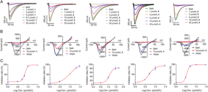
As stated in the introduction, agents with Cav3.1 LVGCC regulatory effects may have promising potential to treat several neurological disorders [18-20]. In the present study, we firstly revealed that four new compounds, saldigitins A–D (1–3, and 5), as well as the known one, salvicanol (4), dose-relatedly blocked Cav3.1 peak currents among their testing concentration range (Fig. 3 and Table S1 in Supporting information). The IC50 values of 1–5 on Cav3.1 are 7.08, 10.09, 11.70, 4.97, and 3.43 µmol/L, respectively. Mibefradil, the positive control, showed stronger activity than 1–5, with an IC50 value of 1.12 µmol/L (Fig. S12 in Supporting information). As shown in Fig. 3, compared to 2, 4, and 5, the blockages of 1 and 3 were more difficult to wash out, suggesting these two ones may have deeper binding sites inside the channel. Saldigitin E (6) showed negligible effect on Cav3.1 at the concentration of 10 µmol/L (Fig. S11 in Supporting information). The structure–function relationships and action characteristics of those active compounds deserve to be elucidated in future.
To the best of our knowledge, compound 1 can be seen as the first diterpenoid featuring an unprecedented 10-methylated 6/7/6 carbon ring system. In comparison with normal icetexanes with a 10-unmethylated 6/7/6 carbon scaffold, 1 should be derived from a distinct biopathway from the normal abietane skeleton undergone the cleavage of rings B/C and then re-cyclization. The 6/7/6-tricyclic core structure of icetexanes were also constructed via natural rearrangement of more normal 6/6/6-fused abietane diterpenoids [1]. These diterpenoids have aroused great attentions from the synthetic communities for their interesting structures as well as significant bioactivities [41-46]. In the bioactive assay, we firstly revealed that four diterpenoids with 6/7/6 carbon skeleton, 1–4, and a 19-nor-15(13→12)-abeo-abietane (5) showed notable inhibitory effects on Cav3.1 LVGCC, which may have promising potential to treat several neurological disorders. In summary, about 70 icetexanes with the normal 6/7/6 skeleton have been identified and synthesized so far [8,41-44], the present study provides new perspectives for the structures and biological activities of the diterpenoids with 6/7/6 carbon ring system.
The authors declare that they have no known competing financial interests or personal relationships that could have appeared to influence the work reported in this paper.
This work was financially supported by the National Natural Science Foundation of China (Nos. 32070392 and 32070393), the Second Tibetan Plateau Scientific Expedition and Research (STEP) Program (Nos. 2019QZKK0502-0303 and 2019QZKK0502-0304), Natural Science Foundation of Yunnan Province (Nos. 202001AS070040 and 202101AV070010), Yunnan Young & Elite Talents Project (No. YNWR-QNBJ-2020-277), and CAS “Light of West China” Program (2021).
Supplementary material associated with this article can be found, in the online version, at doi:
Y.B. Wu, Z.Y. Ni, Q.W. Shi, et al., Chem. Rev. 112 (2012) 5967–6026. doi: 10.1021/cr200058f
Y.B. Dong, S.L. Morris-Natschke, K.H. Lee, Nat. Prod. Rep. 28 (2011) 529–542. doi: 10.1039/c0np00035c
M. Xue, Y.B. Shi, Y. Chui, et al., Nat. Prod. Res. Dev. 12 (2000) 27–32.
T.A. Munro, M.A. Rizzacasa, B.L. Roth, et al., J. Med. Chem. 48 (2005) 345–348. doi: 10.1021/jm049438q
M. Xiao, L. Wei, L. Li, et al., Org. Chem. 79 (2014) 2746–2750. doi: 10.1021/jo500047q
G. Xu, A.J. Hou, R.R. Wang, et al., Org. Lett. 8 (2006) 4453–4456. doi: 10.1021/ol061609t
G. Xu, A.J. Hou, Y.T. Zheng, et al., Org. Lett. 9 (2007) 291–293. doi: 10.1021/ol062748d
G. Xu, X.W. Yang, C.Y. Wu, et al., Chem. Commun. 48 (2012) 4438–4440. doi: 10.1039/c2cc30405h
C.Y. Wu, Y. Liao, Z.G. Yang, et al., Phytochemistry 106 (2014) 171–177. doi: 10.1016/j.phytochem.2014.07.001
F. Xia, W.Y. Li, X.W. Yang, et al., Org. Lett. 21 (2019) 5670–5674. doi: 10.1021/acs.orglett.9b01527
F. Xia, D.W. Zhang, C.Y. Wu, et al., Org. Chem. Front. 5 (2018) 1262. doi: 10.1039/C7QO01140G
G. Xu, L.Y. Peng, L. Lu, et al., Planta Med. 72 (2006) 84–86. doi: 10.1055/s-2005-873184
Kunming Institute of Botany, Chinese Academy of Sciences, Flora Yunnannica, Science Press, Beijing, 1977, p. 661.
G. Xu, J. Yang, Y.Y. Wang, et al., J. Agric. Food Chem. 58 (2010) 12157–12161. doi: 10.1021/jf103366g
S.J. Wu, H.H. Chan, T.L. Hwang, et al., Tetrahedron Lett. 51 (2010) 4287–4290. doi: 10.1016/j.tetlet.2010.06.048
S.J. Wu, Y.Y. Chan, Molecules 19 (2014) 15521–15534. doi: 10.3390/molecules191015521
J.J. Zhao, S.Y. Li, F. Xia, et al., Nat. Prod. Bioprospect. 11 (2021) 671–678. doi: 10.1007/s13659-021-00307-y
G.W. Zamponi, Nat Rev Drug Discov. 15 (2016) 19–34. doi: 10.1038/nrd.2015.5
E.J. Cheong, H.S. Shin, Physiol. Rev. 93 (2013) 961−992. doi: 10.1152/physrev.00010.2012
K.H. Choi, Expert Opin. Drug Dis. 8 (2013) 919−931. doi: 10.1517/17460441.2013.796926
A. Ulubelen, S. Öksüz, G. Topcu, et al., J. Nat. Prod. 64 (2001) 549–551. doi: 10.1021/np0004956
M. Tada, T. Hara, C. Hara, et al., Phytochemistry 45 (1997) 1474–1477.
Y.Y. Fan, L.S. Gan, H.C. Liu, et al., Org. Lett. 19 (2017) 4580–4580. doi: 10.1021/acs.orglett.7b02181
S.Y. Lee, D.Y. Choi, E.R. Woo, Arch. Pharm. Res. 28 (2005) 909–913. doi: 10.1007/BF02973876
T.D.H. Bugg, Tetrahedron 59 (2003) 7075−7101. doi: 10.1016/S0040-4020(03)00944-X
O. Hayaishi, M. Katagiri, S. Rothberg, J. Am. Chem. Soc. 77 (1955) 5450–5451. doi: 10.1021/ja01625a095
E.M. Simmons, J.R. Yen, R. Sarpong, Org. Lett. 14 (2007) 2705–2708. doi: 10.1021/ol0712428
G. Xu, L.Y. Peng, Y. Zhao, et al., Chem. Pharm. Bull. 53 (2005) 1575–1576. doi: 10.1248/cpb.53.1575
B.M. Fraga, A.G. Gonzalez, J.R. Herrera, et al., Phytochemistry 25 (1985) 269–271. doi: 10.1016/S0031-9422(00)94548-5
M. Bruno, G. Savona, F. Piozzi, et al., Phytochemistry 30 (1991) 2339–2343. doi: 10.1016/0031-9422(91)83645-2
L. Mangoni, R. Caputo, Tetrahedron Lett. 8 (1967) 673–675. doi: 10.1016/S0040-4039(00)90571-9
C. Bustos-Brito, P. Joseph-Nathan, E. Burgueño-Tapia, et al., J. Nat. Prod. 82 (2019) 1207–1216. doi: 10.1021/acs.jnatprod.8b00952
N.M. Tam, L.T. Hieu, N.M. Thong, et al., Chem. Phys. Lett. 777 (2021) 138737. doi: 10.1016/j.cplett.2021.138737
H.W. Luo, J. Ji, M.Y. Wu, et al., Chem. Pharm. Bull. 34 (1986) 3166–3168. doi: 10.1248/cpb.34.3166
X.Y. Yu, S.G. Lin, Z.W. Zhou, et al., Neurosci. Lett. 417 (2007) 261–265. doi: 10.1016/j.neulet.2007.02.079
B. Li, F.D. Niu, Z.W. Lin, et al., Phytochemistry 30 (1991) 3815–3817. doi: 10.1016/0031-9422(91)80121-G
G. Nagy, G. Gunther, I. Mathe, et al., Biochem. Syst. Ecol. 26 (1998) 797–799. doi: 10.1016/S0305-1978(98)00043-X
X.H. Wang, K.F. Bastow, C.M. Sun, et al., J. Med. Chem. 47 (2004) 5816–5819. doi: 10.1021/jm040112r
Y.B. Xue, Y. Wu, H.C. Zhu, et al., Fitoterapia 99 (2014) 204–210. doi: 10.1016/j.fitote.2014.09.022
R. Kasimu, P. Basnet, Y. Tezuka, et al., Chem. Pharm. Bull. 45 (1997) 564–566. doi: 10.1248/cpb.45.564
W. Cao, T.T. Liu, S.T. Yang, et al., J. Nat. Prod. 84 (2021) 2012–2019. doi: 10.1021/acs.jnatprod.1c00310
A. Hymavathi, K. Suresh Babu, V.G.M. Naidu, et al., Bioog. Med. Chem. Lett. 19 (2009) 5727–5731. doi: 10.1016/j.bmcl.2009.08.002
G.J. Zheng, A.K. Kadir, X.F. Zheng, et al. Org. Chem. Front. 7 (2020) 3137–3145. doi: 10.1039/d0qo00735h
D.L. Chem, X.Y. Liu, H. Cheng, et al. Chin. Chem. Lett. 22 (2011) 774–776. doi: 10.1016/j.cclet.2011.01.009
Q.T. Le, L.H. Guo, S.L. Lee, et al., Org. Lett. 22 (2020) 9225–9228. doi: 10.1021/acs.orglett.0c03415
J. Zhang, Y.H. Jin, F.Y.G. Qiu, Org. Lett. 22 (2020) 7415–7418. doi: 10.1021/acs.orglett.0c02309

Figure 3 Inhibitory effect of compounds 1–5 on Cav3.1. (A) Representative Cav3.1 peak current traces elicited by 150 ms depolarization to −30 mV at 4 s intervals from a holding potential (HP) of −100 mV in the absence (control) and presence of different concentrations of 1–5, respectively. (B) Current-voltage (I-V) curves of Cav3.1 in the absence or presence of 1–5 at 10 µmol/L. Cav3.1 currents were evoked from a holding HP of −100 mV by 150 ms depolarization from −80 mV to +60 mV in 10 mV increasement at 4 s intervals. (C) Dose-response relationships of 1–5 for Cav3.1 at HP of −100 mV, respectively. Data points represent mean ± SD of three to five measurements. The solid curve represents a fit to the Hill equation. The IC50 values of 1–5 are 7.08, 10.09, 11.70, 4.97, and 3.43 µmol/L, respectively.
Table 1. 1H (600 MHz) and 13C (150 MHz) NMR spectroscopic data for compounds 1–3, 5, and 6.
|
|
 下载: 导出CSV

 扫一扫看文章

扫一扫关注我们


